SlideShare ist ein Scribd-Unternehmen logo
1 von 1
Conclusions:
Our findings shed light on the interactions of NAGK with dynein and Golgi outposts in dendritic branch points and their roles in dendritic growth. Further investigation is needed into the
molecular mechanisms underlying these interactions.
Cultured rat hippocampal neurons were treated with dithiobis (succinimidyl) propionate
(DSP+), fixed at DIV10 (stage IV) and double-stained with primary antibodies against N-
acetylglucosamine kinase and dynein light-chain roadblock
type 1 (a) or dynein heavy chain (b) or kinesin 5B
(c). The boxed areas of the merged images are enlarged at
the bottom. Colocalized immunopuncta are
indicated by arrowheads. Scale bar: 10 μm. Statistics (d). The numbers
of puncta per dendritic segment (20 μm, n = 20) were counted and
expressed as a percentage of the total (mean±s.d.).**p<0.01.
(a) N-acetylglucosamine kinase-
interacting proteins identified by using the small domain as bait in
yeast two-hybrid screening.
(b) The interacting domains of dynein
light-chain roadblock type 1. The coding region (amino acids
1–96) of dynein light-chain roadblock
type 1 is shown as a bar diagram with the conserved roadblock/LC7
domain. The two positive clones in the yeast two-hybrid selection having a coding region
from 3 or 59 to the C-terminal end are also shown. Full length dynein light-chain roadblock
type 1 (1-end) and kinesin light chain 1 were used as a positive and a negative control,
respectively.
The small domain of NAGK interacts with DYNLRB1 in yeast two-hybrid screen
NAGK-dynein interactions occur at dendritic branch points
NAGK interacts with DYNLRB1 in primary hippocampal neurons
Verification of NAGK-dynein complex interaction by proximity ligation assay
NAGK-dynein complexes colocalize with Golgi ‘outposts’ at dendritic branch
points
N-acetyl-D-glucosamine kinase interacts with
dynein light-chain roadblock type 1 at Golgi
outposts in neuronal dendritic branch points
Md Ariful Islam1
, Syeda Ridita Sharif1
, HyunSook Lee2
, Dae-Hyun Seog3
and Il Soo Moon1,2
1
Department of Anatomy, Dongguk Medical Institute, College of Medicine Dongguk University, Gyeongju, Republic of Korea; 2
Dongguk Medical Institute, College of Medicine
Dongguk University, Gyeongju, Republic of Korea and 3
Departments of Biochemistry, College of Medicine Inje University, Busan, Republic of Korea
N-acetylglucosamine kinase (GlcNAc kinase or NAGK) is a ubiquitously expressed
enzyme in mammalian cells. Recent studies have shown that NAGK has an essential
structural, non-enzymatic role in the upregulation of dendritogenesis. In this study, we
conducted yeast two-hybrid screening to search for NAGK-binding proteins and found a
specific interaction between NAGK and dynein light-chain roadblock type 1 (DYNLRB1).
Immunocytochemistry (ICC) on hippocampal neurons using antibodies against NAGK and
DYNLRB1 or dynein heavy chain showed some colocalization, which was increased by
treating the live cells with a crosslinker. A proximity ligation assay (PLA) of NAGK-dynein
followed by tubulin ICC showed the localization of PLA signals on microtubule fibers at
dendritic branch points. NAGK-dynein PLA combined with ICC for Golgi showed the
colocalization of PLA signals with somal Golgi facing the apical dendrite and with Golgi
outposts in dendritic branch points and distensions. NAGK-Golgi PLA followed by tubulin
or DYNLRB1 ICC showed that PLA signals colocalize with DYNLRB1 at dendritic branch
points and at somal Golgi, indicating a tripartite interaction between NAGK, dynein and
Golgi. Finally, the ectopic introduction of a small peptide derived from the C-terminal amino
acids 74–96 of DYNLRB1 resulted in the stunting of hippocampal neuron dendrites in
culture. Our data indicate that the NAGK-dynein-Golgi tripartite interaction at dendritic
branch points functions to regulate dendritic growth and/or branching.
Abstract
Co-localization of NAGK-Golgi complexes with DYNLRB1 on MT at dendritic
branch points.
Ectopic inclusion of small peptides from DYNLRB1 resulted stunted dendrites
(b) HEK293T cells were fixed, and PLA was performed
using anti-NAGK and anti-dynein light-chain 1 antibodies
(NAGK+DYNLL1, upper panel). Alternatively, the cells
were transfected with Myc-DDK-tagged NAGK plasmids,
and PLA was conducted by using anti-DDK and anti-
DYNLL1 antibodies to show the interaction between
exogenous NAGK and dynein (DDK+DYNLL1, lower
panel). To better depict proximity ligation assay puncta,
phase-contrast images were inverted using Photoshop
software. Scale bar: 10 μm.
(a) Rat hippocampal neurons (DIV2) were fixed, and
proximity ligation assay was performed using mouse anti-
N-acetylglucosamine kinase and rabbit anti-dynein light-
chain roadblock type 1 or anti-dynein heavy chain
antibodies. The PLA was followed by
immunocytochemistry using a mouse anti-tubulin antibody
(green). Boxed areas are enlarged (insets) to show the
colocalization of the NAGK-dynein complex with thinner
microtubule fibers (arrowheads). Scale bar: 10 μm.
(a) N-acetylglucosamine kinase-dynein heavy chain
proximity ligation assay was performed in hippocampal
neurons (stage IV, DIV 2) and followed by tubulin
immunocytochemistry. Proximity ligation assay/phase-
contrast (phase) and proximity ligation assay/
immunocytochemistry merged images are shown. The
PLA puncta (red) are indicated by arrowheads and
with numbers. Scale bar: 10 μm.
(b) PLA puncta were sorted into two categories: one
with ‘established’ branch joints where the microtubule
fiber is already developed (upper panel) and the other
at branch initiation sites where the microtubule is yet to
develop (lower panel). Positions of branches are
indicated with arrowheads.
(c) Statistics. Percentages of NAGK-DHC proximity
ligation assay puncta at branch joints with or without
microtubule in the neonate protrusions are shown by a
pie chart. n = 30, 30 neurons.
N-acetylglucosamine kinase-dynein heavy chain
proximity ligation assay was performed in hippocampal
neurons (developmental stage IV) and was followed by
immunocytochemistry with an anti-TGN38 antibody to
mark Golgi particles. Merged images of proximity
ligation assay/ phase-contrast and proximity ligation
assay/ immunocytochemistry are shown.
(a) PLA signals at soma (1, arrows) and distal branch
points (2, 3, arrowheads) are enlarged to show the
colocalization of NAGK-dynein complex (red) with
Golgi particles (green) oriented to the soma-dendrite
joint (inset 1, arrows) or with the Golgi outpost (green)
at distal branching sites (inset 2, 3, arrowheads).
(b) A high frequency of PLA puncta (red) and Golgi outposts (green) was found at dendritic
distensions. Small branches protruding out of the distensions are marked with arrows.
Dendritic distensions (boxed areas 1, 2) are enlarged (insets) to show the colocalization of
NAGK-dynein complex (red) with Golgi particles (green) marked by arrowheads. Scale bar:
10 μm.
(a) NAGK-TGN38 proximity ligation assay (red dots)
was followed by ICC with an anti-tubulin antibody
(green). The positions of proximity ligation assay
signals at branch points are marked with numbers and
enlarged in insets. Scale bar: 10 μm.
(b) NAGK-GM130 PLA was followed by ICC with an
anti-DYNLRB1 antibody. PLA puncta in the soma (box
i) were enlarged to show the PLA signals facing the
apical dendrite at the somal Golgi apparatus merged
with DYNLRB1 signals. PLA dot positions (red) in
dendrites are enlarged (box ii, 1–3). Scale bar: 10 μm.
(c) Statistics. Pie charts show that ~99% (n = 30 neurons) of NAGK-TGN38 PLA dots were
localized to microtubules and ~ 96% (n = 30 neurons) of NAGK-GM130 PLA signals were
colocalized with DYNLRB1.
Two small peptides are made from DYNLRB1 and named
as ‘DYNLRB1 (59-76)’ and ‘DYNLRB1 (74-96)’ .
(a) Co-transfection of either ‘DYNLRB1 (59-76)’ or
‘DYNLRB1 (74-96)’ peptide with β-galactosidase was
performed and was stained with β-gal staining kit. Neurons
transfected with ‘DYNLRB1 (59-76)’ peptide showed
healthy dendrites. In contrast, neurons transfected with
‘DYNLRB1 (74-96)’ peptide showed short, stunted
dendrites (arrows), while the axon (arrowhead) was
apparently unaffected. Scale bar: 10 μm.
(b) Sholl analysis. The numbers of dendritic intersections were counted at incremental
distances from soma centroids of neurons (n = 30 neurons) transfected with β-gal only
(control), with ‘DYNLRB1 (59-76)’ or with ‘DYNLRB1 (74-96)’ . **p<0.01.
Results

Weitere ähnliche Inhalte

Was ist angesagt?

FINAL poster ORD
FINAL poster ORDFINAL poster ORD
FINAL poster ORDJoe Cameron
 
UTF-8''Final Assessing post-synaptic partners of Dentate Granule Cells in a M...
UTF-8''Final Assessing post-synaptic partners of Dentate Granule Cells in a M...UTF-8''Final Assessing post-synaptic partners of Dentate Granule Cells in a M...
UTF-8''Final Assessing post-synaptic partners of Dentate Granule Cells in a M...Grant Pizzo
 
Hogarth_Cabrales_Strome Research Poster KAS 2014 Rev3 NCH
Hogarth_Cabrales_Strome Research Poster KAS 2014 Rev3 NCHHogarth_Cabrales_Strome Research Poster KAS 2014 Rev3 NCH
Hogarth_Cabrales_Strome Research Poster KAS 2014 Rev3 NCHNathan Hogarth
 
Genome editing & targeting tools
Genome editing & targeting toolsGenome editing & targeting tools
Genome editing & targeting toolsS Rasouli
 
Ectopic Expression of Hawthorn SND1 Gene in Tobacco
Ectopic Expression of Hawthorn SND1 Gene in TobaccoEctopic Expression of Hawthorn SND1 Gene in Tobacco
Ectopic Expression of Hawthorn SND1 Gene in TobaccoAgriculture Journal IJOEAR
 
Animal bt group presentation (1)
Animal bt group presentation (1)Animal bt group presentation (1)
Animal bt group presentation (1)NatashaLeong1
 
Efficient transformation of lactococcus lactis il1403 and generation of knock...
Efficient transformation of lactococcus lactis il1403 and generation of knock...Efficient transformation of lactococcus lactis il1403 and generation of knock...
Efficient transformation of lactococcus lactis il1403 and generation of knock...CAS0609
 
Preimplantation genetic diagnosis_of_marfan_syndrome_using_multiple_displacem...
Preimplantation genetic diagnosis_of_marfan_syndrome_using_multiple_displacem...Preimplantation genetic diagnosis_of_marfan_syndrome_using_multiple_displacem...
Preimplantation genetic diagnosis_of_marfan_syndrome_using_multiple_displacem...t7260678
 
Crispr guides the_future_of_genetic_engineering[1]
Crispr guides the_future_of_genetic_engineering[1]Crispr guides the_future_of_genetic_engineering[1]
Crispr guides the_future_of_genetic_engineering[1]AshishDutta33
 
Gene 151_119 (1994) [SDM of dsDNA]
Gene 151_119 (1994) [SDM of dsDNA]Gene 151_119 (1994) [SDM of dsDNA]
Gene 151_119 (1994) [SDM of dsDNA]Michael Weiner
 
SHSARP paper final
SHSARP paper finalSHSARP paper final
SHSARP paper finalKaylee Racs
 
Characterization in Dvilp 7 gene
Characterization in Dvilp 7 geneCharacterization in Dvilp 7 gene
Characterization in Dvilp 7 geneHunter Kelley
 
Pathema Burkholderia Annotation Jamboree: Prokaryotic Annotation Overview
Pathema Burkholderia Annotation Jamboree: Prokaryotic Annotation OverviewPathema Burkholderia Annotation Jamboree: Prokaryotic Annotation Overview
Pathema Burkholderia Annotation Jamboree: Prokaryotic Annotation OverviewPathema
 
CRISPR – a novel tool for genome editing
CRISPR – a novel tool for genome editingCRISPR – a novel tool for genome editing
CRISPR – a novel tool for genome editingVigneshVikki10
 
Gene editing 1
Gene editing 1Gene editing 1
Gene editing 1ajayveeru
 

Was ist angesagt? (20)

IGEM poster
IGEM posterIGEM poster
IGEM poster
 
FINAL poster ORD
FINAL poster ORDFINAL poster ORD
FINAL poster ORD
 
UTF-8''Final Assessing post-synaptic partners of Dentate Granule Cells in a M...
UTF-8''Final Assessing post-synaptic partners of Dentate Granule Cells in a M...UTF-8''Final Assessing post-synaptic partners of Dentate Granule Cells in a M...
UTF-8''Final Assessing post-synaptic partners of Dentate Granule Cells in a M...
 
Hogarth_Cabrales_Strome Research Poster KAS 2014 Rev3 NCH
Hogarth_Cabrales_Strome Research Poster KAS 2014 Rev3 NCHHogarth_Cabrales_Strome Research Poster KAS 2014 Rev3 NCH
Hogarth_Cabrales_Strome Research Poster KAS 2014 Rev3 NCH
 
Genome editing & targeting tools
Genome editing & targeting toolsGenome editing & targeting tools
Genome editing & targeting tools
 
Ectopic Expression of Hawthorn SND1 Gene in Tobacco
Ectopic Expression of Hawthorn SND1 Gene in TobaccoEctopic Expression of Hawthorn SND1 Gene in Tobacco
Ectopic Expression of Hawthorn SND1 Gene in Tobacco
 
SMU Poster
SMU PosterSMU Poster
SMU Poster
 
Animal bt group presentation (1)
Animal bt group presentation (1)Animal bt group presentation (1)
Animal bt group presentation (1)
 
Efficient transformation of lactococcus lactis il1403 and generation of knock...
Efficient transformation of lactococcus lactis il1403 and generation of knock...Efficient transformation of lactococcus lactis il1403 and generation of knock...
Efficient transformation of lactococcus lactis il1403 and generation of knock...
 
Preimplantation genetic diagnosis_of_marfan_syndrome_using_multiple_displacem...
Preimplantation genetic diagnosis_of_marfan_syndrome_using_multiple_displacem...Preimplantation genetic diagnosis_of_marfan_syndrome_using_multiple_displacem...
Preimplantation genetic diagnosis_of_marfan_syndrome_using_multiple_displacem...
 
1.4 av
1.4 av1.4 av
1.4 av
 
Natalizio_MBOC_2013
Natalizio_MBOC_2013Natalizio_MBOC_2013
Natalizio_MBOC_2013
 
Crispr guides the_future_of_genetic_engineering[1]
Crispr guides the_future_of_genetic_engineering[1]Crispr guides the_future_of_genetic_engineering[1]
Crispr guides the_future_of_genetic_engineering[1]
 
Gene 151_119 (1994) [SDM of dsDNA]
Gene 151_119 (1994) [SDM of dsDNA]Gene 151_119 (1994) [SDM of dsDNA]
Gene 151_119 (1994) [SDM of dsDNA]
 
SHSARP paper final
SHSARP paper finalSHSARP paper final
SHSARP paper final
 
Characterization in Dvilp 7 gene
Characterization in Dvilp 7 geneCharacterization in Dvilp 7 gene
Characterization in Dvilp 7 gene
 
Pathema Burkholderia Annotation Jamboree: Prokaryotic Annotation Overview
Pathema Burkholderia Annotation Jamboree: Prokaryotic Annotation OverviewPathema Burkholderia Annotation Jamboree: Prokaryotic Annotation Overview
Pathema Burkholderia Annotation Jamboree: Prokaryotic Annotation Overview
 
CRISPR – a novel tool for genome editing
CRISPR – a novel tool for genome editingCRISPR – a novel tool for genome editing
CRISPR – a novel tool for genome editing
 
Gene editing 1
Gene editing 1Gene editing 1
Gene editing 1
 
UnERD poster UPDATED
UnERD poster UPDATEDUnERD poster UPDATED
UnERD poster UPDATED
 

Andere mochten auch

Documento final plan de gestion uso de tics colegio simon bolivar duitama
Documento final  plan de gestion uso de tics colegio simon bolivar duitamaDocumento final  plan de gestion uso de tics colegio simon bolivar duitama
Documento final plan de gestion uso de tics colegio simon bolivar duitamaJohanna Tovar
 
Sistemas Operativos
Sistemas OperativosSistemas Operativos
Sistemas OperativosJonatanTd
 
Presentatie Elma Durmisevic Lean Construction Symposium 2015
Presentatie Elma Durmisevic Lean Construction Symposium 2015Presentatie Elma Durmisevic Lean Construction Symposium 2015
Presentatie Elma Durmisevic Lean Construction Symposium 2015EDI Avans Hogeschool
 
Компьютерные Вирусы
Компьютерные ВирусыКомпьютерные Вирусы
Компьютерные Вирусыmarisha20111
 

Andere mochten auch (7)

Documento final plan de gestion uso de tics colegio simon bolivar duitama
Documento final  plan de gestion uso de tics colegio simon bolivar duitamaDocumento final  plan de gestion uso de tics colegio simon bolivar duitama
Documento final plan de gestion uso de tics colegio simon bolivar duitama
 
Sistemas Operativos
Sistemas OperativosSistemas Operativos
Sistemas Operativos
 
Presentatie Elma Durmisevic Lean Construction Symposium 2015
Presentatie Elma Durmisevic Lean Construction Symposium 2015Presentatie Elma Durmisevic Lean Construction Symposium 2015
Presentatie Elma Durmisevic Lean Construction Symposium 2015
 
Магистратура факультета Психология образования
Магистратура факультета Психология образованияМагистратура факультета Психология образования
Магистратура факультета Психология образования
 
Fix your bad powerpoint esp
Fix your bad powerpoint espFix your bad powerpoint esp
Fix your bad powerpoint esp
 
Компьютерные Вирусы
Компьютерные ВирусыКомпьютерные Вирусы
Компьютерные Вирусы
 
Nonchronoreport
NonchronoreportNonchronoreport
Nonchronoreport
 

Ähnlich wie NAGK-dynein-Golgi interaction at Golgi outpost

ShRNA-specific regulation of FMNL2 expression in P19 cells
ShRNA-specific regulation of FMNL2 expression in P19 cellsShRNA-specific regulation of FMNL2 expression in P19 cells
ShRNA-specific regulation of FMNL2 expression in P19 cellsYousefLayyous
 
Genetic Dna And Bioinformatics ( Accession No. Xp Essay
Genetic Dna And Bioinformatics ( Accession No. Xp EssayGenetic Dna And Bioinformatics ( Accession No. Xp Essay
Genetic Dna And Bioinformatics ( Accession No. Xp EssayJessica Deakin
 
Poster_RosanaLopez_SULI_Summer2011_aug8_final_2
Poster_RosanaLopez_SULI_Summer2011_aug8_final_2Poster_RosanaLopez_SULI_Summer2011_aug8_final_2
Poster_RosanaLopez_SULI_Summer2011_aug8_final_2Rosana Lopez
 
GCPALS - Summer Research Poster Template 1
GCPALS - Summer Research Poster Template 1GCPALS - Summer Research Poster Template 1
GCPALS - Summer Research Poster Template 1Sarah Bailey
 
journal.pone.0113435
journal.pone.0113435journal.pone.0113435
journal.pone.0113435Eric Chiou
 
An Investigation Of The Rigor Of Interpretation Rules
An Investigation Of The Rigor Of Interpretation RulesAn Investigation Of The Rigor Of Interpretation Rules
An Investigation Of The Rigor Of Interpretation RulesNick Brown
 
Biology PROBLEM SET #2 Part I To study the control of ge
Biology PROBLEM SET #2 Part I To study the control of geBiology PROBLEM SET #2 Part I To study the control of ge
Biology PROBLEM SET #2 Part I To study the control of geChantellPantoja184
 
Biology problem set #2 part i to study the control of ge
Biology problem set #2 part i to study the control of geBiology problem set #2 part i to study the control of ge
Biology problem set #2 part i to study the control of gessuserf9c51d
 
NIH Poster Brian A. Smith
NIH Poster Brian A. SmithNIH Poster Brian A. Smith
NIH Poster Brian A. SmithBrian A. Smith
 
chromosomal abnormalities by Iqra malik
chromosomal abnormalities by Iqra malik chromosomal abnormalities by Iqra malik
chromosomal abnormalities by Iqra malik hafizaiqramalik
 
Get your DNA epigeneticaly reoriented for better learning experience
Get your DNA epigeneticaly reoriented for better learning experienceGet your DNA epigeneticaly reoriented for better learning experience
Get your DNA epigeneticaly reoriented for better learning experienceROHIT KUMAR
 
Tumor Microenvironment-Mediated Construction and Deconstruction of Extracellu...
Tumor Microenvironment-Mediated Construction and Deconstruction of Extracellu...Tumor Microenvironment-Mediated Construction and Deconstruction of Extracellu...
Tumor Microenvironment-Mediated Construction and Deconstruction of Extracellu...S KANDAKUMAR CUDDALORE
 

Ähnlich wie NAGK-dynein-Golgi interaction at Golgi outpost (20)

Research Article Presentation.pptx
Research Article Presentation.pptxResearch Article Presentation.pptx
Research Article Presentation.pptx
 
ShRNA-specific regulation of FMNL2 expression in P19 cells
ShRNA-specific regulation of FMNL2 expression in P19 cellsShRNA-specific regulation of FMNL2 expression in P19 cells
ShRNA-specific regulation of FMNL2 expression in P19 cells
 
Genetic Dna And Bioinformatics ( Accession No. Xp Essay
Genetic Dna And Bioinformatics ( Accession No. Xp EssayGenetic Dna And Bioinformatics ( Accession No. Xp Essay
Genetic Dna And Bioinformatics ( Accession No. Xp Essay
 
Poster_RosanaLopez_SULI_Summer2011_aug8_final_2
Poster_RosanaLopez_SULI_Summer2011_aug8_final_2Poster_RosanaLopez_SULI_Summer2011_aug8_final_2
Poster_RosanaLopez_SULI_Summer2011_aug8_final_2
 
ganzyme PDF.pdf
ganzyme PDF.pdfganzyme PDF.pdf
ganzyme PDF.pdf
 
ganzyme PDF.pdf
ganzyme PDF.pdfganzyme PDF.pdf
ganzyme PDF.pdf
 
GCPALS - Summer Research Poster Template 1
GCPALS - Summer Research Poster Template 1GCPALS - Summer Research Poster Template 1
GCPALS - Summer Research Poster Template 1
 
Reference 1
Reference 1Reference 1
Reference 1
 
journal.pone.0113435
journal.pone.0113435journal.pone.0113435
journal.pone.0113435
 
An Investigation Of The Rigor Of Interpretation Rules
An Investigation Of The Rigor Of Interpretation RulesAn Investigation Of The Rigor Of Interpretation Rules
An Investigation Of The Rigor Of Interpretation Rules
 
Biology PROBLEM SET #2 Part I To study the control of ge
Biology PROBLEM SET #2 Part I To study the control of geBiology PROBLEM SET #2 Part I To study the control of ge
Biology PROBLEM SET #2 Part I To study the control of ge
 
Biology problem set #2 part i to study the control of ge
Biology problem set #2 part i to study the control of geBiology problem set #2 part i to study the control of ge
Biology problem set #2 part i to study the control of ge
 
Austin Neurology & Neurosciences
Austin Neurology & NeurosciencesAustin Neurology & Neurosciences
Austin Neurology & Neurosciences
 
NIH Poster Brian A. Smith
NIH Poster Brian A. SmithNIH Poster Brian A. Smith
NIH Poster Brian A. Smith
 
chromosomal abnormalities by Iqra malik
chromosomal abnormalities by Iqra malik chromosomal abnormalities by Iqra malik
chromosomal abnormalities by Iqra malik
 
Molecular hybridization
Molecular hybridizationMolecular hybridization
Molecular hybridization
 
Symposium 2012
Symposium 2012Symposium 2012
Symposium 2012
 
URCA Poster
URCA PosterURCA Poster
URCA Poster
 
Get your DNA epigeneticaly reoriented for better learning experience
Get your DNA epigeneticaly reoriented for better learning experienceGet your DNA epigeneticaly reoriented for better learning experience
Get your DNA epigeneticaly reoriented for better learning experience
 
Tumor Microenvironment-Mediated Construction and Deconstruction of Extracellu...
Tumor Microenvironment-Mediated Construction and Deconstruction of Extracellu...Tumor Microenvironment-Mediated Construction and Deconstruction of Extracellu...
Tumor Microenvironment-Mediated Construction and Deconstruction of Extracellu...
 

NAGK-dynein-Golgi interaction at Golgi outpost

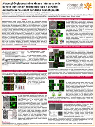
  • 1. Conclusions: Our findings shed light on the interactions of NAGK with dynein and Golgi outposts in dendritic branch points and their roles in dendritic growth. Further investigation is needed into the molecular mechanisms underlying these interactions. Cultured rat hippocampal neurons were treated with dithiobis (succinimidyl) propionate (DSP+), fixed at DIV10 (stage IV) and double-stained with primary antibodies against N- acetylglucosamine kinase and dynein light-chain roadblock type 1 (a) or dynein heavy chain (b) or kinesin 5B (c). The boxed areas of the merged images are enlarged at the bottom. Colocalized immunopuncta are indicated by arrowheads. Scale bar: 10 μm. Statistics (d). The numbers of puncta per dendritic segment (20 μm, n = 20) were counted and expressed as a percentage of the total (mean±s.d.).**p<0.01. (a) N-acetylglucosamine kinase- interacting proteins identified by using the small domain as bait in yeast two-hybrid screening. (b) The interacting domains of dynein light-chain roadblock type 1. The coding region (amino acids 1–96) of dynein light-chain roadblock type 1 is shown as a bar diagram with the conserved roadblock/LC7 domain. The two positive clones in the yeast two-hybrid selection having a coding region from 3 or 59 to the C-terminal end are also shown. Full length dynein light-chain roadblock type 1 (1-end) and kinesin light chain 1 were used as a positive and a negative control, respectively. The small domain of NAGK interacts with DYNLRB1 in yeast two-hybrid screen NAGK-dynein interactions occur at dendritic branch points NAGK interacts with DYNLRB1 in primary hippocampal neurons Verification of NAGK-dynein complex interaction by proximity ligation assay NAGK-dynein complexes colocalize with Golgi ‘outposts’ at dendritic branch points N-acetyl-D-glucosamine kinase interacts with dynein light-chain roadblock type 1 at Golgi outposts in neuronal dendritic branch points Md Ariful Islam1 , Syeda Ridita Sharif1 , HyunSook Lee2 , Dae-Hyun Seog3 and Il Soo Moon1,2 1 Department of Anatomy, Dongguk Medical Institute, College of Medicine Dongguk University, Gyeongju, Republic of Korea; 2 Dongguk Medical Institute, College of Medicine Dongguk University, Gyeongju, Republic of Korea and 3 Departments of Biochemistry, College of Medicine Inje University, Busan, Republic of Korea N-acetylglucosamine kinase (GlcNAc kinase or NAGK) is a ubiquitously expressed enzyme in mammalian cells. Recent studies have shown that NAGK has an essential structural, non-enzymatic role in the upregulation of dendritogenesis. In this study, we conducted yeast two-hybrid screening to search for NAGK-binding proteins and found a specific interaction between NAGK and dynein light-chain roadblock type 1 (DYNLRB1). Immunocytochemistry (ICC) on hippocampal neurons using antibodies against NAGK and DYNLRB1 or dynein heavy chain showed some colocalization, which was increased by treating the live cells with a crosslinker. A proximity ligation assay (PLA) of NAGK-dynein followed by tubulin ICC showed the localization of PLA signals on microtubule fibers at dendritic branch points. NAGK-dynein PLA combined with ICC for Golgi showed the colocalization of PLA signals with somal Golgi facing the apical dendrite and with Golgi outposts in dendritic branch points and distensions. NAGK-Golgi PLA followed by tubulin or DYNLRB1 ICC showed that PLA signals colocalize with DYNLRB1 at dendritic branch points and at somal Golgi, indicating a tripartite interaction between NAGK, dynein and Golgi. Finally, the ectopic introduction of a small peptide derived from the C-terminal amino acids 74–96 of DYNLRB1 resulted in the stunting of hippocampal neuron dendrites in culture. Our data indicate that the NAGK-dynein-Golgi tripartite interaction at dendritic branch points functions to regulate dendritic growth and/or branching. Abstract Co-localization of NAGK-Golgi complexes with DYNLRB1 on MT at dendritic branch points. Ectopic inclusion of small peptides from DYNLRB1 resulted stunted dendrites (b) HEK293T cells were fixed, and PLA was performed using anti-NAGK and anti-dynein light-chain 1 antibodies (NAGK+DYNLL1, upper panel). Alternatively, the cells were transfected with Myc-DDK-tagged NAGK plasmids, and PLA was conducted by using anti-DDK and anti- DYNLL1 antibodies to show the interaction between exogenous NAGK and dynein (DDK+DYNLL1, lower panel). To better depict proximity ligation assay puncta, phase-contrast images were inverted using Photoshop software. Scale bar: 10 μm. (a) Rat hippocampal neurons (DIV2) were fixed, and proximity ligation assay was performed using mouse anti- N-acetylglucosamine kinase and rabbit anti-dynein light- chain roadblock type 1 or anti-dynein heavy chain antibodies. The PLA was followed by immunocytochemistry using a mouse anti-tubulin antibody (green). Boxed areas are enlarged (insets) to show the colocalization of the NAGK-dynein complex with thinner microtubule fibers (arrowheads). Scale bar: 10 μm. (a) N-acetylglucosamine kinase-dynein heavy chain proximity ligation assay was performed in hippocampal neurons (stage IV, DIV 2) and followed by tubulin immunocytochemistry. Proximity ligation assay/phase- contrast (phase) and proximity ligation assay/ immunocytochemistry merged images are shown. The PLA puncta (red) are indicated by arrowheads and with numbers. Scale bar: 10 μm. (b) PLA puncta were sorted into two categories: one with ‘established’ branch joints where the microtubule fiber is already developed (upper panel) and the other at branch initiation sites where the microtubule is yet to develop (lower panel). Positions of branches are indicated with arrowheads. (c) Statistics. Percentages of NAGK-DHC proximity ligation assay puncta at branch joints with or without microtubule in the neonate protrusions are shown by a pie chart. n = 30, 30 neurons. N-acetylglucosamine kinase-dynein heavy chain proximity ligation assay was performed in hippocampal neurons (developmental stage IV) and was followed by immunocytochemistry with an anti-TGN38 antibody to mark Golgi particles. Merged images of proximity ligation assay/ phase-contrast and proximity ligation assay/ immunocytochemistry are shown. (a) PLA signals at soma (1, arrows) and distal branch points (2, 3, arrowheads) are enlarged to show the colocalization of NAGK-dynein complex (red) with Golgi particles (green) oriented to the soma-dendrite joint (inset 1, arrows) or with the Golgi outpost (green) at distal branching sites (inset 2, 3, arrowheads). (b) A high frequency of PLA puncta (red) and Golgi outposts (green) was found at dendritic distensions. Small branches protruding out of the distensions are marked with arrows. Dendritic distensions (boxed areas 1, 2) are enlarged (insets) to show the colocalization of NAGK-dynein complex (red) with Golgi particles (green) marked by arrowheads. Scale bar: 10 μm. (a) NAGK-TGN38 proximity ligation assay (red dots) was followed by ICC with an anti-tubulin antibody (green). The positions of proximity ligation assay signals at branch points are marked with numbers and enlarged in insets. Scale bar: 10 μm. (b) NAGK-GM130 PLA was followed by ICC with an anti-DYNLRB1 antibody. PLA puncta in the soma (box i) were enlarged to show the PLA signals facing the apical dendrite at the somal Golgi apparatus merged with DYNLRB1 signals. PLA dot positions (red) in dendrites are enlarged (box ii, 1–3). Scale bar: 10 μm. (c) Statistics. Pie charts show that ~99% (n = 30 neurons) of NAGK-TGN38 PLA dots were localized to microtubules and ~ 96% (n = 30 neurons) of NAGK-GM130 PLA signals were colocalized with DYNLRB1. Two small peptides are made from DYNLRB1 and named as ‘DYNLRB1 (59-76)’ and ‘DYNLRB1 (74-96)’ . (a) Co-transfection of either ‘DYNLRB1 (59-76)’ or ‘DYNLRB1 (74-96)’ peptide with β-galactosidase was performed and was stained with β-gal staining kit. Neurons transfected with ‘DYNLRB1 (59-76)’ peptide showed healthy dendrites. In contrast, neurons transfected with ‘DYNLRB1 (74-96)’ peptide showed short, stunted dendrites (arrows), while the axon (arrowhead) was apparently unaffected. Scale bar: 10 μm. (b) Sholl analysis. The numbers of dendritic intersections were counted at incremental distances from soma centroids of neurons (n = 30 neurons) transfected with β-gal only (control), with ‘DYNLRB1 (59-76)’ or with ‘DYNLRB1 (74-96)’ . **p<0.01. Results