SlideShare a Scribd company logo
1 of 10
Download to read offline
CLINICAL PARASITOLOGY (Lecture)
PROTOZOANS
DEFINITION OF TERMS:
 Infective stage – refers to the stage of the
parasite that enters the host or the stage that
is present in the parasite’s source of
infection.
 Pathogenic stage – refers to the stage of the
parasite that is responsible for producing the
organ damage in the host leading to the
clinical manifestation.
 Encystation – Process by which
trophozoites differentiate into cyst forms.
 Excystation – Process by which cysts
trophozoites differentiate into trophozoite
forms.
GENERAL PROPERTIES OF PROTOZOA:
 Single-celled eukaryotic organisms that are
spherical to oval or elongated shape.
 Classification of these organisms is mainly
based on the organs of locomotion utilized.
 Not all are parasitic, some are facultative
parasites capable of free-living state.
 Normally reside in soil or water
 Majority of protozoa divide by binary
fission (flagellates, ciliates, and amebae).
Sporozoans through asexual
(merogony/schizogony) and sexual
reproduction.
 Diagnosed through microscopic
examination of body fluids, tissue
specimens, or feces.
 Trophozoite – motile and feeding form
(pathogenic stage)
 Cyst – dormant, non-motile form
(infective stage)
INTESTINAL AND UROGENITAL
PROTOZOA
Subphylum Sarcodina: Entamoeba histolytica
 An intestinal and tissue amoeba
 The only know pathogenic intestinal ameba
 Has 2 stages in its life cycle
 Non-motile cyst (infective stage)
- Found in non-diarrheal or formed
stools.
 Motile trophozoite (pathogenic stage)
-
- Found within the intestinal and
extra-intestinal lesions, and in
diarrheal stools.
COMPARISON OF E. HISTOLYTICA
TROPHOZOITE & CYST FORMS
EPIDEMIOLOGY AND PATHOGENESIS
 Found worldwide but is more common in
tropical countries, especially in areas with
poor sanitation.
 Primarily transmitted by the fecal-oral route
through ingestion of the cyst. (Water is the
major source of infection)
 Sexual transmission may also occur when a
man has unprotected sex with a woman who
has vaginal amoebiasis or through anal
intercourse.
 Excystation happens in the ileum and the
trophozoite colonizes the cecum and colon.
 Trophozoites of E. histolytica secrete
enzymes that cause local necrosis producing
the typical “flask-shaped” ulcer.
 Invasion of the portal circulation may occur
leading to the development of abscess in the
liver.
DISEASE: AMOEBIASIS
1. Acute intestinal amoebiasis – Present as
bloody, mucus-containing diarrhea
(dysentery) accompanied by lower
abdominal discomfort, flatulence (release of
gas), and tenesmus (feeling of incomplete
defecation). Presence of amoeboma (lesion)
in the cecum or in the rectosigmoid area of
the colon in some patients.
2. Extraintestinal amoebiasis – Occurs when
the parasite enters the circulatory system.
The most common is the amoebic liver
abscess (characterized by right upper
quadrant pain, weight loss, fever, and a
merogony is (biology) a
form of asexual
reproduction whereby a
parasitic protozoan
replicates its own nucleus
inside its host's cell and
then induces cell
segmentation;
schizogony is (biology)
asexual reproduction of
protozoans etc
characterized by multiple
divisions of the nucleus and
cell.
tender, enlarged liver.) Other organs that
may become infected include the
pericardium, spleen, skin, lungs (due to liver
abscess) and brain.
3. Asymptomatic carrier state – Occurs
under the following conditions:
a. If the parasite involved is of low-
virulence strain
b. If the parasite load is low
c. If the patient’s immune system is intact
LABORATORY DIAGNOSIS
 Diagnosis of intestinal amoebiasis is
confirmed by finding the trophozoites or
cysts in stool.
 The trophozoites characteristically
contain ingested red blood cells.
 The stool specimen should be examined
within 1 hour of collection to see the
motility of the trophozoites.
 Serologic testing may be useful for the
diagnosis of invasive amoebiasis.
TREATMENT
1. Metronidazole – The drug of choice for
symptomatic intestinal amoebiasis or
hepatic abscess.
2. Tinidazole – An alternative drug for both
intestinal and extraintestinal amoebiasis.
3. Diloxanide furoate, Metronidazole, or
Paromomycin – For asymptomatic carriers
4. Surgical drainage of amoebic liver abscess
may be necessary if there is no
improvement of with medical therapy.
PREVENTION AND CONTROL
1. Observance of good personal hygiene –
most important preventive measure
2. Proper waste disposal – to avoid fecal
contamination of water sources
3. Avoid using human feces (“night soil”) as
fertilizer for crops
 Adequate washing and cooking of
vegetables should be observed
Subphylum Mastigophora: Giardia lamblia
(Giardia intestinalis)
 An intestinal protozoan
 Initially known as Cercomonas intestinalis
 Another name used is Giardia duodenale
 Has 2 stages in its life cycle
 Non-motile cyst (infective stage) –
typically oval and thick-walled with
four nuclei (Fully matured – contains 4
nuclei with 4 median bodies.)
- It divides through binary fission
(Each cyst gives rise to 2
trophozoites during excystation)
 Motile trophozoite (pathogenic stage)
– pear-shaped or teardrop-shaped with
four pairs of flagella (“old man’s face
with glasses”)
- Has a falling leaf like motility
 Possess a sucking disc which is used to
attach itself to the intestinal villi of the
infected human
EPIDEMIOLOGY AND PATHOGENESIS
 Has a worldwide distribution through
contaminated water sources
 Can occur in outbreak related to
contaminated water supplies
 50% of infected individual serve as carriers
 Mammals may act as reservoirs
 Infection is also common among patients
engaging in oral-anal contact
 Primarily transmitted through ingestion of
the cyst from fecally-contaminated waters
 Damage to intestines is due to inflammation
of the duodenal mucosa, leading to diarrhea
with malabsorption of fat and proteins.
 The trophozoite may also infect the
common bile duct and gallbladder
DISEASE: GIARDIASIS
1. Asymptomatic carrier state – infection
with the parasite is usually completely
asymptomatic.
2. Giardiasis (Traveler’s diarrhea) –
Characterized by a non-bloody, foul-
smelling diarrhea accompanied by nausea,
loss of appetite, flatulence, and abdominal
cramps (may persists for weeks or months).
LABORATORY DIAGNOSIS
 Diagnosis is made by the demonstration of
cyst or trophozoite (or both) in fecal
samples
 Only cysts are isolated from the stools
of asymptomatic carriers.
 String test may also be performed
(trophozoites adhere to the string and can be
visualized after withdrawal of the string.
TREATMENT
 METRONIDAZOLE, TINIDAZOLE,
AND NITAZOXANIDE – PRIMARY
CHOICE OF TREATMENT
PREVENTION AND CONTROL
 Avoidance of fecal contamination of water
supplies
 Drinking water should be boiled,
filtered, or iodine-treated especially in
endemic areas.
 Proper waste disposal
 Improvement of personal hygiene (proper
handwashing)
Subphylum Mastigophora: Trichomonas
vaginalis
 Pear-shaped organism with a central nucleus
 Has 4 anterior flagella, and an undulating
membrane
 Exists only in trophozoite form (infective
and pathogenic)
EPIDEMIOLOGY AND PATHOGENESIS
 Causes urogenital infections
 Main mode of transmission is through
sexual intercourse
 Isolated from the urethra and vagina of
infected women and isolated in urethra and
prostate of infected men
 May occasionally be transmitted through
toilet articles and clothing of infected
individuals.
 Infants may be infected as they pass through
the infected birth canal during delivery
 T. vaginalis multiply through binary fission
DISEASE: TRICHOMONIASIS
1. Infection in men – usually asymptomatic
and men serve as the reservoir for infection
in women.
- Symptoms: Prostatitis, urethritis
and other UTI involvement
2. Infection in women – also asymptomatic,
some may present with scant, watery
vaginal discharge
- In severe cases: discharge may be
foul-smelling and greenish-yellow
in color. Accompanied by itching
(pruritus) and a burning sensation
in the vagina. Has a characteristic
“strawberry cervix” and other
common symptoms include dysuria
and increased frequency of
urination.
3. Infection in infants – The infected infant
may infect conjunctivitis or respiratory
infection.
LABORATORY DIAGNOSIS
 Diagnosis is made by finding the
characteristic trophozoite in a wet mount of
vaginal or prostatic secretions, urine, and
urethral discharge.
TREATMENT
 Metronidazole – drug of choice
 All partners should be treated to avoid
“ping pong” infections
PREVENTION AND CONTROL
 Practice safe sex – best way
 Use of condoms
 Health and sex education
 Maintenance of the acidic pH of the vagina
may also be useful
Phylum Ciliophora: Balantidium coli
 Has a primitive mouth called a cytostome,
food vacuoles, and a pair of contractile
vacuoles.
 Has a cyst (infective stage) and a
trophozoite (pathogenic stage)
 The trophozoite invades the mucosal lining
of the terminal ileum, cecum, and colon.
 The largest protozoan to infect humans
 The trophozoite typically exhibit a rotary,
boring motility and contain 2 nuclei
 The cyst also contain 2 nuclei
EPIDEMIOLOGY AND PATHOGENESIS
 Has a worldwide distribution
 The most common and most important
reservoir is the pig (monkeys may
occasionally act as reservoir).
 The main source of infection is water
contaminated with pig feces (MOT: oral-
fecal route)
 No extraintestinal involvement seen
DISEASE: BALANTIDIASIS
 Most infected individuals are asymptomatic
 A dysenteric type of diarrhea resembling
amebic dysentery may occur in patients with
high parasite load.
 Acute infections may manifest with liquid
stools containing pus, blood, and mucus
while chronic infections may manifest with
a tender colon, anemia, wasting (cachexia),
and alternating diarrhea and constipation.
 Extraintestinal infection is rare (may
involve the liver, lungs, mesenteric nodes,
and urogenital tract.
LABORATORY DIAGNOSIS
DIAGNOSIS IS BASED ON FINDING THE
CHARACTERISTIC TROPHOZOITES AND
CYSTS IN STOOL SPECIMENS.
TREATMENT
1. Oxytetracycline and Iodoquinol – current
recommended treatment for patients with
balantidiasis.
2. Metronidazole – alternative treatment
PREVENTION AND CONTROL
 Preventive measures are similar to those for
amoebiasis.
 Maintenance of sanitary hygiene,
proper disposal of pig feces, and boiling
of drinking water.
BLOOD AND TISSUE PROTOZOA
Subphylum Sarcodina: Acanthamoeba (Free-
living Amoeba)
 A minor protozoan pathogen
 Usually causes infection in
immunocompromised patients
 Causes inflammation of the brain substances
and meningeal coverings.
 Found widely in soil, contaminated fresh
water lake, and other water environment.
 Able to survive in cold water
 2 forms: Cyst (Infective) and Trophozoite
(Pathogenic)
EPIDEMIOLOGY AND PATHOGENESIS
 2 ways by which the parasite can be
acquired
 Aspiration or nasal inhalation
 Direct invasion in the eye
 Eye infection occurs primarily in patients
who wear contact lenses.
 water contaminated with parasites is
source of infection
DISEASE
 Granulomatous amebic encephalitis –
infections occur primarily in
immunocompromised individuals.
 Symptoms develop slowly and include
headache, seizures, stiff neck, nausea,
and vomiting.
 may also produce lesions in the
kidneys, pancreas, prostate, and uterus
(rare instances)
 Keratitis – Infection of the cornea of the
eye.
 Symptoms include severe eye pain
and vision problems
LABORATORY DIAGNOSIS
 Diagnosis is made by finding the
characteristic trophozoite or cyst in the CSF
as well as brain tissues and corneal
scrapings.
 Histologic examination of corneal scrapings
may also be done.
TREATMENT
1. Pentamidine, Ketoconazole, or Flucytosine
may be effective in the treatment of
infection (prognosis is poor)
2. For eye and skin, topical miconazole,
chlorhexidine, itraconazole, ketoconazole,
rifampicin, or propamidine may be used.
PREVENTION AND CONTROL
 Adequate boiling of drinking water
 Regular disinfection of contact lenses
 Avoid using homemade non-sterile
saline solution
Subphylum Sarcodina: Naegleria
 A free-living protozoan
 Found worldwide in soil and contaminated
water environment.
 Can survive in thermal spring water.
 Known pathogen worldwide is Naegleria
fowleri (the only amoeba with 3 identified
forms)
 3 forms: Cyst, Trophozoite, and Flagellate
 The trophozoite exhibits a “slug-like”
motility.
 The flagellate form is pear-shaped and
is equipped with 2 flagella (responsible
for jerky or spinning movement)
 The non-motile form is the cyst
 The trophozoite form is the only form
known to exist in humans
EPIDEMIOLOGY AND PATHOGENESIS
 Usually acquired trans nasally when
swimming in contaminated water
 The parasite enters the CNS and produces a
rapidly fatal meningitis and encephalitis
 Can produce infection in healthy individuals
 Can be acquired by inhalation of dust
containing parasite
 The entire life cycle of the parasite occurs
entirely in the external environment
DISEASE
 Asymptomatic infection – Most common
in patients with colonization of the nasal
passages
 Primary amoebic meningoencephalitis –
Colonization of the brain by the amoeboid
trophozoites leading to rapid tissue
destruction. (Patients complain of sore
throat, nausea, vomiting, fever, and
headache.)
LABORATORY DIAGNOSIS
DIAGNOSIS IS BASED ON THE FINDING OF
THE AMOEBOID TROPHOZOITES IN THE CSF
TREATMENT
 Treatment is ineffective because of its
rapidly fatal course.
 Treatment of choice is Amphotericin B in
combination with miconazole, and
rifampicin.
PREVENTION AND CONTROL
 There is no known prevention of Naegleria
infection other than prevention of
contamination of water sources
 Adequate and Frequent chlorination of
swimming pools and hot tubs are
recommended
Subphylum Mastigophora: Hemoflagellates
Leishmania spp.
 The life cycle involves a vector (The female
sandfly: Phlebotomus and Lutzomyia
genera)
 Obligate intracellular parasites
 3 morphologic forms: Amastigote,
Promastigote, and Epimastigote.
 The epimastigotes are found primarily
in the vector
 The amastigotes are the pathogenic and
diagnostic form (found in tissue and
muscles, the CNS within macrophages
and in cells of the reticuloendothelial
system
- The typical amastigote is round to
oval in shape and contains a
nucleus, a basal body structure
called a blepharoblast and a small
parabasal body adjacent to it.
- The blepharoblast and the small
parabasal body is collectively
known as the kinetoplast
 The promastigote is the infective stage
(maybe seen only if a blood sample is
collected and examined immediately upon
transmission)
- The promastigote is long and slender,
with a kinetoplast located in its anterior
end, and a single free flagellum
extending from the anterior portion.
EPIDEMIOLOGY AND PATHOGENESIS
 The parasite has a worldwide distribution.
 Natural reservoirs include rodents, ant
eaters, dogs, and cats.
 In endemic areas, it can be transmitted in a
human-vector-human life cycle.
 There are 3 major strains:
1. Leishmania donovani – Visceral
leishmaniasis
2. Leishmania tropica – Cutaneous
leishmaniasis
3. Leishmania braziliensis –
Mucocutaneous leishmaniasis
LEISHMANIA DONOVANI COMPLEX
 L. donovani is the causative agent of
visceral leishmaniasis
 AKA kala-azar or dumdum fever
 The complex consist of:
 L. donovani chagari – Mainly seen in
Central America and is transmitted by
the Lutzomiya sandfly
 L. donovani donovani – Found in
parts of Africa and Asia and is
transmitted by the Phlebotomus sandfly
 L. donovani infantum – Found mainly
in Mediterranean Europe, near east, and
Africa and is transmitted by the
Phlebotomus sandfly
 The organs of the reticuloendothelial system
are the ones severely affected.
DISEASE: VISCERAL LEISHMANIASIS
(KALA-AZAR/DUMDUM FEVER)
 Incubation period: 2 weeks to 18 months
 The disease begins with intermittent fever,
weakness, and weight loss.
 Massive enlargement of the spleen leading
to hypersplenism and resulting to anemia.
 Hepatomegaly also occurs
 Glomerulonephritis may also occur
 In light-skinned patients, hyperpigmentation
of the skin may be observed
 Involvement of the bone marrow leads to
destruction of cellular components
 Anemia
 Bleeding tendencies
 Increased risk for secondary infection
LABORATORY DIAGNOSIS
 The screening test is called the Montenegro
skin test (similar to tuberculin skin test).
 Definitive diagnosis is done by the
demonstration of the amastigote from
Giemsa-stained slides of specimens from
blood, bone marrow, lymph nodes, and
biopsies of infected areas.
 Culture of blood, bone marrow, and tissues
mat also be done which will show the
promastigote form.
 Serological test are now available
 IFA – indirect fluorescent antibody
 ELISA – enzyme-linked
immunosorbent assay
 DAT – direct agglutination test
TREATMENT
1. Liposomal Amphotericin B (Ambisome) –
Recommended drug of choice
2. Sodium stibogluconate - also found to be
effective but the development of resistance
may occur
3. Gamma interferon with pentavalent
antimony – Have favorable response in
other patients
PREVENTION AND CONTROL
 Control of the vector population
 Use of insect repellants, protective
clothing, and installation of screen may
help
 Prompt treatment of infected humans is
essential to halt the spread
LEISHMANIA BRAZILIENSIS COMPLEX
 L. braziliensis is the causative agent of
mucocutaneous leishmaniasis which
involves skin, cartilage, and mucous
membranes.
 L. braziliensis occur mostly in Brazil and
Central America (primarily in construction
and forestry workers)
 The complex consist of:
1. L. panamensis – Panama and
Columbia
2. L. peruviana – Peruvian Andes
3. L. guyanensis – The Guianas, parts of
Brazil and Venezuela
 Infection is transmitted by sandflies
(Lutzomiya and Psychodopigus) through
skin bite.
DISEASE: MUCOCUTANEOUS
LEISHMANIASIS
 Also called espundia, begins with a papule
at the site of insect bite, then forms
metastatic lesions (junction of the nose and
mouth)
 Disfiguring granulomatous, ulcerating
lesions destroy the nasal cartilage (tapir
nose) but not the bone.
 Death may occur from secondary infections
LABORATORY DIAGNOSIS
 Diagnosis is confirmed by the
demonstration of amastigotes in clinical
specimens.
 Ulcer biopsy for the diagnosis of
mucocutaneous leshmaniasis
 Microscopic examination of Giemsa-
stained ulcer biopsy specimen showing
diagnostic amastigotes
 Culture of infected material may show
promastigotes
 Serologic test may also be done
TREATMENT
1. Sodium stibogluconate – most widely used
drug (resistance may develop)
2. Liposomal Amphotericin B and oral anti-
fungal drugs – alternative drugs
PREVENTION AND CONTROL
 Control of the vector population
 Use of insect repellants, protective
clothing, and installation of screen may
help
 Prompt treatment of infected humans is
essential to halt the spread
LEISHMANIA TROPICA COMPLEX
 Complex consist of:
1. L. tropica
2. L. aethiopica
3. L. major
 The 3 are the causative agent for old world
cutaneous leishmaniasis.
 All 3 are transmitted by the Phlebotomus
sandfly and primarily attacks the human
lymphoid tissue of the skin.
DISEASE: OLD WORLD CUTANEOUS
LEISHMANIASIS
 AKA as Oriental sore, and Baghdad or
Delhi boil.
 Characterized by one or many pus-
containing ulcers that may heal
spontaneously
 The initial lesion is a small, pruritic red
papule at the bite site
 Thick skin plaques with multiple nodules
may develop, especially on the limbs and
face
LABORATORY DIAGNOSIS
 Microscopic examination of Giemsa-stained
slides of fluid aspirated beneath the ulcer
bed is the diagnostic procedure of choice.
 Reveals the typical amastigotes
 Culture of infected material may show
promastigotes
 Serologic test may also be done
TREATMENT
1. Sodium stibogluconate – drug of choice
2. Steroids with application of heat to the
infected lesions may be used.
3. Meglumine antimonite, pentamidine and
oral ketoconazole – Alternative drugs
4. Paromomycin ointment may be helpful in
healing the ulcers
PREVENTION AND CONTROL
 Control of the vector population
 Use of insect repellants, protective
clothing, and installation of screen may
help
 Prompt treatment of infected humans is
essential to halt the spread
Trypanosoma spp.
 Hemoflagellates
 Trypomastigote (diagnostic stage) –
Curved, assuming the letters C, S, or U.
Posteriorly located, with the single large
nucleus anterior to it.
 Visible in the peripheral blood
Trypanosoma cruzi
 Primarily found in South and Central
America
 Transmitted by the reduviid or triatomid
bug (“kissing bug”)
 Transmitted when the feces of the infected
bug is deposited near the bite site and
introduced through scratching
 Other routes of transmission include blood
transfusion, sexual intercourse,
transplacental transmission, and through the
mucous membranes when the bite site is
near the eye or mouth.
 Humans and animals serve as reservoir
hosts.
 Glial cells, Reticuloendothelial cells, and
myocardial cells are the most frequently
affected.
 Primarily seen in rural areas
DISEASE: CHAGAS DISEASE (AMERICAN
TRYPANOSOMIASIS)
 The acute phase begins with a nodule
(chagoma) near the bite site and unilateral
swelling of the eyelid with conjunctivitis
(Romana’s sign).
 Accompanied by fever, chills, malaise,
myalgia, and fatigue. Patients may recover
or enter chronic phase.
 Hepatosplenomegaly, lymphadenopathy,
and myocarditis with cardiac arrythmia
characterize the chronic phase.
 Loss of tone of the colon and esophagus due
to the destruction of Auerbach’s plexus may
lead to abnormal dilatation of organs
(megacolon and megaesophagus)
 CNS involvement may be seen in the form
of meningoencephalitis and cysts.
 Death may occur due to cardiac failure and
arrhythmias
LABORATORY DIAGNOSIS
 Acute disease is diagnosed by finding
trypomastigotes in thick and thin films of
the patient’s blood.
 BM aspiration, Muscle biopsy, Culture on
special medium, Xenodiagnosis – Other
diagnostic methods
 Serologic test may also be done
 Both Xenodiagnosis and Serologic test is
useful in the chronic form
TREATMENT
1. Benznidazole and nifurtimox – Drug of
choice but less effective during the chronic
phase
2. Allopurinol and ketoconazole –
Alternative agents
PREVENTION AND CONTROL
 Protection from the bite of the reduviid bug
 Improvement of housing conditions,
and insect control
 Education regarding the disease and its
transmission
Trypanosoma brucei gambiense and
Trypanosoma brucei rhodesiense
 The 2 species are similar in morphology and
life cycle.
 Tsetse fly (Glossina) as the vector
 Humans – reservoir for T. brucei
gambiense
 Domestic animals and Wild animals –
reservoir for T. brucei rhodesiense
 Trypomastigote – the infective and
pathogenic stage
EPIDEMIOLOGY AND PATHOGENESIS
 Trypomastigotes spread from the skin to the
blood then to the lymph nodes and the brain.
 A demyelinating encephalitis occurs leading
to the characteristic manifestation of the
disease.
 T. gambiense infection (West African or
Gambian Sleeping Sickness) is chronic
 Causes disease along water courses in
West Africa
 T. rhodesiense infection (East African or
Rhodesian Sleeping Sickness) is more
rapidly fatal
 Causes disease mostly in arid regions in
East Africa
 Endemic in sub-Saharan Africa
DISEASE: AFRICAN SLEEPING SICKNESS
 Presence of a chancre (indurated ulcer) at
the site of the insect bite.
 Intermittent weekly fever and
lymphadenopathy then develop.
 Winterbottom’s sign (enlargement of
posterior cervical lymph nodes) is
commonly seen.
 Other manifestation: red rash accompanied
by pruritus, localized edema, and
Kerandel’s sign (delayed pain sensation).
 Encephalitis is characterized by headache,
insomnia, and mood changes.
 Muscle tremors, slurred speech, and apathy
progressing to somnolence (sleeping
sickness) and coma.
 T. rhodesiense is more virulent than T.
gambiense
 Shorter incubation period
 CNS involvement occurs early
 A rapid and fulminating disease may
follow with the parasite spreading in
the blood.
 Death is usually within 9-12 months
(due to glomerulonephritis and
myocarditis)
LABORATORY DIAGNOSIS
 Microscopic exam of Giemsa-stained slides
(blood, lymph node aspirations, and CSF)
will reveal trypomastigote during early
stage of the disease.
 Aspiration of the chancre or enlarged lymph
node may also reveal the parasite
 The presence in the serum and/or CSF (CNS
involvement) of IgM is considered
diagnostics
TREATMENT
 Melarsoprol, suramine, pentamidine, and
eflornithine – treatment of both East African
and West African sleeping sickness
 Choice of drug depends on: pregnancy
status, age, and the stage of the disease
PREVENTION AND CONTROL
 Protection against the tsetse fly
 Use of netting, and protective clothing
 Use of fly traps and insecticides
 Clearing the forest around the village
Subphylum Apicomplexa: Plasmodium spp.
 Causative agent of Malaria
 Has 5 species:
1. Plasmodium vivax
2. Plasmodium ovale
3. Plasmodium falciparum
4. Plasmodium malariae
5. Plasmodium knowlesi
 The sexual cycle (sporogony) occurs
primarily in mosquitoes, and the asexual
cycle (schizogony) occurs in humans (IH)
 Sporozoites (infective stage) enters the body
from the saliva of a biting mosquito which
is taken up by liver cells (exoerythrocytic
phase)
 P. vivax and P. ovale produce a latent form
(hypnozoite or sleeping form) in the liver,
causing relapse.
 Merozoites (pathogenic stage) are released
from the liver and infect the RBCs
(erythrocytic phase).
 Some merozoites then develop into
microgametocytes (male gametocytes) and
macrogametocytes (female gametocytes)
and then ingested by feeding mosquitoes.
COMPARISON OF DIFFERENT
TROPHOZOITE FORMS
EPIDEMIOLOGY AND PATHOGENESIS
 Infection of plasmodia occur worldwide
 Primarily in tropical and subtropical areas
(Asia, Africa, and Central and South
America)
 69% of cases in the Philippines is due to P.
falciparum while the remaining 31% is due
to P. vivax
 The primary vector is Anopheles
flavirostris, which breeds in clear, slow-
flowing streams
 Main mode of transmission of malaria is the
bite of female mosquito vector.
 Can also be transmitted through blood
transfusion (transfusion malaria), IV needles
(“main-line malaria), and transplacental
transmission (congenital malaria).
 Most pathologic findings result from the
destruction of RBCs.
 P. falciparum and P. knowlesi can infect
both young and old RBCs leading to high
levels of parasitemia.
 P. vivax and P. ovale mainly infect young
RBCs while P. malariae infects old RBCs.
 P. knowlesi is a natural parasite of macaque
monkeys (RBC infected is of normal
morphology)
DISEASE: MALARIA
 Paroxysm of malaria is divided into 3
stages: Cold stage, Hot stage, and the
sweating stage.
 Considered partially as allergic
response to the schizonts and antigens
released following the release of
merozoites.
 A malarial paroxysm presents with abrupt
onset of chills (rigors) accompanied by
headache, muscle pain (myalgia), and joint
pains (arthralgia).
 Patients usually feel well in between febrile
episodes.
 Splenomegaly is often present and anemia is
prominent.
 Timing of fever cycle:
 P. malariae – 72 hours in which
symptoms recur every 4th day (quartan
malaria)
 P. vivax, P. ovale, and P. falciparum –
recur every 3rd day (tertian malaria)
 P. knowlesi – 24 hour erythrocyte cycle
(quotidian malaria)
 P. falciparum causes malignant tertian
malaria since it causes severe infection
which is potentially life threatening due to
extensive brain (cerebral malaria) and
kidney damage.
 “black water fever” due to the dark
color of the urine from extensive
kidney damage
 P. ovale and P. vivax cause benign tertian
malaria (relapse)
 P. knowlesi – resembles infection of other
malarial parasite (severity is due to high
parasitemia)
LABORATORY DIAGNOSIS
 Examination of Giemsa-stained or Wright-
stained thick and thin blood smears.
 Thick for quantitative count of the
bacteria
 Thin for species identification of the
various Plasmodium
 The best time to take blood films is midway
between paroxysms of chills and fevers or
before the onset of the fever
 P. falciparum – show characteristic
crescent-shaped or banana-shaped
gametocytes
 P. malariae and P. knowlesi – rosette
schizont is characteristic and diagnostic
TREATMENT
 Chloroquine or parenteral quinine – drug
of choice for acute malarial infection
 Chloroquine does not affect hynozoites
(P. vivax and P. ovale)
 Primaquine – for hypnozoites treatment
 Mefloquine+artesunate,
artemetherlumafrantine, atovaquone-
proguanil, quinine, quinidine,
pyrimethamine-sulfadoxine (Fansidar),
and doxycycline – for treatment of
chloroquine-resistant strains of P.
falciparum
 Artemisin-based combination therapies
(ACTs) – for uncomplicated malaria and
chloroquine-resistant P. vivax malaria
 Artesunate (+amodiaquine, +mefloquine,
or +sulfadoxine-pyrimethamine) – drug of
choice for severe malaria
 P. knowlesi is managed similar to P.
falciparum due to its potential to produce
severe infection
PREVENTION AND CONTROL
 Chemoprophylaxis (mefloquine or
doxycycline) of travelers going to endemic
areas
 Chloroquine 2 weeks before and
continued for 6 weeks –
chemoprophylaxis for travelers going to
areas where other plasmodia are found
(followed by 2-week course of
primaquine if exposure is high)
 Avoidance of the bite of the vector
 Use of mosquito net, window screens,
protective clothing, and insect
repellants
 Reduction of mosquito population
 Use of insecticide sprays, drainage of
stagnant water
Phylum Apicomplexa: Toxoplasma gondii
 The definitive host is the domestic cat or
other felines
 Humans and other mammals serve as
intermediate host
 Develop in the cat’s intestine and passes
through the bloodstream. Passed in the cat’s
feces and mature into infective oocysts in
the external environment.
 Infection in humans begins with the
ingestion of oocysts (infective form) in
undercooked meat or from contact with cat
feces
 Tachyzoites (rapidly multiplying forms)
responsible for the initial infection
 Bradyzoites (shorter, slow-growing forms)
are seen in chronic infections
EPIDEMIOLOGY AND PATHOGENESIS
 Occurs worldwide and is usually sporadic
but outbreaks may occur
 Individuals who are immunocompromised
are most likely to develop severe disease
 Can be mainly transmitted in 2 ways:
1. Ingestion of improperly cooked meat of
animals that serve as intermediate host
2. Ingestion of oocysts from contaminated
water
 Transplacental transmission may occur with
sever consequences to the fetus
 Sharing of IV needles and Blood transfusion
are less common modes of transmission
DISEASE: TOXOPLASMOSIS
 Infection in immunocompetent
individuals – Usually asymptomatic. Acute
infection may manifest non-specific
symptoms such as chills, fever, headache,
and fatigue (may be accompanied by
inflammation of lymph nodes). Chronic
infection may manifest with lymphadenitis,
hepatitis, myocarditis, and
encephalomyelitis.
 Congenital infection – Occurs in infants
born to mothers who were infected during
pregnancy. Infection in the 1st trimester
may lead to miscarriage, stillbirth, or severe
infections. Infection in the last trimester,
symptoms may not develop until months to
years. Chorioretinitis with or without
blindness is the most common
manifestation.
 Infection in immunocompromised hosts –
Usually with neurological symptoms similar
to patients with encephalopathy,
meningoencephalitis, or brain tumor.
Reactivation of latent toxoplasma is
common. Other sites: lungs, eyes, and
testes.
LABORATORY DIAGNOSIS
 Demonstration of high antibody titers
through immunofluorescence assay is
essential for diagnosis.
 Microscopic examination of Giemsa-stained
preparations will show crescent-shaped
trophozoites during acute infection.
 Cysts may be seen in the tissues
 Ultrasonography and amniocentesis with
PCR analysis of the amniotic fluid (method
of choice) – For prenatal diagnosis
TREATMENT
 Infection in immunocompetent host is
usually self-limiting.
 High-dose of pyrimethamine plus
sulfadiazine – Regimen of choice for
immunocompromised patients for an
indefinite period.
 Clindamycin plus pyrimethamine –
alternative regimen for those who develop
symptoms for drug toxicity
 Clindamycin or spiramycin – for pregnant
women
PREVENTION AND CONTROL
 The most effective preventive measure is
through adequate cooking of meat.
 Cats should not be fed raw meat
 Pregnant women should refrain from
eating undercooked meat and avoid
contact with cats and litter boxes

More Related Content

Similar to Protozoans.pdf

Similar to Protozoans.pdf (20)

E histolytica gr_19_09
E histolytica gr_19_09E histolytica gr_19_09
E histolytica gr_19_09
 
Protozoa amoeba
Protozoa amoebaProtozoa amoeba
Protozoa amoeba
 
Balantidium coli
Balantidium coliBalantidium coli
Balantidium coli
 
Intestinal and Urogenital Protozoa
Intestinal and Urogenital ProtozoaIntestinal and Urogenital Protozoa
Intestinal and Urogenital Protozoa
 
HCM 124 lecture 5 .ppt
HCM 124 lecture 5 .pptHCM 124 lecture 5 .ppt
HCM 124 lecture 5 .ppt
 
Parasitology.pptx
Parasitology.pptxParasitology.pptx
Parasitology.pptx
 
Parasitology 2020
Parasitology 2020Parasitology 2020
Parasitology 2020
 
balantidiumcoli-140225040255-phpapp01 2.pdf
balantidiumcoli-140225040255-phpapp01 2.pdfbalantidiumcoli-140225040255-phpapp01 2.pdf
balantidiumcoli-140225040255-phpapp01 2.pdf
 
Parasitology.pdf
Parasitology.pdfParasitology.pdf
Parasitology.pdf
 
Entamoeba histolytica
Entamoeba histolytica Entamoeba histolytica
Entamoeba histolytica
 
Essay para
Essay paraEssay para
Essay para
 
Balantidium Coli
Balantidium ColiBalantidium Coli
Balantidium Coli
 
Protozoa I
Protozoa IProtozoa I
Protozoa I
 
Protozoa
ProtozoaProtozoa
Protozoa
 
Intestinal Flagellates notes 2014
Intestinal Flagellates notes 2014Intestinal Flagellates notes 2014
Intestinal Flagellates notes 2014
 
1. Entamoeba histolytica
1. Entamoeba histolytica1. Entamoeba histolytica
1. Entamoeba histolytica
 
Parasitology Review 2019
Parasitology Review 2019Parasitology Review 2019
Parasitology Review 2019
 
Parasitology 2024 | Microbes with Morgan
Parasitology 2024 | Microbes with MorganParasitology 2024 | Microbes with Morgan
Parasitology 2024 | Microbes with Morgan
 
Amoebiasis Nursing Management
Amoebiasis Nursing ManagementAmoebiasis Nursing Management
Amoebiasis Nursing Management
 
Introprotozoa
IntroprotozoaIntroprotozoa
Introprotozoa
 

Recently uploaded

Call Girls Hsr Layout Whatsapp 7001305949 Independent Escort Service
Call Girls Hsr Layout Whatsapp 7001305949 Independent Escort ServiceCall Girls Hsr Layout Whatsapp 7001305949 Independent Escort Service
Call Girls Hsr Layout Whatsapp 7001305949 Independent Escort Servicenarwatsonia7
 
College Call Girls Hyderabad Sakshi 9907093804 Independent Escort Service Hyd...
College Call Girls Hyderabad Sakshi 9907093804 Independent Escort Service Hyd...College Call Girls Hyderabad Sakshi 9907093804 Independent Escort Service Hyd...
College Call Girls Hyderabad Sakshi 9907093804 Independent Escort Service Hyd...delhimodelshub1
 
Low Rate Call Girls In Bommanahalli Just Call 7001305949
Low Rate Call Girls In Bommanahalli Just Call 7001305949Low Rate Call Girls In Bommanahalli Just Call 7001305949
Low Rate Call Girls In Bommanahalli Just Call 7001305949ps5894268
 
Soft Toric contact lens fitting (NSO).pptx
Soft Toric contact lens fitting (NSO).pptxSoft Toric contact lens fitting (NSO).pptx
Soft Toric contact lens fitting (NSO).pptxJasmin Modi
 
Call Girls South Delhi 9999965857 Cheap and Best with original Photos
Call Girls South Delhi 9999965857 Cheap and Best with original PhotosCall Girls South Delhi 9999965857 Cheap and Best with original Photos
Call Girls South Delhi 9999965857 Cheap and Best with original Photosparshadkalavatidevi7
 
Russian Call Girls Ajmeri Gate | 9711199171 | High Profile -New Model -Availa...
Russian Call Girls Ajmeri Gate | 9711199171 | High Profile -New Model -Availa...Russian Call Girls Ajmeri Gate | 9711199171 | High Profile -New Model -Availa...
Russian Call Girls Ajmeri Gate | 9711199171 | High Profile -New Model -Availa...sandeepkumar69420
 
Book Call Girls in Hosur - 7001305949 | 24x7 Service Available Near Me
Book Call Girls in Hosur - 7001305949 | 24x7 Service Available Near MeBook Call Girls in Hosur - 7001305949 | 24x7 Service Available Near Me
Book Call Girls in Hosur - 7001305949 | 24x7 Service Available Near Menarwatsonia7
 
Call Girls Hyderabad Krisha 9907093804 Independent Escort Service Hyderabad
Call Girls Hyderabad Krisha 9907093804 Independent Escort Service HyderabadCall Girls Hyderabad Krisha 9907093804 Independent Escort Service Hyderabad
Call Girls Hyderabad Krisha 9907093804 Independent Escort Service Hyderabaddelhimodelshub1
 
FAMILY in sociology for physiotherapists.pptx
FAMILY in sociology for physiotherapists.pptxFAMILY in sociology for physiotherapists.pptx
FAMILY in sociology for physiotherapists.pptxMumux Mirani
 
Call Girls in Adil Nagar 7001305949 Free Delivery at Your Door Model
Call Girls in Adil Nagar 7001305949 Free Delivery at Your Door ModelCall Girls in Adil Nagar 7001305949 Free Delivery at Your Door Model
Call Girls in Adil Nagar 7001305949 Free Delivery at Your Door ModelCall Girls Lucknow
 
Russian Call Girl Chandapura Dommasandra Road - 7001305949 Escorts Service 50...
Russian Call Girl Chandapura Dommasandra Road - 7001305949 Escorts Service 50...Russian Call Girl Chandapura Dommasandra Road - 7001305949 Escorts Service 50...
Russian Call Girl Chandapura Dommasandra Road - 7001305949 Escorts Service 50...narwatsonia7
 
Call Girls Ghaziabad 9999965857 Cheap and Best with original Photos
Call Girls Ghaziabad 9999965857 Cheap and Best with original PhotosCall Girls Ghaziabad 9999965857 Cheap and Best with original Photos
Call Girls Ghaziabad 9999965857 Cheap and Best with original Photosparshadkalavatidevi7
 
EMS and Extrication: Coordinating Critical Care
EMS and Extrication: Coordinating Critical CareEMS and Extrication: Coordinating Critical Care
EMS and Extrication: Coordinating Critical CareRommie Duckworth
 
Air-Hostess Call Girls Shanti Nagar - Call 7001305949 Rs-3500 with A/C Room C...
Air-Hostess Call Girls Shanti Nagar - Call 7001305949 Rs-3500 with A/C Room C...Air-Hostess Call Girls Shanti Nagar - Call 7001305949 Rs-3500 with A/C Room C...
Air-Hostess Call Girls Shanti Nagar - Call 7001305949 Rs-3500 with A/C Room C...narwatsonia7
 
independent Call Girls Sarjapur Road - 7001305949 with real photos and phone ...
independent Call Girls Sarjapur Road - 7001305949 with real photos and phone ...independent Call Girls Sarjapur Road - 7001305949 with real photos and phone ...
independent Call Girls Sarjapur Road - 7001305949 with real photos and phone ...narwatsonia7
 
Call Girls Laxmi Nagar 9999965857 Cheap and Best with original Photos
Call Girls Laxmi Nagar 9999965857 Cheap and Best with original PhotosCall Girls Laxmi Nagar 9999965857 Cheap and Best with original Photos
Call Girls Laxmi Nagar 9999965857 Cheap and Best with original Photosparshadkalavatidevi7
 
Disaster Management Cycle (DMC)| Ms. Pooja Sharma , Department of Hospital A...
Disaster Management Cycle (DMC)| Ms. Pooja Sharma , Department of  Hospital A...Disaster Management Cycle (DMC)| Ms. Pooja Sharma , Department of  Hospital A...
Disaster Management Cycle (DMC)| Ms. Pooja Sharma , Department of Hospital A...Era University , Lucknow
 
Russian Call Girls Mohan Nagar | 9711199171 | High Profile -New Model -Availa...
Russian Call Girls Mohan Nagar | 9711199171 | High Profile -New Model -Availa...Russian Call Girls Mohan Nagar | 9711199171 | High Profile -New Model -Availa...
Russian Call Girls Mohan Nagar | 9711199171 | High Profile -New Model -Availa...sandeepkumar69420
 
Gurgaon iffco chowk 🔝 Call Girls Service 🔝 ( 8264348440 ) unlimited hard sex ...
Gurgaon iffco chowk 🔝 Call Girls Service 🔝 ( 8264348440 ) unlimited hard sex ...Gurgaon iffco chowk 🔝 Call Girls Service 🔝 ( 8264348440 ) unlimited hard sex ...
Gurgaon iffco chowk 🔝 Call Girls Service 🔝 ( 8264348440 ) unlimited hard sex ...callgirlsinsaket2024
 
2024 HCAT Healthcare Technology Insights
2024 HCAT Healthcare Technology Insights2024 HCAT Healthcare Technology Insights
2024 HCAT Healthcare Technology InsightsHealth Catalyst
 

Recently uploaded (20)

Call Girls Hsr Layout Whatsapp 7001305949 Independent Escort Service
Call Girls Hsr Layout Whatsapp 7001305949 Independent Escort ServiceCall Girls Hsr Layout Whatsapp 7001305949 Independent Escort Service
Call Girls Hsr Layout Whatsapp 7001305949 Independent Escort Service
 
College Call Girls Hyderabad Sakshi 9907093804 Independent Escort Service Hyd...
College Call Girls Hyderabad Sakshi 9907093804 Independent Escort Service Hyd...College Call Girls Hyderabad Sakshi 9907093804 Independent Escort Service Hyd...
College Call Girls Hyderabad Sakshi 9907093804 Independent Escort Service Hyd...
 
Low Rate Call Girls In Bommanahalli Just Call 7001305949
Low Rate Call Girls In Bommanahalli Just Call 7001305949Low Rate Call Girls In Bommanahalli Just Call 7001305949
Low Rate Call Girls In Bommanahalli Just Call 7001305949
 
Soft Toric contact lens fitting (NSO).pptx
Soft Toric contact lens fitting (NSO).pptxSoft Toric contact lens fitting (NSO).pptx
Soft Toric contact lens fitting (NSO).pptx
 
Call Girls South Delhi 9999965857 Cheap and Best with original Photos
Call Girls South Delhi 9999965857 Cheap and Best with original PhotosCall Girls South Delhi 9999965857 Cheap and Best with original Photos
Call Girls South Delhi 9999965857 Cheap and Best with original Photos
 
Russian Call Girls Ajmeri Gate | 9711199171 | High Profile -New Model -Availa...
Russian Call Girls Ajmeri Gate | 9711199171 | High Profile -New Model -Availa...Russian Call Girls Ajmeri Gate | 9711199171 | High Profile -New Model -Availa...
Russian Call Girls Ajmeri Gate | 9711199171 | High Profile -New Model -Availa...
 
Book Call Girls in Hosur - 7001305949 | 24x7 Service Available Near Me
Book Call Girls in Hosur - 7001305949 | 24x7 Service Available Near MeBook Call Girls in Hosur - 7001305949 | 24x7 Service Available Near Me
Book Call Girls in Hosur - 7001305949 | 24x7 Service Available Near Me
 
Call Girls Hyderabad Krisha 9907093804 Independent Escort Service Hyderabad
Call Girls Hyderabad Krisha 9907093804 Independent Escort Service HyderabadCall Girls Hyderabad Krisha 9907093804 Independent Escort Service Hyderabad
Call Girls Hyderabad Krisha 9907093804 Independent Escort Service Hyderabad
 
FAMILY in sociology for physiotherapists.pptx
FAMILY in sociology for physiotherapists.pptxFAMILY in sociology for physiotherapists.pptx
FAMILY in sociology for physiotherapists.pptx
 
Call Girls in Adil Nagar 7001305949 Free Delivery at Your Door Model
Call Girls in Adil Nagar 7001305949 Free Delivery at Your Door ModelCall Girls in Adil Nagar 7001305949 Free Delivery at Your Door Model
Call Girls in Adil Nagar 7001305949 Free Delivery at Your Door Model
 
Russian Call Girl Chandapura Dommasandra Road - 7001305949 Escorts Service 50...
Russian Call Girl Chandapura Dommasandra Road - 7001305949 Escorts Service 50...Russian Call Girl Chandapura Dommasandra Road - 7001305949 Escorts Service 50...
Russian Call Girl Chandapura Dommasandra Road - 7001305949 Escorts Service 50...
 
Call Girls Ghaziabad 9999965857 Cheap and Best with original Photos
Call Girls Ghaziabad 9999965857 Cheap and Best with original PhotosCall Girls Ghaziabad 9999965857 Cheap and Best with original Photos
Call Girls Ghaziabad 9999965857 Cheap and Best with original Photos
 
EMS and Extrication: Coordinating Critical Care
EMS and Extrication: Coordinating Critical CareEMS and Extrication: Coordinating Critical Care
EMS and Extrication: Coordinating Critical Care
 
Air-Hostess Call Girls Shanti Nagar - Call 7001305949 Rs-3500 with A/C Room C...
Air-Hostess Call Girls Shanti Nagar - Call 7001305949 Rs-3500 with A/C Room C...Air-Hostess Call Girls Shanti Nagar - Call 7001305949 Rs-3500 with A/C Room C...
Air-Hostess Call Girls Shanti Nagar - Call 7001305949 Rs-3500 with A/C Room C...
 
independent Call Girls Sarjapur Road - 7001305949 with real photos and phone ...
independent Call Girls Sarjapur Road - 7001305949 with real photos and phone ...independent Call Girls Sarjapur Road - 7001305949 with real photos and phone ...
independent Call Girls Sarjapur Road - 7001305949 with real photos and phone ...
 
Call Girls Laxmi Nagar 9999965857 Cheap and Best with original Photos
Call Girls Laxmi Nagar 9999965857 Cheap and Best with original PhotosCall Girls Laxmi Nagar 9999965857 Cheap and Best with original Photos
Call Girls Laxmi Nagar 9999965857 Cheap and Best with original Photos
 
Disaster Management Cycle (DMC)| Ms. Pooja Sharma , Department of Hospital A...
Disaster Management Cycle (DMC)| Ms. Pooja Sharma , Department of  Hospital A...Disaster Management Cycle (DMC)| Ms. Pooja Sharma , Department of  Hospital A...
Disaster Management Cycle (DMC)| Ms. Pooja Sharma , Department of Hospital A...
 
Russian Call Girls Mohan Nagar | 9711199171 | High Profile -New Model -Availa...
Russian Call Girls Mohan Nagar | 9711199171 | High Profile -New Model -Availa...Russian Call Girls Mohan Nagar | 9711199171 | High Profile -New Model -Availa...
Russian Call Girls Mohan Nagar | 9711199171 | High Profile -New Model -Availa...
 
Gurgaon iffco chowk 🔝 Call Girls Service 🔝 ( 8264348440 ) unlimited hard sex ...
Gurgaon iffco chowk 🔝 Call Girls Service 🔝 ( 8264348440 ) unlimited hard sex ...Gurgaon iffco chowk 🔝 Call Girls Service 🔝 ( 8264348440 ) unlimited hard sex ...
Gurgaon iffco chowk 🔝 Call Girls Service 🔝 ( 8264348440 ) unlimited hard sex ...
 
2024 HCAT Healthcare Technology Insights
2024 HCAT Healthcare Technology Insights2024 HCAT Healthcare Technology Insights
2024 HCAT Healthcare Technology Insights
 

Protozoans.pdf

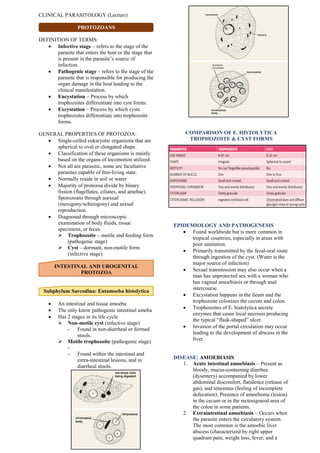
  • 1. CLINICAL PARASITOLOGY (Lecture) PROTOZOANS DEFINITION OF TERMS:  Infective stage – refers to the stage of the parasite that enters the host or the stage that is present in the parasite’s source of infection.  Pathogenic stage – refers to the stage of the parasite that is responsible for producing the organ damage in the host leading to the clinical manifestation.  Encystation – Process by which trophozoites differentiate into cyst forms.  Excystation – Process by which cysts trophozoites differentiate into trophozoite forms. GENERAL PROPERTIES OF PROTOZOA:  Single-celled eukaryotic organisms that are spherical to oval or elongated shape.  Classification of these organisms is mainly based on the organs of locomotion utilized.  Not all are parasitic, some are facultative parasites capable of free-living state.  Normally reside in soil or water  Majority of protozoa divide by binary fission (flagellates, ciliates, and amebae). Sporozoans through asexual (merogony/schizogony) and sexual reproduction.  Diagnosed through microscopic examination of body fluids, tissue specimens, or feces.  Trophozoite – motile and feeding form (pathogenic stage)  Cyst – dormant, non-motile form (infective stage) INTESTINAL AND UROGENITAL PROTOZOA Subphylum Sarcodina: Entamoeba histolytica  An intestinal and tissue amoeba  The only know pathogenic intestinal ameba  Has 2 stages in its life cycle  Non-motile cyst (infective stage) - Found in non-diarrheal or formed stools.  Motile trophozoite (pathogenic stage) - - Found within the intestinal and extra-intestinal lesions, and in diarrheal stools. COMPARISON OF E. HISTOLYTICA TROPHOZOITE & CYST FORMS EPIDEMIOLOGY AND PATHOGENESIS  Found worldwide but is more common in tropical countries, especially in areas with poor sanitation.  Primarily transmitted by the fecal-oral route through ingestion of the cyst. (Water is the major source of infection)  Sexual transmission may also occur when a man has unprotected sex with a woman who has vaginal amoebiasis or through anal intercourse.  Excystation happens in the ileum and the trophozoite colonizes the cecum and colon.  Trophozoites of E. histolytica secrete enzymes that cause local necrosis producing the typical “flask-shaped” ulcer.  Invasion of the portal circulation may occur leading to the development of abscess in the liver. DISEASE: AMOEBIASIS 1. Acute intestinal amoebiasis – Present as bloody, mucus-containing diarrhea (dysentery) accompanied by lower abdominal discomfort, flatulence (release of gas), and tenesmus (feeling of incomplete defecation). Presence of amoeboma (lesion) in the cecum or in the rectosigmoid area of the colon in some patients. 2. Extraintestinal amoebiasis – Occurs when the parasite enters the circulatory system. The most common is the amoebic liver abscess (characterized by right upper quadrant pain, weight loss, fever, and a merogony is (biology) a form of asexual reproduction whereby a parasitic protozoan replicates its own nucleus inside its host's cell and then induces cell segmentation; schizogony is (biology) asexual reproduction of protozoans etc characterized by multiple divisions of the nucleus and cell.
  • 2. tender, enlarged liver.) Other organs that may become infected include the pericardium, spleen, skin, lungs (due to liver abscess) and brain. 3. Asymptomatic carrier state – Occurs under the following conditions: a. If the parasite involved is of low- virulence strain b. If the parasite load is low c. If the patient’s immune system is intact LABORATORY DIAGNOSIS  Diagnosis of intestinal amoebiasis is confirmed by finding the trophozoites or cysts in stool.  The trophozoites characteristically contain ingested red blood cells.  The stool specimen should be examined within 1 hour of collection to see the motility of the trophozoites.  Serologic testing may be useful for the diagnosis of invasive amoebiasis. TREATMENT 1. Metronidazole – The drug of choice for symptomatic intestinal amoebiasis or hepatic abscess. 2. Tinidazole – An alternative drug for both intestinal and extraintestinal amoebiasis. 3. Diloxanide furoate, Metronidazole, or Paromomycin – For asymptomatic carriers 4. Surgical drainage of amoebic liver abscess may be necessary if there is no improvement of with medical therapy. PREVENTION AND CONTROL 1. Observance of good personal hygiene – most important preventive measure 2. Proper waste disposal – to avoid fecal contamination of water sources 3. Avoid using human feces (“night soil”) as fertilizer for crops  Adequate washing and cooking of vegetables should be observed Subphylum Mastigophora: Giardia lamblia (Giardia intestinalis)  An intestinal protozoan  Initially known as Cercomonas intestinalis  Another name used is Giardia duodenale  Has 2 stages in its life cycle  Non-motile cyst (infective stage) – typically oval and thick-walled with four nuclei (Fully matured – contains 4 nuclei with 4 median bodies.) - It divides through binary fission (Each cyst gives rise to 2 trophozoites during excystation)  Motile trophozoite (pathogenic stage) – pear-shaped or teardrop-shaped with four pairs of flagella (“old man’s face with glasses”) - Has a falling leaf like motility  Possess a sucking disc which is used to attach itself to the intestinal villi of the infected human EPIDEMIOLOGY AND PATHOGENESIS  Has a worldwide distribution through contaminated water sources  Can occur in outbreak related to contaminated water supplies  50% of infected individual serve as carriers  Mammals may act as reservoirs  Infection is also common among patients engaging in oral-anal contact  Primarily transmitted through ingestion of the cyst from fecally-contaminated waters  Damage to intestines is due to inflammation of the duodenal mucosa, leading to diarrhea with malabsorption of fat and proteins.  The trophozoite may also infect the common bile duct and gallbladder DISEASE: GIARDIASIS 1. Asymptomatic carrier state – infection with the parasite is usually completely asymptomatic. 2. Giardiasis (Traveler’s diarrhea) – Characterized by a non-bloody, foul- smelling diarrhea accompanied by nausea, loss of appetite, flatulence, and abdominal cramps (may persists for weeks or months). LABORATORY DIAGNOSIS  Diagnosis is made by the demonstration of cyst or trophozoite (or both) in fecal samples  Only cysts are isolated from the stools of asymptomatic carriers.  String test may also be performed (trophozoites adhere to the string and can be visualized after withdrawal of the string. TREATMENT  METRONIDAZOLE, TINIDAZOLE, AND NITAZOXANIDE – PRIMARY CHOICE OF TREATMENT PREVENTION AND CONTROL  Avoidance of fecal contamination of water supplies  Drinking water should be boiled, filtered, or iodine-treated especially in endemic areas.  Proper waste disposal  Improvement of personal hygiene (proper handwashing)
  • 3. Subphylum Mastigophora: Trichomonas vaginalis  Pear-shaped organism with a central nucleus  Has 4 anterior flagella, and an undulating membrane  Exists only in trophozoite form (infective and pathogenic) EPIDEMIOLOGY AND PATHOGENESIS  Causes urogenital infections  Main mode of transmission is through sexual intercourse  Isolated from the urethra and vagina of infected women and isolated in urethra and prostate of infected men  May occasionally be transmitted through toilet articles and clothing of infected individuals.  Infants may be infected as they pass through the infected birth canal during delivery  T. vaginalis multiply through binary fission DISEASE: TRICHOMONIASIS 1. Infection in men – usually asymptomatic and men serve as the reservoir for infection in women. - Symptoms: Prostatitis, urethritis and other UTI involvement 2. Infection in women – also asymptomatic, some may present with scant, watery vaginal discharge - In severe cases: discharge may be foul-smelling and greenish-yellow in color. Accompanied by itching (pruritus) and a burning sensation in the vagina. Has a characteristic “strawberry cervix” and other common symptoms include dysuria and increased frequency of urination. 3. Infection in infants – The infected infant may infect conjunctivitis or respiratory infection. LABORATORY DIAGNOSIS  Diagnosis is made by finding the characteristic trophozoite in a wet mount of vaginal or prostatic secretions, urine, and urethral discharge. TREATMENT  Metronidazole – drug of choice  All partners should be treated to avoid “ping pong” infections PREVENTION AND CONTROL  Practice safe sex – best way  Use of condoms  Health and sex education  Maintenance of the acidic pH of the vagina may also be useful Phylum Ciliophora: Balantidium coli  Has a primitive mouth called a cytostome, food vacuoles, and a pair of contractile vacuoles.  Has a cyst (infective stage) and a trophozoite (pathogenic stage)  The trophozoite invades the mucosal lining of the terminal ileum, cecum, and colon.  The largest protozoan to infect humans  The trophozoite typically exhibit a rotary, boring motility and contain 2 nuclei  The cyst also contain 2 nuclei EPIDEMIOLOGY AND PATHOGENESIS  Has a worldwide distribution  The most common and most important reservoir is the pig (monkeys may occasionally act as reservoir).  The main source of infection is water contaminated with pig feces (MOT: oral- fecal route)  No extraintestinal involvement seen DISEASE: BALANTIDIASIS  Most infected individuals are asymptomatic  A dysenteric type of diarrhea resembling amebic dysentery may occur in patients with high parasite load.  Acute infections may manifest with liquid stools containing pus, blood, and mucus while chronic infections may manifest with a tender colon, anemia, wasting (cachexia), and alternating diarrhea and constipation.
  • 4.  Extraintestinal infection is rare (may involve the liver, lungs, mesenteric nodes, and urogenital tract. LABORATORY DIAGNOSIS DIAGNOSIS IS BASED ON FINDING THE CHARACTERISTIC TROPHOZOITES AND CYSTS IN STOOL SPECIMENS. TREATMENT 1. Oxytetracycline and Iodoquinol – current recommended treatment for patients with balantidiasis. 2. Metronidazole – alternative treatment PREVENTION AND CONTROL  Preventive measures are similar to those for amoebiasis.  Maintenance of sanitary hygiene, proper disposal of pig feces, and boiling of drinking water. BLOOD AND TISSUE PROTOZOA Subphylum Sarcodina: Acanthamoeba (Free- living Amoeba)  A minor protozoan pathogen  Usually causes infection in immunocompromised patients  Causes inflammation of the brain substances and meningeal coverings.  Found widely in soil, contaminated fresh water lake, and other water environment.  Able to survive in cold water  2 forms: Cyst (Infective) and Trophozoite (Pathogenic) EPIDEMIOLOGY AND PATHOGENESIS  2 ways by which the parasite can be acquired  Aspiration or nasal inhalation  Direct invasion in the eye  Eye infection occurs primarily in patients who wear contact lenses.  water contaminated with parasites is source of infection DISEASE  Granulomatous amebic encephalitis – infections occur primarily in immunocompromised individuals.  Symptoms develop slowly and include headache, seizures, stiff neck, nausea, and vomiting.  may also produce lesions in the kidneys, pancreas, prostate, and uterus (rare instances)  Keratitis – Infection of the cornea of the eye.  Symptoms include severe eye pain and vision problems LABORATORY DIAGNOSIS  Diagnosis is made by finding the characteristic trophozoite or cyst in the CSF as well as brain tissues and corneal scrapings.  Histologic examination of corneal scrapings may also be done. TREATMENT 1. Pentamidine, Ketoconazole, or Flucytosine may be effective in the treatment of infection (prognosis is poor) 2. For eye and skin, topical miconazole, chlorhexidine, itraconazole, ketoconazole, rifampicin, or propamidine may be used. PREVENTION AND CONTROL  Adequate boiling of drinking water  Regular disinfection of contact lenses  Avoid using homemade non-sterile saline solution Subphylum Sarcodina: Naegleria  A free-living protozoan  Found worldwide in soil and contaminated water environment.  Can survive in thermal spring water.  Known pathogen worldwide is Naegleria fowleri (the only amoeba with 3 identified forms)  3 forms: Cyst, Trophozoite, and Flagellate  The trophozoite exhibits a “slug-like” motility.  The flagellate form is pear-shaped and is equipped with 2 flagella (responsible for jerky or spinning movement)  The non-motile form is the cyst  The trophozoite form is the only form known to exist in humans
  • 5. EPIDEMIOLOGY AND PATHOGENESIS  Usually acquired trans nasally when swimming in contaminated water  The parasite enters the CNS and produces a rapidly fatal meningitis and encephalitis  Can produce infection in healthy individuals  Can be acquired by inhalation of dust containing parasite  The entire life cycle of the parasite occurs entirely in the external environment DISEASE  Asymptomatic infection – Most common in patients with colonization of the nasal passages  Primary amoebic meningoencephalitis – Colonization of the brain by the amoeboid trophozoites leading to rapid tissue destruction. (Patients complain of sore throat, nausea, vomiting, fever, and headache.) LABORATORY DIAGNOSIS DIAGNOSIS IS BASED ON THE FINDING OF THE AMOEBOID TROPHOZOITES IN THE CSF TREATMENT  Treatment is ineffective because of its rapidly fatal course.  Treatment of choice is Amphotericin B in combination with miconazole, and rifampicin. PREVENTION AND CONTROL  There is no known prevention of Naegleria infection other than prevention of contamination of water sources  Adequate and Frequent chlorination of swimming pools and hot tubs are recommended Subphylum Mastigophora: Hemoflagellates Leishmania spp.  The life cycle involves a vector (The female sandfly: Phlebotomus and Lutzomyia genera)  Obligate intracellular parasites  3 morphologic forms: Amastigote, Promastigote, and Epimastigote.  The epimastigotes are found primarily in the vector  The amastigotes are the pathogenic and diagnostic form (found in tissue and muscles, the CNS within macrophages and in cells of the reticuloendothelial system - The typical amastigote is round to oval in shape and contains a nucleus, a basal body structure called a blepharoblast and a small parabasal body adjacent to it. - The blepharoblast and the small parabasal body is collectively known as the kinetoplast  The promastigote is the infective stage (maybe seen only if a blood sample is collected and examined immediately upon transmission) - The promastigote is long and slender, with a kinetoplast located in its anterior end, and a single free flagellum extending from the anterior portion. EPIDEMIOLOGY AND PATHOGENESIS  The parasite has a worldwide distribution.  Natural reservoirs include rodents, ant eaters, dogs, and cats.  In endemic areas, it can be transmitted in a human-vector-human life cycle.  There are 3 major strains: 1. Leishmania donovani – Visceral leishmaniasis 2. Leishmania tropica – Cutaneous leishmaniasis 3. Leishmania braziliensis – Mucocutaneous leishmaniasis LEISHMANIA DONOVANI COMPLEX  L. donovani is the causative agent of visceral leishmaniasis  AKA kala-azar or dumdum fever  The complex consist of:  L. donovani chagari – Mainly seen in Central America and is transmitted by the Lutzomiya sandfly  L. donovani donovani – Found in parts of Africa and Asia and is transmitted by the Phlebotomus sandfly  L. donovani infantum – Found mainly in Mediterranean Europe, near east, and Africa and is transmitted by the Phlebotomus sandfly  The organs of the reticuloendothelial system are the ones severely affected. DISEASE: VISCERAL LEISHMANIASIS (KALA-AZAR/DUMDUM FEVER)  Incubation period: 2 weeks to 18 months  The disease begins with intermittent fever, weakness, and weight loss.  Massive enlargement of the spleen leading to hypersplenism and resulting to anemia.  Hepatomegaly also occurs  Glomerulonephritis may also occur  In light-skinned patients, hyperpigmentation of the skin may be observed  Involvement of the bone marrow leads to destruction of cellular components  Anemia  Bleeding tendencies  Increased risk for secondary infection
  • 6. LABORATORY DIAGNOSIS  The screening test is called the Montenegro skin test (similar to tuberculin skin test).  Definitive diagnosis is done by the demonstration of the amastigote from Giemsa-stained slides of specimens from blood, bone marrow, lymph nodes, and biopsies of infected areas.  Culture of blood, bone marrow, and tissues mat also be done which will show the promastigote form.  Serological test are now available  IFA – indirect fluorescent antibody  ELISA – enzyme-linked immunosorbent assay  DAT – direct agglutination test TREATMENT 1. Liposomal Amphotericin B (Ambisome) – Recommended drug of choice 2. Sodium stibogluconate - also found to be effective but the development of resistance may occur 3. Gamma interferon with pentavalent antimony – Have favorable response in other patients PREVENTION AND CONTROL  Control of the vector population  Use of insect repellants, protective clothing, and installation of screen may help  Prompt treatment of infected humans is essential to halt the spread LEISHMANIA BRAZILIENSIS COMPLEX  L. braziliensis is the causative agent of mucocutaneous leishmaniasis which involves skin, cartilage, and mucous membranes.  L. braziliensis occur mostly in Brazil and Central America (primarily in construction and forestry workers)  The complex consist of: 1. L. panamensis – Panama and Columbia 2. L. peruviana – Peruvian Andes 3. L. guyanensis – The Guianas, parts of Brazil and Venezuela  Infection is transmitted by sandflies (Lutzomiya and Psychodopigus) through skin bite. DISEASE: MUCOCUTANEOUS LEISHMANIASIS  Also called espundia, begins with a papule at the site of insect bite, then forms metastatic lesions (junction of the nose and mouth)  Disfiguring granulomatous, ulcerating lesions destroy the nasal cartilage (tapir nose) but not the bone.  Death may occur from secondary infections LABORATORY DIAGNOSIS  Diagnosis is confirmed by the demonstration of amastigotes in clinical specimens.  Ulcer biopsy for the diagnosis of mucocutaneous leshmaniasis  Microscopic examination of Giemsa- stained ulcer biopsy specimen showing diagnostic amastigotes  Culture of infected material may show promastigotes  Serologic test may also be done TREATMENT 1. Sodium stibogluconate – most widely used drug (resistance may develop) 2. Liposomal Amphotericin B and oral anti- fungal drugs – alternative drugs PREVENTION AND CONTROL  Control of the vector population  Use of insect repellants, protective clothing, and installation of screen may help  Prompt treatment of infected humans is essential to halt the spread LEISHMANIA TROPICA COMPLEX  Complex consist of: 1. L. tropica 2. L. aethiopica 3. L. major  The 3 are the causative agent for old world cutaneous leishmaniasis.  All 3 are transmitted by the Phlebotomus sandfly and primarily attacks the human lymphoid tissue of the skin. DISEASE: OLD WORLD CUTANEOUS LEISHMANIASIS  AKA as Oriental sore, and Baghdad or Delhi boil.  Characterized by one or many pus- containing ulcers that may heal spontaneously  The initial lesion is a small, pruritic red papule at the bite site  Thick skin plaques with multiple nodules may develop, especially on the limbs and face
  • 7. LABORATORY DIAGNOSIS  Microscopic examination of Giemsa-stained slides of fluid aspirated beneath the ulcer bed is the diagnostic procedure of choice.  Reveals the typical amastigotes  Culture of infected material may show promastigotes  Serologic test may also be done TREATMENT 1. Sodium stibogluconate – drug of choice 2. Steroids with application of heat to the infected lesions may be used. 3. Meglumine antimonite, pentamidine and oral ketoconazole – Alternative drugs 4. Paromomycin ointment may be helpful in healing the ulcers PREVENTION AND CONTROL  Control of the vector population  Use of insect repellants, protective clothing, and installation of screen may help  Prompt treatment of infected humans is essential to halt the spread Trypanosoma spp.  Hemoflagellates  Trypomastigote (diagnostic stage) – Curved, assuming the letters C, S, or U. Posteriorly located, with the single large nucleus anterior to it.  Visible in the peripheral blood Trypanosoma cruzi  Primarily found in South and Central America  Transmitted by the reduviid or triatomid bug (“kissing bug”)  Transmitted when the feces of the infected bug is deposited near the bite site and introduced through scratching  Other routes of transmission include blood transfusion, sexual intercourse, transplacental transmission, and through the mucous membranes when the bite site is near the eye or mouth.  Humans and animals serve as reservoir hosts.  Glial cells, Reticuloendothelial cells, and myocardial cells are the most frequently affected.  Primarily seen in rural areas DISEASE: CHAGAS DISEASE (AMERICAN TRYPANOSOMIASIS)  The acute phase begins with a nodule (chagoma) near the bite site and unilateral swelling of the eyelid with conjunctivitis (Romana’s sign).  Accompanied by fever, chills, malaise, myalgia, and fatigue. Patients may recover or enter chronic phase.  Hepatosplenomegaly, lymphadenopathy, and myocarditis with cardiac arrythmia characterize the chronic phase.  Loss of tone of the colon and esophagus due to the destruction of Auerbach’s plexus may lead to abnormal dilatation of organs (megacolon and megaesophagus)  CNS involvement may be seen in the form of meningoencephalitis and cysts.  Death may occur due to cardiac failure and arrhythmias LABORATORY DIAGNOSIS  Acute disease is diagnosed by finding trypomastigotes in thick and thin films of the patient’s blood.  BM aspiration, Muscle biopsy, Culture on special medium, Xenodiagnosis – Other diagnostic methods  Serologic test may also be done  Both Xenodiagnosis and Serologic test is useful in the chronic form TREATMENT 1. Benznidazole and nifurtimox – Drug of choice but less effective during the chronic phase 2. Allopurinol and ketoconazole – Alternative agents PREVENTION AND CONTROL  Protection from the bite of the reduviid bug  Improvement of housing conditions, and insect control  Education regarding the disease and its transmission Trypanosoma brucei gambiense and Trypanosoma brucei rhodesiense  The 2 species are similar in morphology and life cycle.  Tsetse fly (Glossina) as the vector  Humans – reservoir for T. brucei gambiense  Domestic animals and Wild animals – reservoir for T. brucei rhodesiense  Trypomastigote – the infective and pathogenic stage
  • 8. EPIDEMIOLOGY AND PATHOGENESIS  Trypomastigotes spread from the skin to the blood then to the lymph nodes and the brain.  A demyelinating encephalitis occurs leading to the characteristic manifestation of the disease.  T. gambiense infection (West African or Gambian Sleeping Sickness) is chronic  Causes disease along water courses in West Africa  T. rhodesiense infection (East African or Rhodesian Sleeping Sickness) is more rapidly fatal  Causes disease mostly in arid regions in East Africa  Endemic in sub-Saharan Africa DISEASE: AFRICAN SLEEPING SICKNESS  Presence of a chancre (indurated ulcer) at the site of the insect bite.  Intermittent weekly fever and lymphadenopathy then develop.  Winterbottom’s sign (enlargement of posterior cervical lymph nodes) is commonly seen.  Other manifestation: red rash accompanied by pruritus, localized edema, and Kerandel’s sign (delayed pain sensation).  Encephalitis is characterized by headache, insomnia, and mood changes.  Muscle tremors, slurred speech, and apathy progressing to somnolence (sleeping sickness) and coma.  T. rhodesiense is more virulent than T. gambiense  Shorter incubation period  CNS involvement occurs early  A rapid and fulminating disease may follow with the parasite spreading in the blood.  Death is usually within 9-12 months (due to glomerulonephritis and myocarditis) LABORATORY DIAGNOSIS  Microscopic exam of Giemsa-stained slides (blood, lymph node aspirations, and CSF) will reveal trypomastigote during early stage of the disease.  Aspiration of the chancre or enlarged lymph node may also reveal the parasite  The presence in the serum and/or CSF (CNS involvement) of IgM is considered diagnostics TREATMENT  Melarsoprol, suramine, pentamidine, and eflornithine – treatment of both East African and West African sleeping sickness  Choice of drug depends on: pregnancy status, age, and the stage of the disease PREVENTION AND CONTROL  Protection against the tsetse fly  Use of netting, and protective clothing  Use of fly traps and insecticides  Clearing the forest around the village Subphylum Apicomplexa: Plasmodium spp.  Causative agent of Malaria  Has 5 species: 1. Plasmodium vivax 2. Plasmodium ovale 3. Plasmodium falciparum 4. Plasmodium malariae 5. Plasmodium knowlesi  The sexual cycle (sporogony) occurs primarily in mosquitoes, and the asexual cycle (schizogony) occurs in humans (IH)  Sporozoites (infective stage) enters the body from the saliva of a biting mosquito which is taken up by liver cells (exoerythrocytic phase)  P. vivax and P. ovale produce a latent form (hypnozoite or sleeping form) in the liver, causing relapse.  Merozoites (pathogenic stage) are released from the liver and infect the RBCs (erythrocytic phase).  Some merozoites then develop into microgametocytes (male gametocytes) and macrogametocytes (female gametocytes) and then ingested by feeding mosquitoes.
  • 9. COMPARISON OF DIFFERENT TROPHOZOITE FORMS EPIDEMIOLOGY AND PATHOGENESIS  Infection of plasmodia occur worldwide  Primarily in tropical and subtropical areas (Asia, Africa, and Central and South America)  69% of cases in the Philippines is due to P. falciparum while the remaining 31% is due to P. vivax  The primary vector is Anopheles flavirostris, which breeds in clear, slow- flowing streams  Main mode of transmission of malaria is the bite of female mosquito vector.  Can also be transmitted through blood transfusion (transfusion malaria), IV needles (“main-line malaria), and transplacental transmission (congenital malaria).  Most pathologic findings result from the destruction of RBCs.  P. falciparum and P. knowlesi can infect both young and old RBCs leading to high levels of parasitemia.  P. vivax and P. ovale mainly infect young RBCs while P. malariae infects old RBCs.  P. knowlesi is a natural parasite of macaque monkeys (RBC infected is of normal morphology) DISEASE: MALARIA  Paroxysm of malaria is divided into 3 stages: Cold stage, Hot stage, and the sweating stage.  Considered partially as allergic response to the schizonts and antigens released following the release of merozoites.  A malarial paroxysm presents with abrupt onset of chills (rigors) accompanied by headache, muscle pain (myalgia), and joint pains (arthralgia).  Patients usually feel well in between febrile episodes.  Splenomegaly is often present and anemia is prominent.  Timing of fever cycle:  P. malariae – 72 hours in which symptoms recur every 4th day (quartan malaria)  P. vivax, P. ovale, and P. falciparum – recur every 3rd day (tertian malaria)  P. knowlesi – 24 hour erythrocyte cycle (quotidian malaria)  P. falciparum causes malignant tertian malaria since it causes severe infection which is potentially life threatening due to extensive brain (cerebral malaria) and kidney damage.  “black water fever” due to the dark color of the urine from extensive kidney damage  P. ovale and P. vivax cause benign tertian malaria (relapse)  P. knowlesi – resembles infection of other malarial parasite (severity is due to high parasitemia) LABORATORY DIAGNOSIS  Examination of Giemsa-stained or Wright- stained thick and thin blood smears.  Thick for quantitative count of the bacteria  Thin for species identification of the various Plasmodium  The best time to take blood films is midway between paroxysms of chills and fevers or before the onset of the fever  P. falciparum – show characteristic crescent-shaped or banana-shaped gametocytes  P. malariae and P. knowlesi – rosette schizont is characteristic and diagnostic TREATMENT  Chloroquine or parenteral quinine – drug of choice for acute malarial infection  Chloroquine does not affect hynozoites (P. vivax and P. ovale)  Primaquine – for hypnozoites treatment  Mefloquine+artesunate, artemetherlumafrantine, atovaquone- proguanil, quinine, quinidine, pyrimethamine-sulfadoxine (Fansidar), and doxycycline – for treatment of chloroquine-resistant strains of P. falciparum  Artemisin-based combination therapies (ACTs) – for uncomplicated malaria and chloroquine-resistant P. vivax malaria  Artesunate (+amodiaquine, +mefloquine, or +sulfadoxine-pyrimethamine) – drug of choice for severe malaria  P. knowlesi is managed similar to P. falciparum due to its potential to produce severe infection
  • 10. PREVENTION AND CONTROL  Chemoprophylaxis (mefloquine or doxycycline) of travelers going to endemic areas  Chloroquine 2 weeks before and continued for 6 weeks – chemoprophylaxis for travelers going to areas where other plasmodia are found (followed by 2-week course of primaquine if exposure is high)  Avoidance of the bite of the vector  Use of mosquito net, window screens, protective clothing, and insect repellants  Reduction of mosquito population  Use of insecticide sprays, drainage of stagnant water Phylum Apicomplexa: Toxoplasma gondii  The definitive host is the domestic cat or other felines  Humans and other mammals serve as intermediate host  Develop in the cat’s intestine and passes through the bloodstream. Passed in the cat’s feces and mature into infective oocysts in the external environment.  Infection in humans begins with the ingestion of oocysts (infective form) in undercooked meat or from contact with cat feces  Tachyzoites (rapidly multiplying forms) responsible for the initial infection  Bradyzoites (shorter, slow-growing forms) are seen in chronic infections EPIDEMIOLOGY AND PATHOGENESIS  Occurs worldwide and is usually sporadic but outbreaks may occur  Individuals who are immunocompromised are most likely to develop severe disease  Can be mainly transmitted in 2 ways: 1. Ingestion of improperly cooked meat of animals that serve as intermediate host 2. Ingestion of oocysts from contaminated water  Transplacental transmission may occur with sever consequences to the fetus  Sharing of IV needles and Blood transfusion are less common modes of transmission DISEASE: TOXOPLASMOSIS  Infection in immunocompetent individuals – Usually asymptomatic. Acute infection may manifest non-specific symptoms such as chills, fever, headache, and fatigue (may be accompanied by inflammation of lymph nodes). Chronic infection may manifest with lymphadenitis, hepatitis, myocarditis, and encephalomyelitis.  Congenital infection – Occurs in infants born to mothers who were infected during pregnancy. Infection in the 1st trimester may lead to miscarriage, stillbirth, or severe infections. Infection in the last trimester, symptoms may not develop until months to years. Chorioretinitis with or without blindness is the most common manifestation.  Infection in immunocompromised hosts – Usually with neurological symptoms similar to patients with encephalopathy, meningoencephalitis, or brain tumor. Reactivation of latent toxoplasma is common. Other sites: lungs, eyes, and testes. LABORATORY DIAGNOSIS  Demonstration of high antibody titers through immunofluorescence assay is essential for diagnosis.  Microscopic examination of Giemsa-stained preparations will show crescent-shaped trophozoites during acute infection.  Cysts may be seen in the tissues  Ultrasonography and amniocentesis with PCR analysis of the amniotic fluid (method of choice) – For prenatal diagnosis TREATMENT  Infection in immunocompetent host is usually self-limiting.  High-dose of pyrimethamine plus sulfadiazine – Regimen of choice for immunocompromised patients for an indefinite period.  Clindamycin plus pyrimethamine – alternative regimen for those who develop symptoms for drug toxicity  Clindamycin or spiramycin – for pregnant women PREVENTION AND CONTROL  The most effective preventive measure is through adequate cooking of meat.  Cats should not be fed raw meat  Pregnant women should refrain from eating undercooked meat and avoid contact with cats and litter boxes