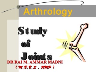
Arthrology (General Lectures). Study of Joints
•Download as PPT, PDF•
13 likes•4,667 views

The document discusses different types of joints in the human body. It describes arthrology as the scientific study of joints and their structure and function. There are three main classifications of joints: fibrous joints which connect bones using connective tissue, cartilaginous joints which connect bones using cartilage, and synovial joints which are freely movable and contained within an articular capsule. Synovial joints can be further classified based on their shape into six primary types: gliding, hinge, pivot, condyloid, saddle, and ball and socket joints. Each joint type allows for different ranges and axes of motion.

Recommended
More Related Content
What's hot
What's hot (20)
Similar to Arthrology (General Lectures). Study of Joints
Similar to Arthrology (General Lectures). Study of Joints (20)
More from DRAM NOTES | DR RAI M. AMMAR MADNI
More from DRAM NOTES | DR RAI M. AMMAR MADNI (20)
Recently uploaded
Recently uploaded (20)
Arthrology (General Lectures). Study of Joints
- 1. Arthrology StudyStudy ofof JointsJointsDR RAI M. AMMAR MADNIDR RAI M. AMMAR MADNI ( M.B.B.S , RMP )( M.B.B.S , RMP )
- 2. GET IN TOUCH AT: www.facebook.com/drraiammar www.twitter.com/drraiammar www.instagram.com/drraiammar www.linkedin.com/in/drraiammar www.themedicall.com/blog/auther/drraiammar/ For Any Book or Notes Visit Our Website: www.allmedicaldata.wordpress.com www.drraiammar.blogspot.com YouTube Channel : https://www.youtube.com/channel/UCu-oR9V3OdFNTJW5yqXWXxA BY:BY: DR RAI M. AMMAR MADNIDR RAI M. AMMAR MADNI ( M.B.B.S , RMP )( M.B.B.S , RMP )
- 3. Arthrology Greek root “arthro” means joint Arthrology The scientific study of joints. Kinesiology The study of the motion of human body.
- 4. Joints (Articulations) ALL MEDICAL DATA Joint, also called articulation is a point of contact between rigid elements of the skeleton, bones. Usually, but not always allow for movements.
- 5. Joints (Articulations) Articulations can be: Bone to bone Bone to cartilage Teeth in bony sockets Hold bones together Usually, but not always allow for movements. Enable resistance to crushing, tearing, and other forces
- 6. Bone and Cartilage Bone as tissue Bones as structures formed from bone, cartilage and other tissues Location of cartilage in skeleton and relation to joints DR RAI AMMAR, Human Anatomy, Mechanics of Movement Fig. 6.1, M&M
- 7. Ligaments Follow us At Facebook • Ligaments connect bone-to-bone or reinforce joints--they are made up of tendinous tissue as well •E.g. knee ligaments Fig. 9.12, M&M
- 8. Human Joints There are 360 joints in the human body
- 9. Hadith Prophet Muhammad (PBUH) said that the human body is made up of 360 joints. The following is the Hadith in Arabic : English translation to it :- Hasan bin Al-Hilwani narrated to us that Abu Tawbah Al-Rabie bin Nafi'a narrated that Muaawiyah Yaani ibn Salam narrated that Zaid narrated that he heard Abu Salam said that Abdallah bin Farookh narrated that he heard Aisha (the wife of the Prophet) say that the Prophet, peace be upon him, said that every man from the people of Adam (i.e., mankind) was created UNTO/UPON/ON 60 and 300 MIFSAL. So whoever glorifies Allah, praises Allah, calls people to Allah, makes supplications to Allah, seeks forgiveness from Allah, removes a stone, needle or bone from the people's path (i.e., removes obstacles from people's paths), and enjoins what is just and forbid what is wrong in the number of those 60 and 300 ALSALAMA, he will then on that day remove himself from Hell Fire.
- 10. Classification of Joints Based on Function Based on Structure
- 11. Classification of JointsClassification of Joints • Based on Function: based on amount of movement • Synarthroses – immovable joints - common in axial skeleton • Amphiarthroses – slightly moveable joints - common in axial skeleton • Diarthroses – freely moveable joints - common in appendicular skeleton
- 12. Joints by Functional Classification Type Movement Example Synarthrosis None (minimal) Sutures, Teeth, Epiphyseal plates, 1st rib and costal cart. Amphiarthrosis Slight Distal Tibia/fibula Intervertebral discs Pubic symphysis Diarthrosis Great Glenohumeral joint Knee joint TemporoMandibular Joint
- 13. Classification of JointsClassification of Joints (By Structure) • Material that binds bones together • Presence or absence of a joint cavity ased on
- 14. Classification of JointsClassification of Joints (By Structure) • Fibrous joints - Generally immovable • Cartilaginous joints - Immovable or slightly moveable • Synovial joints - Freely moveable
- 15. Classification of JointsClassification of Joints (By Structure) Fibrous • Sutures: connected by short strands of dense connective tissue (synarthroses) • Syndesmoses: connected by ligaments (varies) • Gomphosis: peg in socket w/short ligament (synarthroses) Cartilagenous • Symphysis: connected by fibrocartilage (amphiarthroses) • Synchondrosis: connected by hyaline cartilage (synarthroses) Synovial (diarthroses) • Six shapes
- 16. Joints by Structural ClassificationJoints by Structural Classification Structure Type Example Fibrous Sutures Syndesmoses Gomphosis Skull Distal Tibia/fibula Teeth in sockets Cartilagenous Symphysis Synchondrosis Intervertebral discs Epiphyseal plates Synovial 6 Shapes Glenohumeral joint Knee joint TemporoMandibular Joint
- 17. Fibrous Joints Bones united by dense(fibrous) connective tissue Mostly immovable or slightly movable – “Synarthrosis” Lack a joint cavity Types:- sutures, syndesmoses, and gomphoses
- 18. Fibrous Joints Sutures: o Bones are Interlocked by thin layer of dense connective issue and irregular edges between bones. o Allow bone growth so that the skull can expand with brain during childhood Example:- Only the bones of skull Fibrous tissue ossifies in middle age Synostoses (closed sutures)
- 19. Fibrous Joints Sutures (bones of the skull)
- 20. Fibrous Joints Syndesmoses: (amphiarthrosis) Bones are connected exclusively by ligaments Permit slight movement More fibrous connective tissue between the articulating bones The tissue is either arranged as a bundle (ligament) or as a sheet (interosseus membrane) Amount of movement depends on length of fibers
- 21. Syndesmoses Distal Tibiofibular joint – (an immovable synarthrosis
- 22. Syndesmoses Interosseus membrane between Radius and Ulna – (a freely immovable di-arthrosis)
- 23. Fibrous joints (a) (b) Dense fibrous connective tissue Suture line Fibula Tibia Suture Syndesmosis Ligament
- 24. Gomphoses A cone-shaped peg fits into a socket. Functionally classified as synarthrosis Very limited movement allowed The only example:- Articulations of the roots of teeth with the alveolar sockets of maxillae and mandible. The tooth is secured by dense fibrous connective tissue periodontal ligament.
- 25. Gomphoses Tooth in a socket Connecting ligament – the periodontal ligament
- 26. Cartilaginous Joints Articulating bone ends are connected by a plate of cartilage Lack joint cavity and joint capsule Generally contain a fibrocartilaginous disc. Permit limited or no movement; (Amphiarthroses) Two types – o symphyses & o synchondroses
- 27. Symphyses Sympheses (growing together): the ends of the articulating bones are covered with hyaline cartilage. the bones are connected by a broad flat disc of fibrocartilage - resists tension and compression Slightly movable joints that provide strength with flexibility
- 28. Symphyses Examples:- Pubic symphysis junction of the manubrium and sternum, intervertebral joints. o Functionally, amphiarthroses, a slightly movable joint
- 29. Symphyses Intervertebral joints – (Hyaline cartilage) – also present as articular cartilage
- 31. SynchondrosesSynchondroses Temporary growth joints Bones are connected by hyaline cartilage Functionally, “synarthrodial” Examples:- o The epiphyseal plate that connects epiphysis and diaphysis of a growing bone. o Joint between the first rib and manubrium of the sternum
- 32. Synchondroses Joint between first rib and manubrium
- 33. Synchondroses Hyaline cartilage unites bones o Epiphyseal plate between epiphysis and diaphysis Figure 9.2a
- 35. Synovial Joints
- 36. Synovial Joints Most movable type of joints Functionally Classified as diarthroses. Articulating bones are separated by a joint cavity synovial cavity Synovial fluid is found in the joint cavity – Uniaxial or multiaxial movement.
- 37. General Features of Synovial Joints Articular capsule:– joint cavity is enclosed in a two-layered capsule Fibrous capsule – dense irregular connective tissue – strengthens joint Synovial membrane –areolar connective tissue with elastic fibers. • Lines joint capsule and covers internal joint surfaces • Functions to make synovial fluid
- 38. Synovial JointsSynovial Joints Articular cartilage (hyaline cartilage):- o covers the ends of bones o Absorbs compression Adipose tissue - articular fat pads.
- 39. Synovial Joints Synovial fluid: o A viscous fluid similar to raw egg white o A filtrate of blood - Arises from capillaries in synovial membrane o Consists of hyaluronic acid and interstitial fluid. o Reduces friction by lubricating the joint o Supplies nutrients and removes metabolic wastes. o Contains phagocytic cells. Synovial Joints
- 40. General Features of Synovial Joints Reinforcing ligaments – Often are thickened parts of the fibrous capsule – Sometimes are extracapsular ligaments – located outside the capsule – Sometimes are intracapsular ligaments – located internal to the capsule
- 41. General Features of Synovial Joints Blood Supply:- Have a rich blood supply, Arteries penetrate the ligaments and articular capsule to deliver oxygen and nutrients. Veins remove carbon dioxide and wastes from the joints. The articulating portions receive nourishment from the fluid, rest by blood capillaries. Extensive capillary beds in the synovial membrane produce basis of synovial fluid.
- 42. General Features of Synovial Joints Nerve Supply:- Richly supplied with sensory nerves, Convey information to the brain and spinal cord. o Detect pain o Most monitor how much the capsule is being stretched
- 43. Components of SYNOVIAL JOINTS (SUMMARY) Articular cartilage: hyaline; covers articulating ends of both bones Synovial (joint) cavity: space holding synovial fluid Articular capsule: Made of 2 layers – Fibrous: external, dense CT for strength – Synovial membrane: internal, produces synovial fluid Synovial fluid: viscous; lubricates and nourishes; contained in capsule and articular cartilages Reinforcing ligaments: extracapsular/intracapsular Nerves + vessels: Highly innervated, Highly vascular Meniscus (some): fibrocartilage; improves the fit of 2 bones to increase stability
- 44. Friction-reducing structures Bursae:- • Flattened fibrous sacs, Lined with synovial membranes • filled with synovial fluidreduce friction, • not actually part of the joint Found between ; • skin and bone, • tendons and bones, • muscles and bones, • ligaments and bones. • Located in the shoulder and knee joints.
- 45. Friction-reducing structures Tendon Sheaths: Elongated , tubelike bursae that wrap around tendons, Found at ; o wrist, o ankle, o fingers and o toes.
- 46. Bursae and tendon sheaths (a) (b) Acromion of scapula Glenoid cavity containing synovial fluid Coracoacromial ligament Subacromial bursa Cavity in bursa containing synovial fluid Synovial membrane Fibrous capsule Humerus Hyaline cartilage Coracoacromial ligament Subacromial bursa Fibrous articular capsule Tendon sheath Tendon of long head of biceps brachii muscle Friction-reducing structures:
- 47. Bursae and tendon sheaths
- 49. A Typical Synovial Joint (b) Periosteum Ligament Joint cavity (contains synovial fluid) Fibrous capsule Synovial membrane Articular (hyaline) cartilage Articular capsule (a)
- 50. A Typical Synovial Joint (b) (b) Periosteum Ligament Joint cavity (contains synovial fluid) Fibrous capsule Synovial membrane Articular (hyaline) cartilage Articular capsule
- 51. Synovial Joints with Articular Discs Some synovial joints contain a Fibrocartilaginous articular disc. o Occur in joints whose articulating bones have somewhat different shapes Examples:- o Knee joint and o Temporomandibular joint (c)
- 52. Types of synovial jointsTypes of synovial joints (Based on shape) PLANE / GLIDING(ARTHODIAL) HING JOINT(GYNGLYMUS) PIVOT JOINT(TROCOID) ELLIPSOIDAL(CONDYLOID) SADDALE(SELLAR) BALL & SOCKET(SPHEROIDAL)
- 53. Types of synovial jointsTypes of synovial joints (Based on shape)
- 54. Types of synovial jointsTypes of synovial joints (Based on shape)
- 55. Types of synovial jointsTypes of synovial joints (Based on shape) Gliding or plane joints :- the articulating surfaces are flat or slightly curved. permit only a little of movement Example are intercarpal joints, intertarsal joints, sternoclavicular joints, acromioclavicular joints, sternocostal joints, vertebrocostal joints. articular processes of vertebrae
- 56. Gliding / plane joints
- 57. Types of synovial jointsTypes of synovial joints (Based on shape) Hinge (uniaxial):- cylindrical end of one bone fits into trough shape of other Movement only in one axis, e.g. coronal, sagittal or vertical axis Examples:- Knee, elbow, ankle, interphalangeal.
- 58. Hinge Joint
- 59. Types of synovial jointsTypes of synovial joints (Based on shape) Pivot Joints (uniaxial) Rounded or pointed surface of one bone articulates with a ring formed partly by another bone and partly by a ligament. Movement only in one axis
- 60. Pivot Joints Examples:- atlas/axis –atlanto- axial joint, superior and inferior radioulnar joint:
- 61. Types of synovial jointsTypes of synovial joints (Based on shape) Condyloid / ellipsoid (Biaxial). A little bit bal&socket like The convex oval-shaped projection of one fits into the oval-shaped depression of another. Movement in two axes which meet at right angle to each other The movements allowed are: flexion/extension,adduction/abduction and circumduction but no rotation.
- 62. Condyloid / ellipsoid Joint Examples:- Radiocarpal (Wrist) joint, metacarpophalangeal (knuckle)
- 63. Types of synovial jointsTypes of synovial joints (Based on shape) Saddle Joints :– (Biaxial) rare, the articular surface of one bone is saddle-shaped and the articular surface of the other fits into the “saddle”. Permit Flexion,abduction,adduction and circumduction
- 64. Examples:- o Carpometacarpal Joint of thumb o Sternoclavicular Joint Saddle Joints
- 65. Types of synovial jointsTypes of synovial joints (Based on shape) Ball-n-Socket Joints:- (Multiaxial) Most freely moving synovial joint The ball-like surface of one bone fitting into a cuplike depression of another bone. Permit flexion and extention,adduction and abduction,medial and lateral rotation, circumduction.
- 66. Ball-n-Socket Joints Examples:- Shoulder (glenohumeral) joint, Hip joints.
- 67. Types of synovial jointsTypes of synovial joints (Based on Function) Uniaxial Joints - move in one plane are called and allow one degree of freedom – i.e., Interphalyngeal joints of fingers, Humero-ulnar (Elbow) joint. Biaxial Joints - Two plane joints with two degrees of freedom – i.e.,Metacarpophalyngeal joints in hand Multiaxial Joints - Three plane joints with three degrees of freedom – i.e, Glenohumeral (Shoulder) joint
- 68. Joints MovementsJoints Movements Joint motions are dictated by; the shape of the bones in the joint and by supporting soft tissue,(muscle attachments, joint capsules and ligaments. Movements are described traditionally by the actual direction the bones move, called Osteokinematic Motion and the axis about which they move.
- 69. Joints MovementsJoints Movements Arthrokinematic Motion :- To consider the movement within the joint itself as it may be different than that of the bone. Osteokinematic Motion:- To study the movement of bone during a particular action at a joint. Example :- the glenohumeral joint – as one flexes or abducts the joint, the head of the humerus will glide inferiorly in the glenoid fossa
- 70. Synovial Joints All joints technically called rotary - one bone in some way rotates on another The moving bone rotates about an imaginary axis called the joint axis The resulting motion of the large bone is the osteokinematic motion. Kinematic chain. Refers to the linkage of joints,bones and muscles as achain of motion Closed chain:- When the end of the chain is against an object, Open chain:-When it is not opposed by the ground or an object, Example:- using the quads to ; 1) squat or 2) extend in free space
- 71. Moving the joints Active motion is produced by muscle contractions Muscles can move the same joint in a variety of ways depending on what is being stabilized Normally, we describe a muscles functions based upon its insertion moving towards the origin when contraction takes place – e.g., elbow flexion Muscle can work in reverse if the distal end is fixed. For example, in doing a chin up, the same muscles are working but the forearm is fixed or held steady and the origin of the elbow flexors moves towards insertion – called Reverse Action
- 72. Moving the joints During normal muscle contractions, the muscle fibers shorten during the activity – called concentric If a muscle lengthens during the contraction as when you perform a squat, is called eccentric (sometimes called negative) Generally, eccentric contractions are antigravity Another example :– the back muscles when you bend forward
- 73. Types of MovementsTypes of Movements Gliding: Side-to-side and back-and- forth movements. Angular movements: there is an increase or decrease in the angle between articulating movements. Includes flexion, extension, lateral flexion, hyperextension.
- 74. Types of MovementsTypes of Movements Abduction: this is the movement of a bone away from the midline. Adduction: this is the movement of bone toward the midline. Circumduction: this is the movement of the distal end of a body part in a circle.
- 75. Types of MovementsTypes of Movements Rotation: a bone revolves around its own longitudinal axis. Pivot and ball-and-socket joints permit rotation. Medial (internal) rotation and lateral (external) rotation. Special movements: elevation, depression, protraction, retraction, inversion, eversion, dorsiflexion, plantar flexion, supination, pronation, opposition.
- 76. Factors affecting ROM atFactors affecting ROM at Synovial JointsSynovial Joints Structure or shape of the articulating bones Strength and tension of ligaments. Arrangement and tension of muscles Apposition of soft parts Hormones Disuse
- 77. Aging and JointsAging and Joints Decreased production of synovial fluid Articular cartilage becomes thinner with age, ligaments shorten and lose flexibility. Genetic factors Males commonly develop degenerative changes in the vertebral column-”hunched back”. Osteoarthritis-occurs over age 70.
- 78. Inflammatory Conditions of JointsInflammatory Conditions of Joints • Bursitis – inflammation of a bursa usually caused by a blow or friction • Tendonitis – inflammation of tendon sheaths • Arthritis – inflammatory or degenerative diseases of joints • Over 100 different types • The most widespread crippling disease in the United States
- 79. Clinical Forms of ArthritisClinical Forms of Arthritis • Osteoarthritis • Most common chronic arthritis • Probably related to normal aging processes • Rheumatoid arthritis • An autoimmune disease – the immune system attacks the joints • Symptoms begin with bilateral inflammation of certain joints • Often leads to deformities
- 81. X-ray of hand affected by arthritis
- 82. X-ray of hand affected by arthritis
- 83. Clinical Forms of ArthritisClinical Forms of Arthritis Slide • Gouty Arthritis •Inflammation of joints is caused by a deposition of urate crystals from the blood •Can usually be controlled with diet
- 85. DR RAI M. AMMAR, Human Anatomy, Mechanics of Movement
- 86. Artificial Hip JointArtificial Hip Joint
- 88. GET IN TOUCH AT: www.facebook.com/drraiammar www.twitter.com/drraiammar www.instagram.com/drraiammar www.linkedin.com/in/drraiammar www.themedicall.com/blog/auther/drraiammar/ For Any Book or Notes Visit Our Website: www.allmedicaldata.wordpress.com www.drraiammar.blogspot.com YouTube Channel : https://www.youtube.com/channel/UCu-oR9V3OdFNTJW5yqXWXxA BY:BY: DR RAI M. AMMAR MADNIDR RAI M. AMMAR MADNI ( M.B.B.S , RMP )( M.B.B.S , RMP )