
Empfohlen
Weitere ähnliche Inhalte
Was ist angesagt?
Was ist angesagt? (20)
Andere mochten auch
Andere mochten auch (20)
Ähnlich wie Basic ap chapter 8 powerpoint 2017
Ähnlich wie Basic ap chapter 8 powerpoint 2017 (20)
Kürzlich hochgeladen
Kürzlich hochgeladen (20)
Basic ap chapter 8 powerpoint 2017
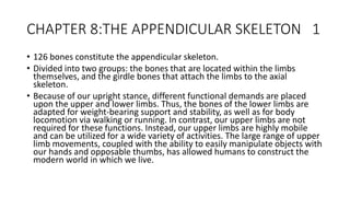
- 1. CHAPTER 8:THE APPENDICULAR SKELETON 1 • 126 bones constitute the appendicular skeleton. • Divided into two groups: the bones that are located within the limbs themselves, and the girdle bones that attach the limbs to the axial skeleton. • Because of our upright stance, different functional demands are placed upon the upper and lower limbs. Thus, the bones of the lower limbs are adapted for weight-bearing support and stability, as well as for body locomotion via walking or running. In contrast, our upper limbs are not required for these functions. Instead, our upper limbs are highly mobile and can be utilized for a wide variety of activities. The large range of upper limb movements, coupled with the ability to easily manipulate objects with our hands and opposable thumbs, has allowed humans to construct the modern world in which we live.
- 2. Pectoral Girdle 2 • The pectoral girdle consists of the clavicle and the scapula, which serve to attach the upper limb to the sternum of the axial skeleton.
- 3. Clavicle 3 • The clavicle: • only long bone that lies in a horizontal position in the body • Clavicle has several important functions. • anchored by muscles from above, it serves as a strut that extends laterally to support the scapula. • This in turn holds the shoulder joint superiorly and laterally from the body trunk, allowing for maximal freedom of motion for the upper limb. • The clavicle also transmits forces acting on the upper limb to the sternum and axial skeleton. • Finally, it serves to protect the underlying nerves and blood vessels as they pass between the trunk of the body and the upper limb.
- 4. Scapula 4 • The scapula is also part of the pectoral girdle and thus plays an important role in anchoring the upper limb to the body. • The scapula is located on the posterior side of the shoulder. • It is surrounded by muscles on both its anterior (deep) and posterior (superficial) sides, and thus does not articulate with the ribs of the thoracic cage.
- 5. Bones of the Upper Limb 5 • The upper limb is divided into three regions. • Arm, Forearm, Hand • 30 bones in each upper limb • Humerus is the single bone of the upper arm • Ulna (medially); Radius (laterally): paired bones of the forearm. • Base of the hand contains eight bones, each called a carpal bone • Palm of the hand is formed by five bones, each called a metacarpal bone. • The fingers and thumb contain a total of 14 bones, each of which is a phalanx bone of the hand
- 6. Humerus 6 • single bone of the upper arm region • The capitulum is the region of the humerus that articulates with the radius as part of the elbow joint.
- 7. ULNA 7 • Ulna is the medial bone of the forearm. • It runs parallel to the radius, which is the lateral bone of the forearm.
- 8. RADIUS 8 • The radius runs parallel to the ulna, on the lateral (thumb) side of the forearm • The radius bone has a head that articulates with the radial notch of the ulna
- 9. Carrying Angle 9 • In the anatomical position, with the elbow fully extended and the palms facing forward, the arm and forearm do not form a straight line. Instead, the forearm deviates laterally by 5–15 degrees from the line of the arm. This deviation is called the carrying angle. It allows the forearm and hand to swing freely or to carry an object without hitting the hip. The carrying angle is larger in females to accommodate their wider pelvis. • Standing in this position, with the palm facing forward, the radius is located laterally.
- 10. Carpal Bones 10 • The wrist and base of the hand are formed by a series of eight small carpal bones • The carpal bones are arranged in two rows, forming a proximal row of four carpal bones and a distal row of four carpal bones.
- 11. Carpal Tunnel 11 • Together, the carpal bones and the flexor retinaculum form a passageway called the carpal tunnel, with the carpal bones forming the walls and floor, and the flexor retinaculum forming the roof of this space • Overuse of the muscle tendons or wrist injury can produce inflammation and swelling within this space. This produces compression of the nerve, resulting in carpal tunnel syndrome, which is characterized by pain or numbness, and muscle weakness in those areas of the hand supplied by this nerve.
- 12. Metacarpal Bones 12 • The palm of the hand contains five elongated metacarpal bones. • These bones lie between the carpal bones of the wrist and the bones of the fingers and thumb. • The distal end also forms the knuckles of the hand, at the base of the fingers
- 13. Phalanx (Phalanges) 13 • The fingers and thumb contain 14 bones, each of which is called a phalanx bone (plural = phalanges). • The thumb ( pollex) is digit number 1 and has two phalanges, a proximal phalanx, and a distal phalanx bone. • Digits 2 (index finger) through 5 (little finger) have three phalanges each, called the proximal, middle, and distal phalanx bones. • An interphalangeal joint is one of the articulations between adjacent phalanges of the digits
- 14. Appendicular System: Fractures of Upper Limb Bones 14 • Due to our constant use of the hands and the rest of our upper limbs, an injury to any of these areas will cause a significant loss of functional ability. Many fractures result from a hard fall onto an outstretched hand. These injuries are especially common in elderly people whose bones are weakened due to osteoporosis. • Falls onto the hand or elbow, or direct blows to the arm, can result in fractures of the humerus. • In children, a fall onto the tip of the elbow frequently results in a distal humerus fracture. • Another frequent injury following a fall onto an outstretched hand is a Colles fracture (“col-lees”) of the distal radius.
- 15. Fractures Upper Limb Bones: 15 • There is the danger of bone necrosis and subsequent degenerative joint disease of the wrist. • Commonly fractured carpal bone is the scaphoid, often resulting from a fall onto the hand. Deep pain at the lateral wrist may yield an initial diagnosis of a wrist sprain, but a radiograph taken several weeks after the injury, after tissue swelling has subsided, will reveal the fracture.
- 16. The Pelvic Girdle and Pelvis 16 • The pelvic girdle (hip girdle) is formed by a single bone. • The hip bone or coxal bone (coxal = “hip”), which serves as the attachment point for each lower limb. Each hip bone, in turn, is firmly joined to the axial skeleton via its attachment to the sacrum of the vertebral column. The right and left hip bones also converge anteriorly to attach to each other. The bony pelvis is the entire structure formed by the two hip bones, the sacrum, and, attached inferiorly to the sacrum, the coccyx. • Unlike the bones of the pectoral girdle, which are highly mobile to enhance the range of upper limb movements, the bones of the pelvis are strongly united to each other to form a largely immobile, weight-bearing structure. • This is important for stability because it enables the weight of the body to be easily transferred laterally from the vertebral column, through the pelvic girdle and hip joints, and into either lower limb whenever the other limb is not bearing weight. • Thus, the immobility of the pelvis provides a strong foundation for the upper body as it rests on top of the mobile lower limbs.
- 17. Hip Bone 17 • The hip bone, or coxal bone, forms the pelvic girdle portion of the pelvis. The paired hip bones are the large, curved bones that form the lateral and anterior aspects of the pelvis. • Each adult hip bone is formed by three separate bones that fuse together during the late teenage years. These bony components are the ilium, ischium, and pubis. • These names are retained and used to define the three regions of the adult hip bone.
- 18. Ilium 18 • The ilium is the fan-like, superior region that forms the largest part of the hip bone. • It is firmly united to the sacrum at the largely immobile sacroiliac joint. • The ischium forms the posteroinferior region of each hip bone. It supports the body when sitting. The pubis forms the anterior portion of the hip bone. The pubis curves medially, where it joins to the pubis of the opposite hip bone at a specialized joint called the pubic symphysis.
- 19. Ischium 19 • Ischium • The ischium forms the posterolateral portion of the hip bone • The large, roughened area of the inferior ischium is the ischial tuberosity. This serves as the attachment for the posterior thigh muscles and also carries the weight of the body when sitting. • You can feel the ischial tuberosity if you wiggle your pelvis against the seat of a chair. • Projecting superiorly and anteriorly from the ischial tuberosity is a narrow segment of bone called the ischial ramus. • The slightly curved posterior margin of the ischium above the ischial tuberosity is the lesser sciatic notch. • The bony projection separating the lesser sciatic notch and greater sciatic notch is the ischial spine.
- 20. Pelvis 20 • The pelvis consists of four bones: the right and left hip bones, the sacrum, and the coccyx. • The pelvis has several important functions. Its primary role is to support the weight of the upper body when sitting and to transfer this weight to the lower limbs when standing. It serves as an attachment point for trunk and lower limb muscles, and also protects the internal pelvic organs. • When standing in the anatomical position, the pelvis is tilted anteriorly. In this position, the anterior superior iliac spines and the pubic tubercles lie in the same vertical plane, and the anterior (internal) surface of the sacrum faces forward and downward.
- 21. Female and Male Pelvis 21 • The differences between the adult female and male pelvis relate to function and body size. • In general, the bones of the male pelvis are thicker and heavier, adapted for support of the male’s heavier physical build and stronger muscles. The greater sciatic notch of the male hip bone is narrower and deeper than the broader notch of females. • Because the female pelvis is adapted for childbirth, it is wider than the male pelvis, as evidenced by the distance between the anterior superior iliac spines. The ischial tuberosities of females are also farther apart, which increases the size of the pelvic outlet. Because of this increased pelvic width, the subpubic angle is larger in females (greater than 80 degrees) than it is in males (less than 70 degrees). • The female sacrum is wider, shorter, and less curved, and the sacral promontory projects less into the pelvic cavity, thus giving the female pelvic inlet (pelvic brim) a more rounded or oval shape compared to males. • The lesser pelvic cavity of females is also wider and more shallow than the narrower, deeper, and tapering lesser pelvis of males.
- 22. Female and Male Pelvis 22 Female pelvis Male pelvis Pelvic weight Bones of the pelvis are lighter and thinner Bones of the pelvis are thicker and heavier Pelvic inlet shape Pelvic inlet has a round or oval shape Pelvic inlet is heart-shaped Lesser pelvic cavity shape Lesser pelvic cavity is shorter and wider Lesser pelvic cavity is longer and narrower Subpubic angle Subpubic angle is greater than 80 degrees Subpubic angle is less than 70 degrees Pelvic outlet shape Pelvic outlet is rounded and larger Pelvic outlet is smaller
- 23. Bones of the Lower Limb 23 • Like the upper limb, the lower limb is divided into three regions. • The thigh is that portion of the lower limb located between the hip joint and knee joint. • The leg is specifically the region between the knee joint and the ankle joint. Distal to the ankle is the foot. • The lower limb contains 30 bones. These are the femur, patella, tibia, fibula, tarsal bones, metatarsal bones, and phalanges. • he femur is the single bone of the thigh. • The patella is the kneecap and articulates with the distal femur. • The tibia is the larger, weight-bearing bone located on the medial side of the leg. • The fibula is the thin bone of the lateral leg. • The bones of the foot are divided into three groups. • The posterior portion of the foot is formed by a group of seven bones, each of which is known as a tarsal bone. • The mid-foot contains five elongated bones, each of which is a metatarsal bone. • The toes contain 14 small bones, each of which is a phalanx bone of the foot.
- 24. Femur 24 • The femur, or thigh bone, is the single bone of the thigh region. • It is the longest and strongest bone of the body, and accounts for approximately one-quarter of a person’s total height. • The rounded, proximal end is the head of the femur, which articulates with the acetabulum of the hip bone to form the hip joint. • The narrowed region below the head is the neck of the femur. This is a common area for fractures of the femur
- 25. Patella 25 • The patella (kneecap) is largest sesamoid bone of the body. • A sesamoid bone is a bone that is incorporated into the tendon of a muscle where that tendon crosses a joint. • The sesamoid bone articulates with the underlying bones to prevent damage to the muscle tendon due to rubbing against the bones during movements of the joint. • The patella does not articulate with the tibia.
- 26. Tibia 26 • The tibia (shin bone) is the medial bone of the leg and is larger than the fibula, with which it is paired. • The tibia is the main weight- bearing bone of the lower leg and the second longest bone of the body, after the femur. • The medial side of the tibia is located immediately under the skin, allowing it to be easily palpated down the entire length of the medial leg.
- 27. Fibula 27 • The fibula is the slender bone located on the lateral side of the leg. • The fibula does not bear weight. • t serves primarily for muscle attachments and thus is largely surrounded by muscles. • Only the proximal and distal ends of the fibula can be palpated.
- 28. Tarsal Bones 28 • The posterior half of the foot is formed by seven tarsal bones. • The most superior bone is the talus. This has a relatively square-shaped, upper surface that articulates with the tibia and fibula to form the ankle joint.
- 29. Metatarsals 29 • The anterior half of the foot is formed by the five metatarsal bones, which are located between the tarsal bones of the posterior foot and the phalanges of the toes. • These elongated bones are numbered 1–5, starting with the medial side of the foot. The first metatarsal bone is shorter and thicker than the others. • The second metatarsal is the longest.
- 30. Phalanges 30 • The toes contain a total of 14 phalanx bones (phalanges), arranged in a similar manner as the phalanges of the fingers. • The toes are numbered 1–5, starting with the big toe ( hallux). • The big toe has two phalanx bones, the proximal and distal phalanges. • The remaining toes all have proximal, middle, and distal phalanges. • A joint between adjacent phalanx bones is called an interphalangeal joint • Phalanges are found in both the toes and the fingers.
- 31. Arches of the Foot 31 • When the foot comes into contact with the ground during walking, running, or jumping activities, the impact of the body weight puts a tremendous amount of pressure and force on the foot. • During running, the force applied to each foot as it contacts the ground can be up to 2.5 times your body weight. The bones, joints, ligaments, and muscles of the foot absorb this force, thus greatly reducing the amount of shock that is passed superiorly into the lower limb and body. • The arches of the foot play an important role in this shock-absorbing ability. When weight is applied to the foot, these arches will flatten somewhat, thus absorbing energy. • When the weight is removed, the arch rebounds, giving “spring” to the step. • The arches also serve to distribute body weight side to side and to either end of the foot.
- 32. Transverse Arch of the Foot 32 • The foot has a transverse arch, a medial longitudinal arch, and a lateral longitudinal arch. • The transverse arch forms the medial-lateral curvature of the mid-foot. • This arch helps to distribute body weight from side to side within the foot, thus allowing the foot to accommodate uneven terrain.
- 33. Longitudinal Arch 33 • The longitudinal arches run down the length of the foot. • The lateral longitudinal arch is relatively flat, whereas the medial longitudinal arch is larger. • These ligaments have elasticity, which allows them to stretch somewhat during weight bearing, thus allowing the longitudinal arches to spread. • The stretching of these ligaments stores energy within the foot, rather than passing these forces into the leg. Contraction of the foot muscles also plays an important role in this energy absorption. • When the weight is removed, the elastic ligaments recoil and pull the ends of the arches closer together. This recovery of the arches releases the stored energy and improves the energy efficiency of walking. • Stretching of the ligaments that support the longitudinal arches can lead to pain. This can occur in overweight individuals, with people who have jobs that involve standing for long periods of time (such as a waitress), or walking or running long distances. • If stretching of the ligaments is prolonged, excessive, or repeated, it can result in a gradual lengthening of the supporting ligaments, with subsequent depression or collapse of the longitudinal arches, particularly on the medial side of the foot. • This condition is called pes planus (“flat foot” or “fallen arches”).
- 34. Congenital Clubfoot 34 • Clubfoot, also known as talipes, is a congenital (present at birth) disorder of unknown cause and is the most common deformity of the lower limb. • It affects the foot and ankle, causing the foot to be twisted inward at a sharp angle, like the head of a golf club. • Clubfoot has a frequency of about 1 out of every 1,000 births, and is twice as likely to occur in a male child as in a female child. • In 50 percent of cases, both feet are affected.
- 35. At birth, children with a clubfoot have the heel turned inward and the anterior foot twisted so that the lateral side of the foot is facing inferiorly, commonly due to ligaments or leg muscles attached to the foot that are shortened or abnormally tight. These pull the foot into an abnormal position, resulting in bone deformities. Other symptoms may include bending of the ankle that lifts the heel of the foot and an extremely high foot arch. Due to the limited range of motion in the affected foot, it is difficult to place the foot into the correct position. Additionally, the affected foot may be shorter than normal, and the calf muscles are usually underdeveloped on the affected side. Despite the appearance, this is not a painful condition for newborns. However, it must be treated early to avoid future pain and impaired walking ability. Although the cause of clubfoot is idiopathic (unknown), evidence indicates that fetal position within the uterus is not a contributing factor. Genetic factors are involved, because clubfoot tends to run within families. Cigarette smoking during pregnancy has been linked to the development of clubfoot, particularly in families with a history of clubfoot Previously, clubfoot required extensive surgery. Today, 90 percent of cases are successfully treated without surgery using new corrective casting techniques. The best chance for a full recovery requires that clubfoot treatment begin during the first 2 weeks after birth. Corrective casting gently stretches the foot, which is followed by the application of a holding cast to keep the foot in the proper position. This stretching and casting is repeated weekly for several weeks. In severe cases, surgery may also be required, after which the foot typically remains in a cast for 6 to 8 weeks. After the cast is removed following either surgical or nonsurgical treatment, the child will be required to wear a brace part-time (at night) for up to 4 years. In addition, special exercises will be prescribed, and the child must also wear special shoes. Close monitoring by the parents and adherence to postoperative instructions are imperative in minimizing the risk of relapse .Despite these difficulties, treatment for clubfoot is usually successful, and the child will grow up to lead a normal, active life. Numerous examples of individuals born with a clubfoot who went on to successful careers include Dudley Moore (comedian and actor), Damon Wayans (comedian and actor), Troy Aikman, Mia Hamm and Kristi Yamaguchi. 36