SlideShare a Scribd company logo
1 of 57
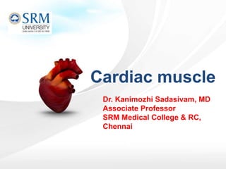
Dr. Kanimozhi Sadasivam, MD
Associate Professor
SRM Medical College & RC,
Chennai
Cardiac muscle
Learning objectives
• Define the terms; Rhythmicity, Excitability, Conductivity and
Contractility.
• Describe cardiac syncytium.
• Outline the normal pathway of the cardiac impulse.
• Describe the excitation-contraction coupling in cardiac muscles and
compare it to excitation-contraction coupling in skeletal muscles.
• Compare and contrast action potential in sino-atrial node and
ventricular muscle.
• Explain the significance of the plateau and refractory period in
ventricular muscle action potential.
The Heart
• Heart is a muscular organ that pumps blood throughout
the circulatory system
• It is situated in between two lungs in the
mediastinum
• It is made up of four chambers, two atria and two
ventricles
• The musculature of ventricles is thicker than that
of atria. Force of contraction of heart depends upon
the muscles
The Heart: Coverings
 Pericardium – a double serous membrane
 Visceral pericardium
 Next to heart
 Parietal pericardium
 Outside layer
 Serous fluid fills the space between the layers of
pericardium
The Heart: Heart Wall
 Three layers
 Epicardium
 Outside layer
 This layer is the parietal
pericardium
 Connective tissue layer
 Myocardium
 Middle layer
 Mostly cardiac muscle
 Endocardium
 Inner layer
 Endothelium
The Heart: Chambers
Slide 11.6Copyright © 2003 Pearson Education, Inc. publishing as Benjamin Cummings
 Right and left side act as separate pumps
 Four chambers
 Atria
 Receiving chambers
 Right atrium
 Left atrium
 Ventricles
 Discharging chambers
 Right ventricle
 Left ventricle
The Heart: Valves
Slide 11.8Copyright © 2003 Pearson Education, Inc. publishing as Benjamin Cummings
 Allow blood to flow in only one direction
 Four valves
 Atrioventricular valves – between atria and ventricles
 Bicuspid valve (left)
 Tricuspid valve (right)
 Semilunar valves between ventricle and artery
 Pulmonary semilunar valve
 Aortic semilunar valve
THE CARDIAC MUSCLE
• Myocardium has three types of muscle fibers:
• i. Muscle fibers which form contractile unit of
heart (99%)
• ii. Muscle fibers which form pacemaker
• iii. Muscle fibers which form conductive system
8
• Striated and resemble the skeletal muscle fibre
• Cardiac muscle fibre is bound by sarcolemma. It has a
centrally placed nucleus. Myofibrils are embedded in
the sarcoplasm.
• Sarcomere of the cardiac muscle has all the contractile
proteins, namely actin, myosin, troponin and
tropomyosin.
• Sarcotubular system in cardiac muscle is slightly different
to that of skeletal muscle.
Muscle Fibres which Form the Contractile
unit
9
Sarcotubular system in cardiac & skeletal muscle
Cardiac muscle Skeletal muscle
Location of T tubules At Z line At A-I junction
Diameter of T tubules More (5times) Less
L tubules Narrow tubular cistern Large dilated cistern
Association of T tubule (
Tubule & cistern)
Diad (1 Tubule &
1cistern)
Triad (1 Tubule &
2cistern)
Sarcomeric organisation Less regular More regular
• Exhibit branching
• Adjacent cardiac cells are joined end to end by specialized
structures known as intercalated discs
• Within intercalated discs there are two types of junctions
– Desmosomes
– Gap junctions that allow action potential to spread from one
cell to adjacent cells
• Heart function as syncytium
when one cardiac cell undergoes an action potential, the electrical
impulse spreads to all other cells that are joined by gap junctions
so they become excited and contract as a single functional
syncytium
Atrial syncytium and ventricular syncytium
Figure 10.10a
13
Structure of Cardiac Muscle Cell
Orientation of cardiac muscle fibres:
• Unlike skeletal muscles, cardiac
muscles have to contract in
• more than one direction.
• Cardiac muscle cells are
• striated, meaning they will only
contract along their long axis.
• In order to get contraction in
• two axis, the fires wrap
• around.
Muscle Fibres which Form the Pacemaker
• Some of the muscle fibres of heart are modified into a specialized
structure known as pacemaker.
• These muscle fibres forming the pacemaker have less striation.
• They are named pacemaker cells or P cells.
• Sino-atrial (SA) node forms the pacemaker in human heart.
Muscle Fibres which Form Conductive
System
• Conductive system of the heart is formed by
modified cardiac muscle fibres
• Impulses from SA node are transmitted to the
atria directly. However, the impulses are
transmitted to ventricles through various
components of conducting system
Conducting system of heart
• Electrical
– Excitability (Bathmotropic action)
– Auto rhythmicity
– Conductivity (Dromotropic action)
• Mechanical
– Contractility (Inotropic action)
– Refractory period
– Staircase / treppe effect
Action potential- the change in electrical potential associated
with the passage of an impulse along the membrane of a
muscle cell or nerve cell.
1. Autorhythmicity
 myogenic (independent of nerve supply)
 due to the specialized excitatory & conductive
system of the heart

intrinsic ability of self-excitation
(waves of depolarization)

cardiac impulses
Definition: the ability of the heart to initiate its beat
continuously and regularly without external stimulation
21
Have two important functions
1. Act as a pacemaker (set the rhythm of electrical
excitation)
2. Form the conductive system (network of
specialized cardiac muscle fibers that provide a
path for each cycle of cardiac excitation to
progress through the heart)
Autorythmic fibers
Forms 1% of the cardiac muscle fibers
22
 Sinoatrial node (SA node)
Specialized region in right atrial wall
near opening of superior
vena cava.
 Atrioventricular node (AV node)
Small bundle of specialized
cardiac cells located at base of
right atrium near septum
 Bundle of His (atrioventricular bundle)
Cells originate at AV node and
enters interventricular septum
Divides to form right and left
bundle branches which travel
down septum, curve around tip
of ventricular chambers, travel
back toward atria along outer
walls
 Purkinje fibers
Small, terminal fibers that extend from
bundle of His and spread
throughout ventricular myocardium
Locations of autorhythmic cells
Mechanism of Autorhythmicity
 Autorhythmic cells do not have
stable resting membrane potential
(RMP)
 Natural leakiness to Na & Ca
spontaneous and gradual
depolarization
 Unstable resting membrane
potential (= pacemaker potential)
 Gradual depolarization reaches
threshold (-40 mv) 
spontaneous AP generation
24
Prepotential / pacemaker potential/ Diastolic
potential
Rate of generation of AP at different sites of the heart
RATE
(Times/min)
SITE
70 - 80SA node
40 - 60AV node
20 - 35AV bundle, bundle
branches,& Purkinje
fibres
SA node acts as heart pacemaker because it has the fastest rate of generating
action potential
Nerve impulses from autonomic nervous system and hormones modify the
timing and strength of each heart beat but do not establish the fundamental
rhythm.
26
• Non-SA nodal tissues are latent pacemakers that can take
over (at a slower rate), should the normal pacemaker (SA node )
fail 29
Demonstration of Properties of Cardiac
Muscle
• The properties of cardiac muscle are demonstrated
using a quiescent heart.
• A quiescent heart is a heart which has stopped
beating but is still alive.
• Such a preparation can be obtained by tying a
Stannius Ligature in the frog’s heart.
Autorhymicity- effect of Stannius Ligature in
the frog’s heart
2. Excitability
Definition: The ability of cardiac muscle to respond to
a stimulus of adequate strength & duration by
generating an AP
• AP initiated by SA node travels along
conductive pathway excites atrial &ventricular
muscle fibres
32
Action potential in contractile fibers
AP-contraction relationship:
• AP in skeletal muscle is
very short-lived-AP is basically
over before an increase in
muscle tension can be measured
• AP in cardiac muscle is
very long-lived
– AP has an extra component
,which extends the duration
.
– The contraction is almost
over before the action
potential has finished.
Refractory Period
• It is that period during which a second stimulus fails to
evoke a response.
• Absolute Refractory Period : It is that period during
which a second stimulus however high it is fails to
evoke a response.
• Relative Refractory Period : It is that period during
which a second stimulus evokes a response if it is
sufficiently high.
Refractory period
• Long refractory period (250 msec)
compared to skeletal muscle (3msec)
• During this period membrane is refractory to f
urther stimulation until contraction is
over.
• It lasts longer than muscle contraction,
prevents tetanus
• Gives time to heart to relax after each contract
ion, prevent fatigue
• It allows time for the heart chambers to fill
during diastole before next contraction
Normal Cardiogram
• It is a recording of the mechanical activity of
the heart
• Systole- Contraction Diastole- Relaxation
Extra systole
• It is an extra contraction seen when the second
stimulus falls during the relative refractory period.
• Systole-Down stroke
• Diastole-Up stroke
Compensatory Pause
• When an external stimulus is applied during the
later 2/3 of diastole an extra contraction is
observed.
• This is followed by a compensatory pause.
• Extra systole + Compensatory pause = 2 cardiac
cycles
Extrasystole & compensatory pause
3. Contractility
Definition: ability of cardiac muscle to contract in
response to stimulation
43
All Or None Law
• The response to a threshold stimulus is maximal. If the
stimulus is below threshold there is no response
provided the physiological conditions remain constant
• The cardiac muscle follows the all or none law as a
whole.
• In the case of skeletal muscle, all-or-none law
is applicable only to a single muscle fiber.
Treppe or Stair-case Phenomenon
• When stimuli of same strength are applied at short
intervals, an increase in the height of contraction is
observed.
• This is due to the BENEFICIAL EFFECT - decrease in
viscosity, mild increase in temperature and increase in
the level of calcium ions.
Summation of Sub-minimal Stimuli
When a series of sub-minimal stimuli are applied
to the cardiac muscle, it responds with a
contraction once all the sub –minimal add up to
produce a threshold stimulus.
Excitation-Contraction Coupling in
Cardiac Contractile Cells
Similar to that in
skeletal muscles
46
The cardiac muscle stores much more calcium in its
tubular system than skeletal muscle and is much more
dependent on extracellular calcium than the skeletal
muscle.
An abundance of calcium is bound by the mucopolysaccha
-rides inside the T-tubule.
This calcium is necessary for contraction of cardiac muscle,
and its strength of contraction depends on the calcium
concentration surrounding the cardiac myocytes.
At the initiation of the action potential, the fast sodium
channels open first, followed later by the opening of the
slow calcium channels.
4. Conductivity
Definition: property by which excitation is conducted
through the cardiac tissue
48
Tissue Conduction rate (m/s)
Atrial muscle 0.3
Atrial pathways 1
AV node 0.05
Bundle of His 1
Purkinje system 4
Ventricular muscle 0.3-0.5
49
Thus, the velocity of impulses is maximum in
Purkinje fibers and minimum at AV node
The atrial and ventricular muscles have a relatively rapid
rate of conduction of the cardiac action potential, and the
anterior internodal pathway also has fairly rapid conduction
of the impulse.
However, the A-V bundle myofibrils have a slow rate of
conduction because their sizes are considerably smaller
than the sizes of the normal atrial and ventricular muscle.
Also, their slow conduction is partly caused by diminished
numbers of gap junctions between successive muscle cells
in the conducting pathway, causing a great resistance to
conduction of the excitatory ions from one cell to the next.
Criteria for spread of excitation & efficient
cardiac function
1. Atrial excitation and contraction should be complete
before onset of ventricular contraction- ensures complete
filling of the ventricles during diastole
2. Excitation of cardiac muscle fibres should be coordinated
ensure each heart chamber contracts as a unit 
accomplish efficient pumping-smooth uniform contraction
essential to squeeze out blood
3. Pair of atria & pair of ventricles should be functionally co-
ordinated  both members contract simultaneously
- permits synchronized pumping of blood into pulmonary
& systemic circulation
51
Summary
• In which phase of the ventricular muscle action
potential is the potassium permeability the
highest?
• A) 0
• B) 1
• C) 2
• D) 3
• E) 4
• Which of the following statements about cardiac muscle is most accurate?
• A) The T-tubules of cardiac muscle can store much less
calcium than T-tubules in skeletal muscle
• B) The strength and contraction of cardiac muscle depends on the amount
of calcium surrounding cardiac myocytes
• C) In cardiac muscle the initiation of the action potential
causes an immediate opening of slow calcium channels
• D) Cardiac muscle repolarization is caused by opening of
sodium channels
• E) Mucopolysaccharides inside the T-tubules bind chloride
ions
• Which of the following structures will have
the slowest rate of conduction of the cardiac AP?
• A) Atrial muscle
• B) Anterior internodal pathway
• C) A-V bundle fibers
• D) Purkinje fibers
• E) Ventricular muscle
• What is the membrane potential (threshold level
) at which the S-A node discharges?
• A) −40 mV
• B) −55 mV
• C) −65 mV
• D) −85 mV
• E) −105 mV
• If the ventricular Purkinje fibers become the
pacemaker of the heart, what is the expected HR?
• A) 30/min
• B) 50/min
• C) 65/min
• D) 75/min
• E) 85/min
• What is the resting membrane potential of the
sinus nodal fibers?
• A) −100 mV
• B) −90 mV
• C) −80 mV
• D) −55 mV
• E) −20 mV
Thank you!

More Related Content

What's hot (20)

NEUROMUSCULAR JUNCTION
NEUROMUSCULAR JUNCTIONNEUROMUSCULAR JUNCTION
NEUROMUSCULAR JUNCTION
 
Regulation of heart rate
Regulation of heart rateRegulation of heart rate
Regulation of heart rate
 
Smooth Muscles
Smooth MusclesSmooth Muscles
Smooth Muscles
 
Regulation of respiration
Regulation of respirationRegulation of respiration
Regulation of respiration
 
Mechanism of-respiration
Mechanism of-respirationMechanism of-respiration
Mechanism of-respiration
 
Events of cardiac cycle and cardiac output
Events of cardiac cycle and cardiac outputEvents of cardiac cycle and cardiac output
Events of cardiac cycle and cardiac output
 
DETERMINANTS AND FACTORS AFFECTING CARDIAC OUTPUT
DETERMINANTS AND FACTORS AFFECTING CARDIAC OUTPUTDETERMINANTS AND FACTORS AFFECTING CARDIAC OUTPUT
DETERMINANTS AND FACTORS AFFECTING CARDIAC OUTPUT
 
Lung volume and capacities
Lung volume and capacitiesLung volume and capacities
Lung volume and capacities
 
JUXTA GLOMERULAR apparatus
JUXTA GLOMERULAR apparatus JUXTA GLOMERULAR apparatus
JUXTA GLOMERULAR apparatus
 
Coronary circulation
Coronary circulationCoronary circulation
Coronary circulation
 
Muscle contraction
Muscle contractionMuscle contraction
Muscle contraction
 
Cardiovascular physiology
Cardiovascular physiologyCardiovascular physiology
Cardiovascular physiology
 
Physiology of gas exchange
Physiology of gas exchangePhysiology of gas exchange
Physiology of gas exchange
 
Structure of the Cardiac muscles
Structure of the Cardiac muscles Structure of the Cardiac muscles
Structure of the Cardiac muscles
 
3.9 SMOOTH MUSCLE PHYSIOLOGY
3.9 SMOOTH MUSCLE PHYSIOLOGY3.9 SMOOTH MUSCLE PHYSIOLOGY
3.9 SMOOTH MUSCLE PHYSIOLOGY
 
Regulation of blood pressure
Regulation of blood pressureRegulation of blood pressure
Regulation of blood pressure
 
The pleurae
The pleuraeThe pleurae
The pleurae
 
Blood supply to heart
Blood supply to heartBlood supply to heart
Blood supply to heart
 
Cardiac muscle
Cardiac muscleCardiac muscle
Cardiac muscle
 
Cardiac output
Cardiac outputCardiac output
Cardiac output
 

Similar to Cardiac muscle physiology

Cardiovascular physiology
Cardiovascular physiologyCardiovascular physiology
Cardiovascular physiologyAmen Ullah
 
Basic Exercise physiology
Basic Exercise physiologyBasic Exercise physiology
Basic Exercise physiologyedwinmills39
 
Cvs 1. functional anatomy
Cvs 1.  functional anatomyCvs 1.  functional anatomy
Cvs 1. functional anatomyRameshKumar1814
 
Cardiovascular physiology.1. hussein farouk sakr
Cardiovascular physiology.1. hussein farouk sakrCardiovascular physiology.1. hussein farouk sakr
Cardiovascular physiology.1. hussein farouk sakrHussein Sakr
 
1AA L Cardiovascular system.ppt
1AA L Cardiovascular system.ppt1AA L Cardiovascular system.ppt
1AA L Cardiovascular system.pptMalikMomin3
 
Cardiovascular Physiology.ppt basic scie
Cardiovascular Physiology.ppt basic scieCardiovascular Physiology.ppt basic scie
Cardiovascular Physiology.ppt basic scieHarithamuralidharan3
 
L3 conduction system of the heart
L3 conduction system of the heartL3 conduction system of the heart
L3 conduction system of the heartReach Na
 
Chapter 5b- CVSvvvvvvvvvvvvvvvvvvvvvv.pptx
Chapter 5b- CVSvvvvvvvvvvvvvvvvvvvvvv.pptxChapter 5b- CVSvvvvvvvvvvvvvvvvvvvvvv.pptx
Chapter 5b- CVSvvvvvvvvvvvvvvvvvvvvvv.pptxErmiyasBeletew
 
physiologyofheart-181202084458.pdf
physiologyofheart-181202084458.pdfphysiologyofheart-181202084458.pdf
physiologyofheart-181202084458.pdfVijayKumar592916
 
cardiovascular physiology
 cardiovascular physiology cardiovascular physiology
cardiovascular physiologyaliagr
 
Cardiac muscle (The Guyton and Hall Physiology)
Cardiac muscle (The Guyton and Hall Physiology)Cardiac muscle (The Guyton and Hall Physiology)
Cardiac muscle (The Guyton and Hall Physiology)Maryam Fida
 
biomechanicsofcirculation-190924150904.pdf
biomechanicsofcirculation-190924150904.pdfbiomechanicsofcirculation-190924150904.pdf
biomechanicsofcirculation-190924150904.pdfVeenaMoondra
 
3. Cardiovacular system.pptx
3. Cardiovacular system.pptx3. Cardiovacular system.pptx
3. Cardiovacular system.pptxAbhiDabra
 

Similar to Cardiac muscle physiology (20)

The Myocardium
The MyocardiumThe Myocardium
The Myocardium
 
Cardiovascular physiology
Cardiovascular physiologyCardiovascular physiology
Cardiovascular physiology
 
Cardiac muscle tissue
Cardiac muscle tissueCardiac muscle tissue
Cardiac muscle tissue
 
Basic Exercise physiology
Basic Exercise physiologyBasic Exercise physiology
Basic Exercise physiology
 
Cvs 1. functional anatomy
Cvs 1.  functional anatomyCvs 1.  functional anatomy
Cvs 1. functional anatomy
 
Cardiovascular physiology.1. hussein farouk sakr
Cardiovascular physiology.1. hussein farouk sakrCardiovascular physiology.1. hussein farouk sakr
Cardiovascular physiology.1. hussein farouk sakr
 
HAP I CLASS.pptx
HAP I CLASS.pptxHAP I CLASS.pptx
HAP I CLASS.pptx
 
heart.pptx
heart.pptxheart.pptx
heart.pptx
 
1AA L Cardiovascular system.ppt
1AA L Cardiovascular system.ppt1AA L Cardiovascular system.ppt
1AA L Cardiovascular system.ppt
 
Cardiovascular Physiology.ppt basic scie
Cardiovascular Physiology.ppt basic scieCardiovascular Physiology.ppt basic scie
Cardiovascular Physiology.ppt basic scie
 
L3 conduction system of the heart
L3 conduction system of the heartL3 conduction system of the heart
L3 conduction system of the heart
 
Chapter 5b- CVSvvvvvvvvvvvvvvvvvvvvvv.pptx
Chapter 5b- CVSvvvvvvvvvvvvvvvvvvvvvv.pptxChapter 5b- CVSvvvvvvvvvvvvvvvvvvvvvv.pptx
Chapter 5b- CVSvvvvvvvvvvvvvvvvvvvvvv.pptx
 
Physiology of heart
Physiology of heartPhysiology of heart
Physiology of heart
 
physiologyofheart-181202084458.pdf
physiologyofheart-181202084458.pdfphysiologyofheart-181202084458.pdf
physiologyofheart-181202084458.pdf
 
cardiac.ppt
cardiac.pptcardiac.ppt
cardiac.ppt
 
cardiovascular physiology
 cardiovascular physiology cardiovascular physiology
cardiovascular physiology
 
Cardiac muscle (The Guyton and Hall Physiology)
Cardiac muscle (The Guyton and Hall Physiology)Cardiac muscle (The Guyton and Hall Physiology)
Cardiac muscle (The Guyton and Hall Physiology)
 
biomechanicsofcirculation-190924150904.pdf
biomechanicsofcirculation-190924150904.pdfbiomechanicsofcirculation-190924150904.pdf
biomechanicsofcirculation-190924150904.pdf
 
Biomechanics of circulation
Biomechanics of circulationBiomechanics of circulation
Biomechanics of circulation
 
3. Cardiovacular system.pptx
3. Cardiovacular system.pptx3. Cardiovacular system.pptx
3. Cardiovacular system.pptx
 

Recently uploaded

Clinical Pharmacotherapy of Scabies Disease
Clinical Pharmacotherapy of Scabies DiseaseClinical Pharmacotherapy of Scabies Disease
Clinical Pharmacotherapy of Scabies DiseaseSreenivasa Reddy Thalla
 
CEHPALOSPORINS.pptx By Harshvardhan Dev Bhoomi Uttarakhand University
CEHPALOSPORINS.pptx By Harshvardhan Dev Bhoomi Uttarakhand UniversityCEHPALOSPORINS.pptx By Harshvardhan Dev Bhoomi Uttarakhand University
CEHPALOSPORINS.pptx By Harshvardhan Dev Bhoomi Uttarakhand UniversityHarshChauhan475104
 
Lippincott Microcards_ Microbiology Flash Cards-LWW (2015).pdf
Lippincott Microcards_ Microbiology Flash Cards-LWW (2015).pdfLippincott Microcards_ Microbiology Flash Cards-LWW (2015).pdf
Lippincott Microcards_ Microbiology Flash Cards-LWW (2015).pdfSreeja Cherukuru
 
97111 47426 Call Girls In Delhi MUNIRKAA
97111 47426 Call Girls In Delhi MUNIRKAA97111 47426 Call Girls In Delhi MUNIRKAA
97111 47426 Call Girls In Delhi MUNIRKAAjennyeacort
 
Informed Consent Empowering Healthcare Decision-Making.pptx
Informed Consent Empowering Healthcare Decision-Making.pptxInformed Consent Empowering Healthcare Decision-Making.pptx
Informed Consent Empowering Healthcare Decision-Making.pptxSasikiranMarri
 
SWD (Short wave diathermy)- Physiotherapy.ppt
SWD (Short wave diathermy)- Physiotherapy.pptSWD (Short wave diathermy)- Physiotherapy.ppt
SWD (Short wave diathermy)- Physiotherapy.pptMumux Mirani
 
Hematology and Immunology - Leukocytes Functions
Hematology and Immunology - Leukocytes FunctionsHematology and Immunology - Leukocytes Functions
Hematology and Immunology - Leukocytes FunctionsMedicoseAcademics
 
Apiculture Chapter 1. Introduction 2.ppt
Apiculture Chapter 1. Introduction 2.pptApiculture Chapter 1. Introduction 2.ppt
Apiculture Chapter 1. Introduction 2.pptkedirjemalharun
 
PULMONARY EDEMA AND ITS MANAGEMENT.pdf
PULMONARY EDEMA AND  ITS  MANAGEMENT.pdfPULMONARY EDEMA AND  ITS  MANAGEMENT.pdf
PULMONARY EDEMA AND ITS MANAGEMENT.pdfDolisha Warbi
 
Culture and Health Disorders Social change.pptx
Culture and Health Disorders Social change.pptxCulture and Health Disorders Social change.pptx
Culture and Health Disorders Social change.pptxDr. Dheeraj Kumar
 
SYNDESMOTIC INJURY- ANATOMICAL REPAIR.pptx
SYNDESMOTIC INJURY- ANATOMICAL REPAIR.pptxSYNDESMOTIC INJURY- ANATOMICAL REPAIR.pptx
SYNDESMOTIC INJURY- ANATOMICAL REPAIR.pptxdrashraf369
 
Basic principles involved in the traditional systems of medicine PDF.pdf
Basic principles involved in the traditional systems of medicine PDF.pdfBasic principles involved in the traditional systems of medicine PDF.pdf
Basic principles involved in the traditional systems of medicine PDF.pdfDivya Kanojiya
 
PERFECT BUT PAINFUL TKR -ROLE OF SYNOVECTOMY.pptx
PERFECT BUT PAINFUL TKR -ROLE OF SYNOVECTOMY.pptxPERFECT BUT PAINFUL TKR -ROLE OF SYNOVECTOMY.pptx
PERFECT BUT PAINFUL TKR -ROLE OF SYNOVECTOMY.pptxdrashraf369
 
systemic bacteriology (7)............pptx
systemic bacteriology (7)............pptxsystemic bacteriology (7)............pptx
systemic bacteriology (7)............pptxEyobAlemu11
 
VarSeq 2.6.0: Advancing Pharmacogenomics and Genomic Analysis
VarSeq 2.6.0: Advancing Pharmacogenomics and Genomic AnalysisVarSeq 2.6.0: Advancing Pharmacogenomics and Genomic Analysis
VarSeq 2.6.0: Advancing Pharmacogenomics and Genomic AnalysisGolden Helix
 
Study on the Impact of FOCUS-PDCA Management Model on the Disinfection Qualit...
Study on the Impact of FOCUS-PDCA Management Model on the Disinfection Qualit...Study on the Impact of FOCUS-PDCA Management Model on the Disinfection Qualit...
Study on the Impact of FOCUS-PDCA Management Model on the Disinfection Qualit...MehranMouzam
 
MedDRA-A-Comprehensive-Guide-to-Standardized-Medical-Terminology.pdf
MedDRA-A-Comprehensive-Guide-to-Standardized-Medical-Terminology.pdfMedDRA-A-Comprehensive-Guide-to-Standardized-Medical-Terminology.pdf
MedDRA-A-Comprehensive-Guide-to-Standardized-Medical-Terminology.pdfSasikiranMarri
 
Music Therapy's Impact in Palliative Care| IAPCON2024| Dr. Tara Rajendran
Music Therapy's Impact in Palliative Care| IAPCON2024| Dr. Tara RajendranMusic Therapy's Impact in Palliative Care| IAPCON2024| Dr. Tara Rajendran
Music Therapy's Impact in Palliative Care| IAPCON2024| Dr. Tara RajendranTara Rajendran
 
Big Data Analysis Suggests COVID Vaccination Increases Excess Mortality Of ...
Big Data Analysis Suggests COVID  Vaccination Increases Excess Mortality Of  ...Big Data Analysis Suggests COVID  Vaccination Increases Excess Mortality Of  ...
Big Data Analysis Suggests COVID Vaccination Increases Excess Mortality Of ...sdateam0
 

Recently uploaded (20)

Clinical Pharmacotherapy of Scabies Disease
Clinical Pharmacotherapy of Scabies DiseaseClinical Pharmacotherapy of Scabies Disease
Clinical Pharmacotherapy of Scabies Disease
 
CEHPALOSPORINS.pptx By Harshvardhan Dev Bhoomi Uttarakhand University
CEHPALOSPORINS.pptx By Harshvardhan Dev Bhoomi Uttarakhand UniversityCEHPALOSPORINS.pptx By Harshvardhan Dev Bhoomi Uttarakhand University
CEHPALOSPORINS.pptx By Harshvardhan Dev Bhoomi Uttarakhand University
 
Lippincott Microcards_ Microbiology Flash Cards-LWW (2015).pdf
Lippincott Microcards_ Microbiology Flash Cards-LWW (2015).pdfLippincott Microcards_ Microbiology Flash Cards-LWW (2015).pdf
Lippincott Microcards_ Microbiology Flash Cards-LWW (2015).pdf
 
97111 47426 Call Girls In Delhi MUNIRKAA
97111 47426 Call Girls In Delhi MUNIRKAA97111 47426 Call Girls In Delhi MUNIRKAA
97111 47426 Call Girls In Delhi MUNIRKAA
 
Informed Consent Empowering Healthcare Decision-Making.pptx
Informed Consent Empowering Healthcare Decision-Making.pptxInformed Consent Empowering Healthcare Decision-Making.pptx
Informed Consent Empowering Healthcare Decision-Making.pptx
 
SWD (Short wave diathermy)- Physiotherapy.ppt
SWD (Short wave diathermy)- Physiotherapy.pptSWD (Short wave diathermy)- Physiotherapy.ppt
SWD (Short wave diathermy)- Physiotherapy.ppt
 
Hematology and Immunology - Leukocytes Functions
Hematology and Immunology - Leukocytes FunctionsHematology and Immunology - Leukocytes Functions
Hematology and Immunology - Leukocytes Functions
 
Apiculture Chapter 1. Introduction 2.ppt
Apiculture Chapter 1. Introduction 2.pptApiculture Chapter 1. Introduction 2.ppt
Apiculture Chapter 1. Introduction 2.ppt
 
PULMONARY EDEMA AND ITS MANAGEMENT.pdf
PULMONARY EDEMA AND  ITS  MANAGEMENT.pdfPULMONARY EDEMA AND  ITS  MANAGEMENT.pdf
PULMONARY EDEMA AND ITS MANAGEMENT.pdf
 
Culture and Health Disorders Social change.pptx
Culture and Health Disorders Social change.pptxCulture and Health Disorders Social change.pptx
Culture and Health Disorders Social change.pptx
 
SYNDESMOTIC INJURY- ANATOMICAL REPAIR.pptx
SYNDESMOTIC INJURY- ANATOMICAL REPAIR.pptxSYNDESMOTIC INJURY- ANATOMICAL REPAIR.pptx
SYNDESMOTIC INJURY- ANATOMICAL REPAIR.pptx
 
Basic principles involved in the traditional systems of medicine PDF.pdf
Basic principles involved in the traditional systems of medicine PDF.pdfBasic principles involved in the traditional systems of medicine PDF.pdf
Basic principles involved in the traditional systems of medicine PDF.pdf
 
PERFECT BUT PAINFUL TKR -ROLE OF SYNOVECTOMY.pptx
PERFECT BUT PAINFUL TKR -ROLE OF SYNOVECTOMY.pptxPERFECT BUT PAINFUL TKR -ROLE OF SYNOVECTOMY.pptx
PERFECT BUT PAINFUL TKR -ROLE OF SYNOVECTOMY.pptx
 
systemic bacteriology (7)............pptx
systemic bacteriology (7)............pptxsystemic bacteriology (7)............pptx
systemic bacteriology (7)............pptx
 
VarSeq 2.6.0: Advancing Pharmacogenomics and Genomic Analysis
VarSeq 2.6.0: Advancing Pharmacogenomics and Genomic AnalysisVarSeq 2.6.0: Advancing Pharmacogenomics and Genomic Analysis
VarSeq 2.6.0: Advancing Pharmacogenomics and Genomic Analysis
 
Study on the Impact of FOCUS-PDCA Management Model on the Disinfection Qualit...
Study on the Impact of FOCUS-PDCA Management Model on the Disinfection Qualit...Study on the Impact of FOCUS-PDCA Management Model on the Disinfection Qualit...
Study on the Impact of FOCUS-PDCA Management Model on the Disinfection Qualit...
 
MedDRA-A-Comprehensive-Guide-to-Standardized-Medical-Terminology.pdf
MedDRA-A-Comprehensive-Guide-to-Standardized-Medical-Terminology.pdfMedDRA-A-Comprehensive-Guide-to-Standardized-Medical-Terminology.pdf
MedDRA-A-Comprehensive-Guide-to-Standardized-Medical-Terminology.pdf
 
Music Therapy's Impact in Palliative Care| IAPCON2024| Dr. Tara Rajendran
Music Therapy's Impact in Palliative Care| IAPCON2024| Dr. Tara RajendranMusic Therapy's Impact in Palliative Care| IAPCON2024| Dr. Tara Rajendran
Music Therapy's Impact in Palliative Care| IAPCON2024| Dr. Tara Rajendran
 
Epilepsy
EpilepsyEpilepsy
Epilepsy
 
Big Data Analysis Suggests COVID Vaccination Increases Excess Mortality Of ...
Big Data Analysis Suggests COVID  Vaccination Increases Excess Mortality Of  ...Big Data Analysis Suggests COVID  Vaccination Increases Excess Mortality Of  ...
Big Data Analysis Suggests COVID Vaccination Increases Excess Mortality Of ...
 

Cardiac muscle physiology

  • 1. Dr. Kanimozhi Sadasivam, MD Associate Professor SRM Medical College & RC, Chennai Cardiac muscle
  • 2. Learning objectives • Define the terms; Rhythmicity, Excitability, Conductivity and Contractility. • Describe cardiac syncytium. • Outline the normal pathway of the cardiac impulse. • Describe the excitation-contraction coupling in cardiac muscles and compare it to excitation-contraction coupling in skeletal muscles. • Compare and contrast action potential in sino-atrial node and ventricular muscle. • Explain the significance of the plateau and refractory period in ventricular muscle action potential.
  • 3. The Heart • Heart is a muscular organ that pumps blood throughout the circulatory system • It is situated in between two lungs in the mediastinum • It is made up of four chambers, two atria and two ventricles • The musculature of ventricles is thicker than that of atria. Force of contraction of heart depends upon the muscles
  • 4. The Heart: Coverings  Pericardium – a double serous membrane  Visceral pericardium  Next to heart  Parietal pericardium  Outside layer  Serous fluid fills the space between the layers of pericardium
  • 5. The Heart: Heart Wall  Three layers  Epicardium  Outside layer  This layer is the parietal pericardium  Connective tissue layer  Myocardium  Middle layer  Mostly cardiac muscle  Endocardium  Inner layer  Endothelium
  • 6. The Heart: Chambers Slide 11.6Copyright © 2003 Pearson Education, Inc. publishing as Benjamin Cummings  Right and left side act as separate pumps  Four chambers  Atria  Receiving chambers  Right atrium  Left atrium  Ventricles  Discharging chambers  Right ventricle  Left ventricle
  • 7. The Heart: Valves Slide 11.8Copyright © 2003 Pearson Education, Inc. publishing as Benjamin Cummings  Allow blood to flow in only one direction  Four valves  Atrioventricular valves – between atria and ventricles  Bicuspid valve (left)  Tricuspid valve (right)  Semilunar valves between ventricle and artery  Pulmonary semilunar valve  Aortic semilunar valve
  • 8. THE CARDIAC MUSCLE • Myocardium has three types of muscle fibers: • i. Muscle fibers which form contractile unit of heart (99%) • ii. Muscle fibers which form pacemaker • iii. Muscle fibers which form conductive system 8
  • 9. • Striated and resemble the skeletal muscle fibre • Cardiac muscle fibre is bound by sarcolemma. It has a centrally placed nucleus. Myofibrils are embedded in the sarcoplasm. • Sarcomere of the cardiac muscle has all the contractile proteins, namely actin, myosin, troponin and tropomyosin. • Sarcotubular system in cardiac muscle is slightly different to that of skeletal muscle. Muscle Fibres which Form the Contractile unit 9
  • 10. Sarcotubular system in cardiac & skeletal muscle Cardiac muscle Skeletal muscle Location of T tubules At Z line At A-I junction Diameter of T tubules More (5times) Less L tubules Narrow tubular cistern Large dilated cistern Association of T tubule ( Tubule & cistern) Diad (1 Tubule & 1cistern) Triad (1 Tubule & 2cistern) Sarcomeric organisation Less regular More regular
  • 11. • Exhibit branching • Adjacent cardiac cells are joined end to end by specialized structures known as intercalated discs • Within intercalated discs there are two types of junctions – Desmosomes – Gap junctions that allow action potential to spread from one cell to adjacent cells • Heart function as syncytium when one cardiac cell undergoes an action potential, the electrical impulse spreads to all other cells that are joined by gap junctions so they become excited and contract as a single functional syncytium Atrial syncytium and ventricular syncytium
  • 13. 13 Structure of Cardiac Muscle Cell
  • 14. Orientation of cardiac muscle fibres: • Unlike skeletal muscles, cardiac muscles have to contract in • more than one direction. • Cardiac muscle cells are • striated, meaning they will only contract along their long axis. • In order to get contraction in • two axis, the fires wrap • around.
  • 15. Muscle Fibres which Form the Pacemaker • Some of the muscle fibres of heart are modified into a specialized structure known as pacemaker. • These muscle fibres forming the pacemaker have less striation. • They are named pacemaker cells or P cells. • Sino-atrial (SA) node forms the pacemaker in human heart.
  • 16. Muscle Fibres which Form Conductive System • Conductive system of the heart is formed by modified cardiac muscle fibres • Impulses from SA node are transmitted to the atria directly. However, the impulses are transmitted to ventricles through various components of conducting system
  • 18. • Electrical – Excitability (Bathmotropic action) – Auto rhythmicity – Conductivity (Dromotropic action) • Mechanical – Contractility (Inotropic action) – Refractory period – Staircase / treppe effect
  • 19.
  • 20. Action potential- the change in electrical potential associated with the passage of an impulse along the membrane of a muscle cell or nerve cell.
  • 21. 1. Autorhythmicity  myogenic (independent of nerve supply)  due to the specialized excitatory & conductive system of the heart  intrinsic ability of self-excitation (waves of depolarization)  cardiac impulses Definition: the ability of the heart to initiate its beat continuously and regularly without external stimulation 21
  • 22. Have two important functions 1. Act as a pacemaker (set the rhythm of electrical excitation) 2. Form the conductive system (network of specialized cardiac muscle fibers that provide a path for each cycle of cardiac excitation to progress through the heart) Autorythmic fibers Forms 1% of the cardiac muscle fibers 22
  • 23.  Sinoatrial node (SA node) Specialized region in right atrial wall near opening of superior vena cava.  Atrioventricular node (AV node) Small bundle of specialized cardiac cells located at base of right atrium near septum  Bundle of His (atrioventricular bundle) Cells originate at AV node and enters interventricular septum Divides to form right and left bundle branches which travel down septum, curve around tip of ventricular chambers, travel back toward atria along outer walls  Purkinje fibers Small, terminal fibers that extend from bundle of His and spread throughout ventricular myocardium Locations of autorhythmic cells
  • 24. Mechanism of Autorhythmicity  Autorhythmic cells do not have stable resting membrane potential (RMP)  Natural leakiness to Na & Ca spontaneous and gradual depolarization  Unstable resting membrane potential (= pacemaker potential)  Gradual depolarization reaches threshold (-40 mv)  spontaneous AP generation 24
  • 25. Prepotential / pacemaker potential/ Diastolic potential
  • 26. Rate of generation of AP at different sites of the heart RATE (Times/min) SITE 70 - 80SA node 40 - 60AV node 20 - 35AV bundle, bundle branches,& Purkinje fibres SA node acts as heart pacemaker because it has the fastest rate of generating action potential Nerve impulses from autonomic nervous system and hormones modify the timing and strength of each heart beat but do not establish the fundamental rhythm. 26
  • 27.
  • 28. • Non-SA nodal tissues are latent pacemakers that can take over (at a slower rate), should the normal pacemaker (SA node ) fail 29
  • 29. Demonstration of Properties of Cardiac Muscle • The properties of cardiac muscle are demonstrated using a quiescent heart. • A quiescent heart is a heart which has stopped beating but is still alive. • Such a preparation can be obtained by tying a Stannius Ligature in the frog’s heart.
  • 30. Autorhymicity- effect of Stannius Ligature in the frog’s heart
  • 31. 2. Excitability Definition: The ability of cardiac muscle to respond to a stimulus of adequate strength & duration by generating an AP • AP initiated by SA node travels along conductive pathway excites atrial &ventricular muscle fibres 32
  • 32. Action potential in contractile fibers
  • 33. AP-contraction relationship: • AP in skeletal muscle is very short-lived-AP is basically over before an increase in muscle tension can be measured • AP in cardiac muscle is very long-lived – AP has an extra component ,which extends the duration . – The contraction is almost over before the action potential has finished.
  • 34.
  • 35.
  • 36. Refractory Period • It is that period during which a second stimulus fails to evoke a response. • Absolute Refractory Period : It is that period during which a second stimulus however high it is fails to evoke a response. • Relative Refractory Period : It is that period during which a second stimulus evokes a response if it is sufficiently high.
  • 37. Refractory period • Long refractory period (250 msec) compared to skeletal muscle (3msec) • During this period membrane is refractory to f urther stimulation until contraction is over. • It lasts longer than muscle contraction, prevents tetanus • Gives time to heart to relax after each contract ion, prevent fatigue • It allows time for the heart chambers to fill during diastole before next contraction
  • 38. Normal Cardiogram • It is a recording of the mechanical activity of the heart • Systole- Contraction Diastole- Relaxation
  • 39. Extra systole • It is an extra contraction seen when the second stimulus falls during the relative refractory period. • Systole-Down stroke • Diastole-Up stroke
  • 40. Compensatory Pause • When an external stimulus is applied during the later 2/3 of diastole an extra contraction is observed. • This is followed by a compensatory pause. • Extra systole + Compensatory pause = 2 cardiac cycles
  • 42. 3. Contractility Definition: ability of cardiac muscle to contract in response to stimulation 43 All Or None Law • The response to a threshold stimulus is maximal. If the stimulus is below threshold there is no response provided the physiological conditions remain constant • The cardiac muscle follows the all or none law as a whole. • In the case of skeletal muscle, all-or-none law is applicable only to a single muscle fiber.
  • 43. Treppe or Stair-case Phenomenon • When stimuli of same strength are applied at short intervals, an increase in the height of contraction is observed. • This is due to the BENEFICIAL EFFECT - decrease in viscosity, mild increase in temperature and increase in the level of calcium ions.
  • 44. Summation of Sub-minimal Stimuli When a series of sub-minimal stimuli are applied to the cardiac muscle, it responds with a contraction once all the sub –minimal add up to produce a threshold stimulus.
  • 45. Excitation-Contraction Coupling in Cardiac Contractile Cells Similar to that in skeletal muscles 46
  • 46. The cardiac muscle stores much more calcium in its tubular system than skeletal muscle and is much more dependent on extracellular calcium than the skeletal muscle. An abundance of calcium is bound by the mucopolysaccha -rides inside the T-tubule. This calcium is necessary for contraction of cardiac muscle, and its strength of contraction depends on the calcium concentration surrounding the cardiac myocytes. At the initiation of the action potential, the fast sodium channels open first, followed later by the opening of the slow calcium channels.
  • 47. 4. Conductivity Definition: property by which excitation is conducted through the cardiac tissue 48
  • 48. Tissue Conduction rate (m/s) Atrial muscle 0.3 Atrial pathways 1 AV node 0.05 Bundle of His 1 Purkinje system 4 Ventricular muscle 0.3-0.5 49 Thus, the velocity of impulses is maximum in Purkinje fibers and minimum at AV node
  • 49. The atrial and ventricular muscles have a relatively rapid rate of conduction of the cardiac action potential, and the anterior internodal pathway also has fairly rapid conduction of the impulse. However, the A-V bundle myofibrils have a slow rate of conduction because their sizes are considerably smaller than the sizes of the normal atrial and ventricular muscle. Also, their slow conduction is partly caused by diminished numbers of gap junctions between successive muscle cells in the conducting pathway, causing a great resistance to conduction of the excitatory ions from one cell to the next.
  • 50. Criteria for spread of excitation & efficient cardiac function 1. Atrial excitation and contraction should be complete before onset of ventricular contraction- ensures complete filling of the ventricles during diastole 2. Excitation of cardiac muscle fibres should be coordinated ensure each heart chamber contracts as a unit  accomplish efficient pumping-smooth uniform contraction essential to squeeze out blood 3. Pair of atria & pair of ventricles should be functionally co- ordinated  both members contract simultaneously - permits synchronized pumping of blood into pulmonary & systemic circulation 51
  • 51. Summary • In which phase of the ventricular muscle action potential is the potassium permeability the highest? • A) 0 • B) 1 • C) 2 • D) 3 • E) 4
  • 52. • Which of the following statements about cardiac muscle is most accurate? • A) The T-tubules of cardiac muscle can store much less calcium than T-tubules in skeletal muscle • B) The strength and contraction of cardiac muscle depends on the amount of calcium surrounding cardiac myocytes • C) In cardiac muscle the initiation of the action potential causes an immediate opening of slow calcium channels • D) Cardiac muscle repolarization is caused by opening of sodium channels • E) Mucopolysaccharides inside the T-tubules bind chloride ions
  • 53. • Which of the following structures will have the slowest rate of conduction of the cardiac AP? • A) Atrial muscle • B) Anterior internodal pathway • C) A-V bundle fibers • D) Purkinje fibers • E) Ventricular muscle
  • 54. • What is the membrane potential (threshold level ) at which the S-A node discharges? • A) −40 mV • B) −55 mV • C) −65 mV • D) −85 mV • E) −105 mV
  • 55. • If the ventricular Purkinje fibers become the pacemaker of the heart, what is the expected HR? • A) 30/min • B) 50/min • C) 65/min • D) 75/min • E) 85/min
  • 56. • What is the resting membrane potential of the sinus nodal fibers? • A) −100 mV • B) −90 mV • C) −80 mV • D) −55 mV • E) −20 mV