ANATOMY AND PHYSIOLOGY OF REPRODUCTIVE SYSTEM.pptx
Lumbar plexus (grays anatomy)
1. Lumbar plexus
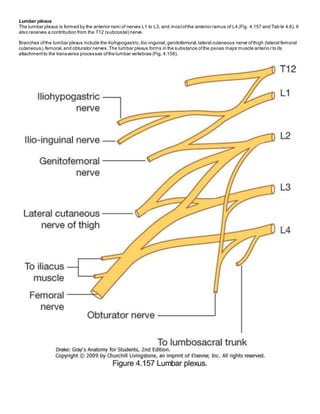
The lumbar plexus is formed by the anterior rami of nerves L1 to L3, and mostofthe anterior ramus of L4 (Fig. 4.157 and Tab le 4.6). It
also receives a contribution from the T12 (subcostal) nerve.
Branches ofthe lumbar plexus include the iliohypogastric,ilio-inguinal,genitofemoral,lateral cutaneous nerve ofthigh (lateral femoral
cutaneous),femoral,and obturator nerves.The lumbar plexus forms in the substance ofthe psoas major muscle anterio r to its
attachmentto the transverse processes ofthe lumbar vertebrae (Fig. 4.158).
2. Therefore, relative to the psoas major muscle,the various branches emerge either:
anterior-genitofemoral nerve;
medial-obturator nerve;or
lateral-iliohypogastric,ilio-inguinal,and femoral nerves,and the lateral cutaneous nerve of the thigh.
Iliohypogastric and ilio-inguinal nerves (L1)
The iliohypogastric and ilio-inguinal nerves arise as a single trunk from the anterior ramus ofnerve L1 (Fig. 4.157).Either before or
soon after emerging from the lateral border of the psoas major muscle,this single trunk divides into the iliohypogastric and the ilio-
inguinal nerves.
Iliohypogastric nerve
The iliohypogastric nerve passes across the anterior surface of the quadratus lumborum muscle,posterior to the kidney. It pierces the
transversus abdominis muscle and continues anteriorlyaround the body between the transversus abdominis and internal oblique
muscles.Above the iliac crest, a lateral cutaneous branch pierces the internal and external oblique muscles to supplythe
posterolateral gluteal skin (Fig.4.159).
3. The remaining partofthe iliohypogastric nerve (the anterior cutaneous branch) continues in an anterior direction,piercing the internal
oblique justmedial to the anterior superior iliac spine as it continues in an obliquelydownward and medial direction.Becoming
cutaneous,justabove the superficial inguinal ring,after piercing the aponeurosis ofthe external oblique,it distributes to the skin in the
pubic region (Fig. 4.159). Throughoutits course,it also s upplies branches to the abdominal musculature.
Ilio-inguinal nerve
The ilio-inguinal nerve is smaller than,and inferior to, the iliohypogastric nerve as it crosses the quadratus lumborum muscle.Its course
is more oblique than that of the iliohypogastric nerve,and it usuallycrosses partof the iliacus muscle on its way to the i liac crest.Near
the anterior end of the iliac crest, it pierces the transversus abdominis muscle,and then pierces the internal oblique muscle and enters
the inguinal canal.
The ilio-inguinal nerve emerges through the superficial inguinal ring,along with the spermatic cord,and provides cutaneous innervati on
to the upper medial thigh,the root of the penis,and the anterior surface of the scrotum in men,or the mons pubis and labium majus in
women (Fig.4.159). Throughoutits course,it also supplies branches to the abdominal musculature.
Genitofemoral nerve (L1 and L2)
4. The genitofemoral nerve arises from the anterior rami ofthe nerves L1 and L2 (Fig. 4.157).It passes downward in the substanceofthe
psoas major muscle until itemerges on the anterior surface of psoas major.It then descends on the surface of the muscle,in a
retroperitoneal position,passing posterior to the ureter. It eventually divides into genital and femoral branches.
The genital branch continues downward and enters the inguinal canal through the deep inguinal ring.It continues through the canal
and:
in men,innervates the cremasteric muscle and terminates on the skin in the upper anterior part of the scrotum;and
in women,accompanies the round ligamentofthe uterus and terminates on the skin of the mons pubis and labium majus.
5. The femoral branch descends on the lateral side ofthe external iliac artery and passes posterior to the inguinal ligament,entering the
femoral sheath lateral to the femoral artery. It pierces the anterior layer of the femoral sheath and the fascia lata to supp lythe skin of
the upper anterior thigh (Fig. 4.159).
Lateral cutaneous nerve of thigh (L2 and L3)
The lateral cutaneous nerve of thigh arises from the anterior rami of nerves L2 and L3 (Fig. 4.157). It emerges from the late ral border of
the psoas major muscle,passing obliquelydownward across the iliacus muscle toward the anterior superior iliac spine (Fig.4.159).It
passes posterior to the inguinal ligamentand enters the thigh.
The lateral cutaneous nerve of thigh supplies the skin on the anterior and lateral thigh to the level of the knee (Fig. 4.159).
Obturator nerve (L2 to L4)
The obturator nerve arises from the anterior rami of the nerves L2 to L4 (Fig. 4.157). It descends in the psoas major muscle,emerging
from its medial side near the pelvic brim (Fig. 4.158).
The obturator nerve continues posterior to the common iliac vessels, passes across the lateral wall of the pelvic cavity, and enters the
obturator canal, through which the obturator nerve gains access to the medial compartmentofthe thigh.
In the area of the obturator canal,the obturator nerve divides into anterior and posterior branches. On entering the medial
compartmentofthe thigh,the two branches are separated bythe obturator externus and adductor brevis muscles.Throughouttheir
course through the medial compartment,these two branches supply:
articular branches to the hip joint;
muscular branches to obturator externus,pectineus,adductor longus,gracilis,adductor brevis,and adductor magnus muscles;
cutaneous branches to the medial aspectofthe thigh;and
in association with the saphenous nerve,cutaneous branches to the medial aspectofthe upper part of the leg,and articular
branches to the knee joint (Fig. 4.159).
Femoral nerve (L2 to L4)
The femoral nerve arises from the anterior rami of nerves L2 to L4 (Fig. 4.157). It descends through the substance ofthe psoas major
muscle,emerging from the lower lateral border of the psoas major (Fig.4.158). Continuing its descent,the femoral nerve lie s between
the lateral border of the psoas major and the anterior surface of the iliacus muscle.It is deep to the iliacus fascia and lateral to the
femoral artery as it passes posterior to the inguinal ligamentand enters the anterior compartmentofthe thigh. Upon enterin g the thigh,
it immediatelydivides into multiple branches.
Cutaneous branches ofthe femoral nerve include:
medial and intermediate cutaneous nerves supplying the skin on the anterior surface of the thigh; and
the saphenous nerve supplying the skin on the medial surface ofthe leg (Fig. 4.159).
Muscular branches innervate the iliacus,pectineus,sartorius,rectus femoris,vastus medialis,vastus intermedius,and vastu s lateralis
muscles.Articular branches supplythe hip and knee joints.
6. Nerves
Somatic plexuses
Sacral and coccygeal plexuses
The sacral and coccygeal plexuses are situated on the posterolateral wall ofthe pelvic cavity and generallyoccur in the pla ne between
the muscles and blood vessels.Theyare formed by the ventral rami of S1 to Co, with a significantcontribution from L4 and L5, which
enter the pelvis from the lumbar plexus (Fig. 5.59). Nerves from these mainlysomatic plexuses contribute to the innervation of the lower
limb and muscles ofthe pelvis and perineum.Cutaneous branches supplyskin over the medial side ofthe foot, the posterior aspectof
the lower limb,and mostof the perineum.
Sacral plexus
The sacral plexus on each side is formed by the anterior rami of S1 to S4, and the lumbosacral trunk (L4 and L 5) (Fig. 5.60). The
plexus is formed in relation to the anterior surface of the piriformis muscle,which is partof the posterolateral pelvic wal l.Sacral
7. contributions to the plexus pass outof the anterior sacral foramina and course laterallyand inferio rly on the pelvic wall.The
lumbosacral trunk,consisting ofpart of the anterior ramus ofL4 and all of the anterior ramus ofL5, courses verticallyinto the pelvic
cavity from the abdomen by passing immediatelyanterior to the sacro-iliac joint.
Gray rami communicantes from ganglia ofthe sympathetic trunk connectwith each of the anterior rami and carry postganglionic
sympathetic fibers destined for the periphery to the somatic nerves (Fig.5.61). In addition,special visceral nerves (pelvic splanchnic
nerves) originating from S2 to S4 deliver preganglionic parasympathetic fibers to the pelvic part of the prevertebral plexus (Fig. 5.62).
Each anterior ramus has ventral and dorsal divisions thatcombine with similar divisions from other levels to form terminal nerves (see
Fig. 5.60). The anterior ramus ofS4 has only a ventral division.
Branches ofthe sacral plexus include the sciatic nerve and gluteal nerves,which are major nerves of the lower limb,and the pudendal
nerve, which is the nerve of the perineum (Table 5.4).Numerous smaller branches supplythe pelvic wall, floor, and lower limb.
8. Most nerves originating from the sacral plexus leave the pelvic cavity by passing through the greater sciatic foramen inferio r to
piriformis muscle,and enter the gluteal region of the lower limb.Other nerves leave the pelvic cavity using differentroutes;a few ne rves
do not leave the pelvic cavity and course directly into the muscles in the pelvic cavity. Finally, two nerves that leave the pelvic cavity
through the greater sciatic foramen loop around the ischial spine and sacrospinous ligamentand pass mediallythrough the les ser
sciatic foramen to supplystructures in the perineum and lateral pelvic wall.
9. Sciatic nerve
The sciatic nerve is the largestnerve of the body and carries contributions from L4 to S3 (Figs. 5.59 and 5.60). It:
forms on the anterior surface of the piriformis muscle and leaves the pelvic cavity through the greater sciatic foramen inferior
to piriformis;
passes through the gluteal region into the thigh,where it divides into its two major branches,the common fibular nerve
(common peroneal nerve) and the tibial nerve-dorsal divisions ofL4,L5, S1, and S2 are carried in the common fibular partof
the nerve and the ventral divisions ofL4, L5, S1, S2, and S3 are carried in the tibial part;
innervates muscles in the posterior compartmentofthe thigh and muscles in the leg and foot; and
carries sensoryfibers from the skin of the foot and lateral leg.
Pudendal nerve
S2 to S4 (Figs. 5.59 and 5.60). It:
leaves the pelvic cavity through the greater sciatic foramen,inferior to the piriformis muscle,and enters the gluteal regio n;
courses into the perineum byimmediatelypassing around the sacrospinous ligament,where the ligamentjoins the ischial
spine,and through the lesser sciatic foramen (this course takes the nerve out of the pelvic cavity, around the peripheral
attachmentof the pelvic floor, and into the perineum);
is accompanied throughoutits course bythe internal pudendal vessels;and
innervates skin and skeletal muscles ofthe perineum,including the external anal and external urethral sphincters.
Other branches ofthe sacral plexus
Other branches ofthe sacral plexus include:
motor branches to muscles ofthe gluteal region,pelvic wall,and pelvic floor (superior and inferior gluteal nerves,nerve to
obturator internus and superior gemellus,nerve to quadratus femoris and inferior gemellus,nerve to piriformis,nerves to
levator ani); and
sensorynerves to skin over the inferior gluteal region and posterior aspects ofthe thigh and upper leg (perforating cutaneo us
nerve and posterior cutaneous nerve of the thigh) (Figs.5.59 and 5.60).
The superior gluteal nerve, formed by branches from the dorsal divisions ofL4 to S1, leaves the pelvic cavity through the greater
sciatic foramen superior to piriformis muscle and supplies muscles in the gluteal region-gluteus medius, gluteus minimus, and
tensor fasciae latae (tensor of fascia lata) muscles.
The inferior gluteal nerve, formed by branches from the dorsal divisions ofL5 to S2, leaves the pelvic cavity through the greater
sciatic foramen inferior to the piriformis muscle and supplies the gluteus maximus,the largestmuscle in the gluteal region.
Both superior and inferior gluteal nerves are accompanied bycorresponding arteries.
The nerve to the obturator internus and the associated superior gemellus muscle originates from the ventral divisions ofL5 to S2
and leaves the pelvic cavity through the greater sciatic foramen inferior to the piriformis muscle.Like the pudendal nerve,it passes
around the ischial spine and through the lesser sciatic foramen to enter the perineum and supplythe obturator internus muscl e from the
medial side ofthe muscle,inferior to the attachmentof the levator ani muscle.
The nerve to the quadratus femoris muscle and the inferior gemellus muscle,and the posterior cutaneous nerve of the thigh
(posterior femoral cutaneous nerve) also leave the pelvic cavity through the greater sciatic foramen inferior to the piriformis muscle
and course to muscles and skin,respectively,in the lower limb.
10. Unlike mostofthe other nerves originating from the sacral plexus,which leave the pelvic cavity through the greater sciatic foramen
either above or below the piriformis muscle,the perforating cutaneous nerve leaves the pelvic cavity by penetrating directly through
the sacrotuberousligamentand then courses to skin over the inferior aspectof the buttocks.
The nerve to the piriformis and a number of small nerves to the levator ani and coccygeus muscles originate from the sacral plexus
and pass directlyinto their target muscles withoutleaving the pelvic cavity.
The obturator nerve (L2 to L4) is a branch of the lumbar plexus.It passes inferiorlyalong the posterior abdominal wall within the psoas
muscle,emerges from the medial surface ofthe psoas,passes posteriorlyto the common iliac arteryand mediallyto the internal artery
at the pelvic inlet, and then courses along the lateral pelvic wall. It leaves the pelvic cavity by traveling through the obturator canal and
supplies the adductor region of the thigh.
Coccygeal plexus
The small coccygeal plexus has a minor contribution from S4 and is form ed mainlyby the anterior rami of S5 and Co, which originate
inferiorly to the pelvic floor. They penetrate the coccygeus muscle to enter the pelvic cavity and join with the anterior ram us ofS4 to
form a single trunk,from which small anococcygealnerves originate (Table 5.4). These nerves penetrate the muscle and the overlying
sacrospinous and sacrotuberous ligaments and pass superficiallyto innervate skin in the anal triangle of the perineum.