
Recommended
More Related Content
What's hot
What's hot (20)
Similar to Physiology of the visual pathway & cerebral integration
Similar to Physiology of the visual pathway & cerebral integration (20)
Recently uploaded
Recently uploaded (20)
Physiology of the visual pathway & cerebral integration
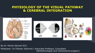
- 1. By Dr. Henok Samuel (R1) Moderator: Dr.Tiliksew Teshome ( Associate Professor, Consultant Ophthalmologist and Vitreoretinal surgeon) PHYSIOLOGY OF THE VISUAL PATHWAY & CEREBRAL INTEGRATION
- 2. Contents Introduction Recap on anatomy of visual pathway Axonal conduction Role of myelin in axonal conduction Axonal transport Optic nerve injury and regeneration Retinotopic Organization and visual field defect Summary References 2
- 3. Introduction The role of the central visual pathways is to process and integrate visual information that travels to the brain by means of the optic nerves. Visual processing poses an enormous computational challenge for the brain, which has evolved highly organized and efficient neural systems to meet these demands. In primates, approximately 55% of the cortex is specialized for visual processing (compared to 3% for auditory processing and 11% for somatosensory processing) 3
- 4. Introduction When light reaches the retina, its energy is converted by retinal photoreceptors into an electrochemical signal that is then relayed by neurons. 4
- 5. Retina Ganglion cells RGC axons begin at the RGC bodies in the inner layer of the retina. The RGCs receive input from bipolar cells and amacrine cells, and their axons form nerve fiber layer. 5
- 6. Retina Ganglion cells 10 types of RGC found (two of them are well studied) Eighty percent of ganglion cells are Midget cells, 10% are Parasol cells, and 10% are other types. 6
- 7. Retina Ganglion cells 7 LARGE PARASOL RGC - Project to the magnocellular / M cell system in LGN. -for low spatial resolution, fast temporal resolution, stereopsis, contrast sensitivity. - high receptive field & more for peripheral vision. SMALL MIDGET RGC -Project to the parvocellular / p cell system in LGN. -for high spatial resolution but has low contrast sensitivity -many in number & more for macular vision “K” (koniocellular) pathway - more recently identified, small bistratified ganglion cells
- 8. Retina Ganglion cells At any given retinal eccentricity, parasol cells have a larger receptive field and lower spatial resolution than midget cells due to their broad dendritic arborization. Parasol ganglion cells have spatially opponent center surround organization, allowing edge detection, but they lack spectrally opponent organization; in essence, these cells are color-blind. The anatomical features of parasol cells underlie their specialization for low spatial resolution, motion detection, and coarse stereopsis. 8
- 9. Cont… Foveal ganglion cells send axons directly to the temporal aspect of the optic disc in the papillomacular bundle. The remaining temporal ganglion cell nerve axons are arranged on either side of the horizontal raphe and form arcuate bundles that course above and below the fovea, and finally enter the superior and inferior portions of the optic nerve. Finally, axons originating nasal to the disc enter the nasal portion of the optic nerve. 9
- 10. Optic Nerve Head The ganglion cell axons form the optic nerve after they leave the eye. And The optic disc or optic nerve head is the point of exit for ganglion cell axons leaving the eye. There are no rods or cones overlying the optic disc, it corresponds to a small physiological blind spot in each eye. The optic disc in a normal human eye carries from 1 to 1.2 million neurons from the eye towards the brain 10
- 11. Optic Nerve Each optic nerve is comprised of approximately 1.2 million retinal ganglion cell axons (in constrast to the acoustic nerve, for example, which has only 31 000 axons). The intraocular segment of the optic nerve head (the optic disc) is typically located 3–4 mm nasal to the fovea and is 1 mm thick. 11
- 12. Optic nerve Intraocular Intraorbital Intracanalicular Intracranial 12
- 13. Retinotopic Organization of Optic Nerve • In the proximal third of the optic nerve the positions of ganglion cell axons are rearranged. • Macular ganglion cell axons which initially lie temporally move to the nerve’s center. • Peripheral temporal fibers become positioned temporally, both superior and inferior to the macular fibers. • Finally, nasal fibers remain in the nasal portion of the optic nerve. 13
- 14. Optic Chiasm Is the site of decussation for axons from the optic nerve. It lies in the subarachnoid space of the suprasellar cistern surrounded by meningeal sheaths and cerebrospinal fluid 10 mm above the pituitary, which rests in the sella turcica within the sphenoid bone The chiasm lies within the circle of Willis. 14
- 15. Relations with neighboring structures 15
- 16. Relations with neighboring structures • The exact location of the chiasm with respect to the sella is variable. • 16
- 17. Arrangement of Nerve fibres in Optic chiasm The lower (inferior) fibers (subserving the superior visual field) traverse the chiasma low and anteriorly - are first to cross. The upper nasal fibers traverse the chiasma high and posteriorly. macular fibers tend to cross decussate in the posterior most part of chiasm which is related to supraoptic recess. 17
- 18. Optic Tract Flattened cylindrical band that travel posteriolaterally from angle of chiasma. Between tuber cinereum and anterior perforated substance upto lateral geniculate body. Each tract contains uncrossed temporal fibres and crossed nasal fibres . 18
- 19. Retinotopy of optic tract Macular fibers (crossed & uncrossed) occupy dorsolateral aspect of optic tract. Upper peripheral fibers (crossed & uncrossed) medially situated Lower peripheral fibers laterally situated 19
- 20. Lateral Geniculate Body (LGB) Elevation produced by lateral geniculate nucleus in which most optic tract fibers end. Axons of ganglion cells of retina synapse with dendrites of LGB cells - 3rd order neurons begins Primary termination of OT fibers Each LGN receives input from the contralateral visual field. 20
- 21. Lateral Geniculate Body Dorsal nucleus Ventral nucleus (rudimentary) 6 laminae (alternating grey & white matter) Axons from the ipsilateral eye –2, 3, 5 Axons from the contralateral eye - 1, 4,6 21
- 22. Lateral Geniculate Body Large magnocellular neurons (M cells) - 1 and 2 layer-Y ganglion cells - perception of movement, gross depth, and small differences in brightness Small parvocellular neurons (P cells) 3,4,5,6 layer- X ganglion cells Colour perception, texture shape &fine depth Koniocellular cells (K cells or interlaminar cells) Short-wavelength "blue" cones 22
- 23. Retinotopic arrangement of LGB • Macular fibres – posterior 2/3 of LGB • Upper retinal fibres – medial half of anterior 1/3 of LGB • Lower retinal fibres – lateral half of anterior 1/3 of LGB 23
- 24. Geniculocalcarine Tract (optic radiations) Axons of LGN neurons travel to primary visual cortex (Area 17) via the geniculocalcarine tract located in the retrolenticular and sublenticular portions of the internal capsule. Axons from upper visual fields take a looping course into the temporal lobe on the way to visual cortex. (=Meyer’s loop) Axons from lower visual fields take a more direct route to visual cortex. Macular fibers are in an intermediate location in the optic radiation 24
- 25. Superior colliculus Pretectum Pulvinar Hypothalamic nuclei AOS 25 Extra geniculate targets of the retinal projections
- 26. Superior Colliculus 26 Approximately 10 percent of all retinal ganglion cells project to the SC. Cells in SC have ill-defined "On" "Off" regions, therefore will respond to most visual stimulus regardless of shape, color, or orientation. SC cells seem most interested in location of visual stimulus, not identity of visual stimulus, therefore, SC has often been referred to as the "where" system, rather than the "what" system. SC appears to have important role in directing ballistic eye movements toward a visual stimulus in the periphery Most of the fibers projecting to SC are from the M type of RGC, therefore, messages often get to SC more quickly than to LGN.
- 27. 27 A group of small midbrain nuclei, is just rostral to the SC. Involved with the control of the pupillary light reflex by means of a projection to the Edinger–Westphal nucleus of the oculomotor complex. The pretectal complex
- 28. The pulvinar is the largest nuclear mass in the primate thalamus and receives a projection from the small-caliber fibers from the optic nerve and the SC. It projects to several visual cortical areas, including V1, and extrastriate, parietal areas. Pathway that can bypass the LGN to get to V1 and may play a role in processing form vision. Integrates neural signals associated with eye, hand and arm movements and may receive signals associated with saccadic eye movements. 28 Pulvinar nucleus of the thalamus
- 29. The suprachiasmatic nucleus receives a sparse projection from fibers that leave the dorsal surface of the optic chiasma and has been implicated in the synchronization of circadian rhythms. The paraventricular and supraoptic nuclei are likely also involved with the regulation of the light–dark cycle for neuroendocrine functions The AOS plays an important role in optokinetic nystagmus (OKN) in which slow compensatory and pursuit type eye movements alternate with fast saccadic-type eye movements in response to viewing prolonged large field motion 29 Other targets RGC
- 30. Primary Visual Cortex (Area 17) Located on either side of & within the calcarine fissure. Upper fields project to the lingual gyrus. Lower fields project to the cuneus. Macular representation is most caudal in Area 17. Peripheral field representation is in the rostral 2/3rds of Area 17. Lesions of Area 17 result in blindness in the contralateral visual field. 30
- 31. Cont… • Dorsal stream – Occipito-parietal stream spatial, Motion perception – action- “where” or “how to” • Ventral stream – Occipito-temporal stream object perception – identification – “what” stream 31
- 32. Cont… 32
- 33. Cont… 33
- 34. Cont… 34
- 35. Visual Cortex • 35 What/Where Pathways Evidence from Neuropsychology Visual agnosia: Inability to identify objects and/or people Caused by damage to inferior (lower) temporal lobe Disruption of the “what” pathway Visual neglect: Inability to see objects in the left visual field Caused by damage to right parietal lobe Disruption of the “where” pathway
- 36. Deficits in Motion Perception: Akinetopsia • 36 Deficits in Color Perception - Achromatopsia
- 37. Visual Cortex Like RGCs and cells of LGN, cells of the PVC have receptive fields which monitor a restricted zone in the retina. Cortical magnification: the disproportionally large amount of cortical tissue dedicated to processing the foveal retina compared to the peripheral retina. Number of RGCs connected to photoreceptors in fovea vs. RGCs connected to photoreceptors in periphery. Remember degree of convergence? 37
- 38. 38 Orientation specificity: cells in PVC differentially respond to stimuli of varying orientations. Simple cells: Well defined excitatory region; location important. Complex cells: less well defined excitatory region; location less important. Hyper-complex: length of contour important Oblique effect Visual Cortex
- 39. 39 Direction specificity: cells in PVC also differentially respond to movement in different directions. Simple cells: slower movement. Complex cells: faster movement Visual Cortex
- 40. • Binocular cells: Two receptive fields, one for each eye. Stronger response to one eye (ocular dominance). • Cells give a most vigorous response when a stimulus of the same size, shape, and orientation is located in a particular position in 3-d space across the two retinae. • Cells are important for the visual system's use of the depth cue called retinal disparity. 40 Visual Cortex
- 41. 41 Cell columns: in the PVC cells (s, c, hc) are arranged in layers by their preferred orientation. Cell hyper columns: the layers of orientation specific cells are arranged such that an orderly incrementing of orientation change exists across layers until a complete cycle has been achieve. Include both eyes. Layer 4: input layer, monocular cells. Blobs: non-orientation specific cells for color processing. Visual Cortex
- 42. Axonal conduction Retinal ganglion cell electrophysiology and synaptic transmission RGCs receive synaptic input from bipolar and amacrine cells, which primarily use glutamate as the major excitatory neurotransmitter. Axonal conduction Action potentials RGC axons transmit information via action potentials, which are all-or-nothing spikes of electrical activity. This is in contrast to most intercellular communication within the retina, where graded potentials are used to transmit information 42
- 43. 43 Mechanism of axonal conduction AT REST(Non depolarization state) Negative voltage in the axon ( negative resting potential) Due to flow of K down its concentration gradient ( from inside to outside) which create a leak current giving negative potential Leak current continue until concentration gradient becomes equal which forms K equilibrium potential At rest flow of Na down its concentration gradient (from outside to inside) occur giving Na equilibrium potential but its much more smaller than K flow
- 44. 44 DEPOLARIZATION Occurs when voltage sensitive Na channels activated More Na enter the cell so axon becomes positive (depolarized) REPOLARIZATION Its the return of membrane potential to resting state & its necessary for another AP to be transmitted down the axon Due to closure of voltage gated Na channels & transient opening of voltage gated K channels Mechanism of axonal conduction
- 45. Role of myelin in AP conduction It’s a multilaminated fatty structured produced by oligodendrocytes which insulates each axon It increase the speed & efficacy of AP conduction by these mechanisms; DECREASE CAPACITANCE (ion needed to change voltage) - less Na needed to depolarize axons INCREASE RESISTANCE (the resistance for ion leakage) - less leakage of charge across the membrane The above two decrease amount of ion flux needed to change voltage across the membrane 45
- 46. Node of Ranvier Its myelin free space in the axon containing many Na channels so AP jump from one node to the other speeding up AP conduction Such type of conduction is called salutatory conduction 46 Role of myelin in AP conduction
- 47. Axonal transport Axonal transport occurs in two directions, orthograde (away from the cell body and towards the brain), and retrograde (towards the cell body and away from the brain). Orthograde Axonal Transport Transport away from the cell body & toward the brain Has two types; 47
- 48. 1. Slow axonal transport; two classes Slower class ( 0.2 – 1 mm/day ) - for cytoskeletal proteins (tubulin & neuro filament) More rapid class (2 – 8 mm / day) - for actin , myosin , enzymes 2. Fast axonal transport ( 90 – 350 mm/day) - for neurotransmitters to axon terminal - continue after transection of the cell body from the axon 48 Axonal transport…
- 49. Introduction Retrograde Axonal Transport Transport toward the cell body and away from the brain - Occur as half velocity of the fast axonal transport - For neurotrophic molecules & growth factors which induce transcription & translation of gens for axonal survival & growth. • 49
- 50. Optic nerve injury Optic nerve injury from the various forms of optic neuropathy affect the RGC by the following mechanisms; 1. Blockage of axonal transport -build up of excess retrograde transported molecules -lack of neurotrophic factors for cell survival 2. Demyelination 3. Excitotoxicity from excess glutamate 4. free radical formation If the pathology not treated the final common pathway is RGC death by apoptosis 50
- 51. Axon repair It’s a mechanism for protecting RGC & enhancing their survival after an insult on the axons by the following ways; Remyelination -using steroids & immune modulators in demyelinating diseases Neuroprotection -Blocking excitotoxicity from glutamate -Administer neurotrophic factors -Inhibition of apoptosis at its various stages 51
- 52. Axonal regeneration No axonal regeneration after optic nerve injury but there is an early sprouting called abortive regeneration which latter dies Mechanisms preventing axon regeneration 1. Glial associated inhibitory signal - Active molecules from astrocyte & oligodendrocyte prevent Regeneration( myelin & proteoglycan) - Glial cells unable to secrete trophic signals after injury • 52
- 53. Axonal regeneration… 2. Neuron intrinsic limitation - Due to gradual increment of gens which limit axonal growth & gradual decrement of gens important for rapid growth • 53
- 54. Retinotopic Organization & Visual field defect Visual field • It is tested monocularly, with the patient looking straight ahead at a fixation point. • 54
- 55. Cont… Inversion and reversal of the field are caused by the optical system of the eye The superior field is imaged on the inferior retina The inferior field on the superior retina; The nasal field is imaged on the temporal retina The temporal field on the nasal retina 55
- 56. Cont… The nasal part of the field for one eye is the same as the temporal part of the field seen by the other eye, with the exception of the far temporal periphery, which is called the temporal crescent The temporal crescent is imaged on the nasal retina of one eye but not on the temporal retina of the other eye. Within each temporal field is an absolute scotoma, the physiologic blind spot. 56
- 57. Retinal nerve fiber layer Papillomacular bundle Central /centrocecal/paracentral scotoma Temporal retinal fiber Arcuate visual field defects Nasal retinal fiber Temporal wedge shaped defects Nasal retinal + temporal fiber defect Altitudinal defect Disorder of rod Ring scotoma, Disorder of cones Central scotoma 57
- 58. Introduction • The role of the central visual pathways is to process and integrate visual information that travels to the brain by means of the optic nerves 58
- 59. LESIONS OF VISUAL PATHWAY 59
- 60. LESIONS OF OPTIC NERVE : Causes: 1. Optic atrophy 2. Indirect optic neuropathy 3. Acute optic neuritis 4. Traumatic avulsion of optic nerve. Characterised by: - Complete blindness in affected eye - loss of both direct on ipsilateral & consensual light reflex on contralateral side. 60
- 61. Describe the visual field defect ? • 61 Bitemporal Heteronymous Hemianopia Paralysis of pupillaryreflex (usually lead to partial descending optic atrophy) MIDDLE CHIASMAL LESIONS: CENTRAL LESIONS OF CHIASMA (SAGITTAL) Causes: Suprasellar aneurysm Tumors of pituitary gland Craniopharyngioma Suprasellar meningioma & glioma of 3rd ventricle. Third ventricular dilatation due to obstructive hydrocepha
- 62. Describe the visual field defect ? • 62 Junctional scotoma: lesion at junction of optic nerve and chiasm ANTERIOR CHIASMAL LESIONS: • Ipsilateral optic neuropathy a central scotoma and a defect involving the contralateral superotemporal fieldalso known as a junctional scotoma • Abolition of direct light reflex on affected side & consensual light reflex on contralateral side. • Near reflex intact.
- 63. POSTERIOR CHIASMAL SYNDROME macular fibers cross more posteriorly in the chiasm paracentral bitemporal field loss Posterior lesions may also involve the optic tract and cause a contralateral homonymous hemianopia. 63
- 64. Describe the visual field defect ? • 64 POSTERIOR CHIASMAL SYNDROME macular fibers cross more posteriorly in the chiasm paracentral bitemporal field loss Posterior lesions may also involve the optic tract and cause a contralateral homonymous hemianopia.
- 65. Describe the visual field defect ? • 65 Left incongruous homonymous hemianopia Contralateral hemianopic pupillary reaction( wernicke’s reaction) LESIONS OF OPTIC TRACT : Causes: 1. Syphilitic meningitis/ Gumm 2. Tuberculous meningitis 3. Tumors of optic thalamus 4. Aneurysm of posterior cerebral arteries.
- 66. Describe the visual field defect ? • 66 Left Homonymous Hemianopia
- 67. Describe the visual field defect ? 67 Left homonymous hemianopia with macular sparing
- 68. Describe the visual field defect ? • 68 Right superior quadrantanopia >> temoporal lobe lesion LESIONS OF OPTIC RADIATIONS : Causes: • Vascular occlusion(Anterior choroidal artery, middle cerebral or posterior cerebral artery) • Primary & secondary tumors • Trauma Characterized by : No Optic Atrophy Normal pupillary reactions
- 69. Describe the visual field defect ? • 69 Left inferior quadrantanopia >> parietal lobe lesion LESIONS OF OPTIC RADIATIONS : Causes: • Vascular occlusion(Anterior choroidal artery, middle cerebral or posterior cerebral artery) • Primary & secondary tumors • Trauma Characterized by : No Optic Atrophy Normal pupillary reactions
- 70. Describe the visual field defect ? • 70 Right homonymous hemianopia Congruous homonymous macular defect Head injury/gun shot injury leading to lesions of tip of occipital cortex
- 71. Describe the visual field defect ? • 71 Enlarged Blind Spot
- 72. Describe the visual field defect ? • 72
- 73. Describe the visual field defect ? • (A) Posterior choridal artery occlusion leads to homonymous horizontal sectroanopia. (B) Anterior choridal artery occlusion causes sector-sparing homonymous hemianopia
- 74. References Duanes clinical ophtalmology 2012 Anatomy of the eye, R. Snell 2nd edition BCSC Fundamentals and principles of ophthalmology , AAO 2016-2017 wolff’s anatomy of the eye and orbit, 8th edition ADELER’S physiology of the eye 11th edition Clinical Anatomy and physiology 3rd edition Clinical neuro-ophthalmology – Thomas duane and Edward jaegar 74
- 75. Thank You!
Editor's Notes
- © Copyright Showeet.com
- Although the eye is responsible for transducing patterns of light energy into neuronal signals, it is the brain that is ultimately responsible for visual perception and cognition.
- . The corneal epithelium and stroma are transparent to permit passage of light without distortion (Maurice, 1970). • The ciliary muscles dynamically adjust the shape of the lens in order to focus light optimally from varying distances upon the retina (accommodation). The total amount of light reaching the retina is controlled by regulation of the pupil aperture. To arrive at the photoreceptors, light must first pass through transparent inner layers of the neurosensory retina, comprised of the nerve fiber layer, ganglion cells, amacrine cells, and bipolar cells . • Ultimately, the visual image becomes projected upside-down and backwards on to the retina (Fishman, 1973)
- . project their axon towards the vitreous, whereupon they turn approximately 90° and project towards the optic nerve head in the nerve fiber layer.
- There are three main types of ganglion cell, each with specialized functions in the detection of visual inputs The different types of ganglion cells comprise separate pathways that are named for their targets in the LGN.
- Midget cells have cone-opponent receptive fields, allowing spectral selectivity along the red–green or blue–yellow axes. These struct arrangts afford midget cells the properties of an extremely small receptive field, with specialization for high spatial acuity, color vision, and fine stereopsis. What does all this mean? M's are more able to pick up the mare fact that something is present and are faster at communicating that. P's are more able to do fine detailed analysis of what that something is.
- The nerve fiber layer is not quite radially arranged around the optic nerve head, as axons near the center of our visual field course away from the fovea, and then towards the optic disk, entering in the superior and inferior portions of the disk . This interesting anatomy prevents axons from crossing the high-sensitivity fovea, where they might otherwise scatter light and degrade visual acuity. The axons from more peripheral RGCs are more superficial (vitread) to those arising from less peripheral ganglion cells. There is strict segregation of those fibers arising from RGCs located superior to the temporal horizontal meridian (raphe) and those fibers arising from RGCs located inferior to the horizontal raphe. Because of this segregation, visual field defects corresponding to injury to RGC axons typically have stereotyped patterns, e.g. superior or inferior nasal steps, temporal wedges, or arcuate scotomas. These are called nerve fiber bundle defects
- The optic disc represents the beginning of the optic nerve and is the point where the axons of retinal ganglion cells come together. Axons of ganglion cells in the retina of the corresponding eye • Outgrowth of diencephalon, so is a CNS tract & not a „true‟ cranial nerve. • Myelinated by oligodendrocytes The optic disc is also the entry point for the major blood vessels that supply the retina.
- Peripheral fibers deep in Retina superficially in opticnerve Fibers close to opticnerve head superficial in retina central in optic nerve The optic nerve travels posteriorly through the lamina cribrosa to exit the back of the globe, where it abruptly increases in diameter from 3 to 4 mm. In order to accommodate the rotations of the globe, intraorbital segment of the optic - 25 and 30 mm in length, at least 5 mm longer than the distance from the globe to the orbital apex. Upon passing through the lamina cribrosa, the optic nerve becomes invested with meninges and becomes myelinated.
- Upon exiting the orbit, the optic nerve enters the optic canal, within the lesser wing of the sphenoid bone, for approximately 6 mm. The intracanalicular optic nerve rises at a 45 angle and then exits the optic canal, where it continues in its intracranial portion for approximately 17 mm before reaching the chiasma.
- The chiasm, which has a dumbbell shape when viewed in coronal section,
- ANTERIORLY : Anterior cerebral artery and their communicating artery POSTERIORLY: Tuber cinerium, Hypophyseal stalk, Pituitary body, mamillary body, posterior perforated substance SUPERIORLY: 3rd ventricle INFERIORLY: Pituitary gland LATERALLY: Extracavernous part of ICA Anterior perforated substance
- normal….directly superior to sella turcica. --prefixed….anterior to sella turcica. --post fixed…..posterior to sella turcica.
- Partial crossing of optic nerve axons in the OC is essential to binocular vision Axons from temporal fields (fibers coming from the nasal retina (approximately 53% of total fibers) cross So are first affected in the tumor of pituitary producing upper temporal quadrantic field defect – these fibers form convex loops in the terminal part of optic nerve and then cross opposite side and occupy lower quadrant Why do half the fibers cross? Nasal half of one eye, and temporal half of other eye, monitor same visual field, therefore, fibers emanating from same areas monitoring same physical space are combined.
- Each tract is ~ 5mm long. The fibers exiting from the chiasm proceed -circumferentially around the diencephalon -lateral to the hypothalamus -in contact with the ambient cistern . - Represent contralateral & ipsilateral hemi field of each eye. Just prior to the LGN, the fibers involved in the pupillary pathways exit. The ipRGCs also project to the suprachiasmatic nucleus of the hypothalamus
- Although the eye is responsible for transducing patterns of light energy into neuronal signals, it is the brain that is ultimately responsible for visual perception and cognition.
- Function: 1) LGN contains cells with similar receptive field characteristics as RGC, except that "off" portions of cells exert even stronger inhibitory affect. This serves to accentuate to an even greater extent the "edges" and "borders" already identified by RGCs. 2) Parvocellular layers contain cells with receptive fields which respond differentially to color. These are called color opponent cells, most are red/green or blue/yellow color opponent. These cells receive most of their input from the P type of RGC. 3) Magnocellular layers are color blind and receive input mostly from M type of RGCs. 4) Both Magnocellular cells and Parvocellular cells can signal movement, but Magno cells respond to fast movement, Parvo respond to slow movement. 5) LGN may also receive "top-down" visual processing biases due to its interconnections with visual cortex. 6) Interconnections with RAS may serve as “volume control” for visual inputs.
- Although the eye is responsible for transducing patterns of light energy into neuronal signals, it is the brain that is ultimately responsible for visual perception and cognition.
- MEYERS LOOP(inferior retinal fibers)-pass through temporal lobe looping around inferior horn of lateral ventricle
- Major targets of the retinal ganglion cell axons the accessory optic system (AOS) (including the nucleus of the optic tract (NOT) and the dorsal, medial, and terminal nuclei. Approximately 90 percent of all retinal ganglion cells project to the LGN, which is laminated and displays retinotopic organization. Each LGN layer receives input from a specific eye and class of ganglion cell
- SC is the more phylogenetically primitive projection site, in some creatures such as fish and frogs, SC is major site of visual info processing. SC is a midbrain structure which, in conjunction with the cortical frontal eye fields and the brainstem reticular formation, is involved in the generation of visually guided saccadic eye movements The majority of retinal axons that terminate in the SC are small caliber, originate from ganglion cells with small dendritic fields, and do not project to other retinal targets. So what does all this mean? Ans: it appears that SC is very important for regulating visually guided reflexive behaviours, for example, the SC would quickly signal the presence of an object coming toward the head from the visual periphery. SC only cares that there is something there, LGN will figure later what it was!
- Receives signals from a group of small-diameter retinal ganglion cells with large receptive fields. The pupillary light reflex demonstrates a consensual response primarily resulting from crossed and uncrossed optic nerve fibers that enter each pretectal complex which in turn sends a bilateral projection to the Edinger–Westphal nucleus.
- It has been shown that the pulvinar, which suggests its role is also one of formulating reference frames for hand-eye coordination
- Different names for primary visual cortex: • Brodmann’s area 17 • V1 • primary visual cortex • striate cortex (“striped” cortex)
- Although the eye is responsible for transducing patterns of light energy into neuronal signals, it is the brain that is ultimately responsible for visual perception and cognition.
- Although the eye is responsible for transducing patterns of light energy into neuronal signals, it is the brain that is ultimately responsible for visual perception and cognition.
- however, the number of cells responding to input from the foveal area of retina far exceeds the number responding to input from more peripheral regions. This is known as cortical magnification. Cortical magnification stands to reason if you consider the number of RGCs connected to photoreceptors in fovea vs. RGCs connected to photoreceptors in periphery. Remember degree of convergence?
- This was not true of LGN cells or RGC cells, both of which contain circular receptive fields insensitive to orientation. Receptive fields in PVC cells tend to be more oblong shaped.
- One cell may respond vigorously if a visual stimulus moves from left to right across the receptive field, but not respond as much if movement is from right to left, other cells do just the opposite (also up/down).
- Seeing vs. Acting distinction. Segregation but not complete separation.
- RGCs have both NMDA and non-NMDA ionotropic glutamate receptors, as well as metabotropic receptors. Other neurotransmitters modulating RGC activity include GABA, acetylcholine, and aspartate (acting on NMDA receptors), as well as serotonin and dopamine. Within the retina itself the levels of glutamate are modulated by Müller cells, have glutamate transporters, and which contain the enzyme glutamine synthetase, converting glutamate to the amino acid glutamine.
- Conduction down individual axons occurs via the same biophysical mechanisms which occur in any myelinated axon At rest, the inside of an axon is at a negative voltage with respect to the outside of the axon. This negative resting potential primarily results from the higher concentration of potassium within the axon leak current out of the axon results in a negative potential within the axon. Outward flux of potassium continues until the charge separation becomes too great, and can no longer be driven by the concentration difference between inside and outside the axon
- During axonal conduction, depolarization of a section of membrane induces opening of voltage sensitive sodium channels located within adjacent membrane. These allow much greater amounts of sodium to enter the axon, and the positive sodium ions cause the axon to become more positive (i.e., depolarized).
- Together, these properties decrease the amount of ionic flux needed to achieve changes in voltage across the membrane, saving on the Na+-K+-ATPase activity and thus energy needed to maintain ionic homeostasis after conduction.
- Conduction in a myelinated axon, called saltatory conduction, becomes much faster, as depolarization “jumps” from one node to the next. The clustering of sodium channels into nodes, as well as an important developmental switch in sodium channel isoforms, are also induced by oligodendrocytes.
- The entire length of the retinal ganglion cell axon must be maintained by transporting proteins and other subcellular constituents. The rates of transport of specifc moieties may vary as a function of stage of optic nerve development, suggesting a developmental regulation of axonal transport. Different rates of axonal transport may be related to differences in the motor proteins interacting with microtubules, e.g. kinesin208 for fast transport and dynein for slow transport.
- Fast axon transport continues despite transection of the axon distal to the cell body, suggesting that intraaxonal components are suffcient for the process to occur. Slow axonal transport does not continue after transection of the axon distal to the cell body, unlike fast axonal transport
- And is the means by which endocytosed substances from the synapse, such as released neurotransmitter, may be returned to the cell body.
- optic neuropathy is damage to the optic nerve from any cause The optic nerve is commonly involved in disease, resulting in optic neuropathy. Optic neuropathies are major causes of visual loss. The most common optic neuropathy is that associated with glaucoma (i.e, glaucomatous optic neuropathy). The inflammatory optic neuropathies are most commonly seen in young or middle-aged adults, and the most common is optic neuritis associated with the demyelinating disease multiple sclerosis. Ischemic optic neuropathy usually affects older adults. The most common type is non arteritic anterior ischemic optic neuropathy, a disorder of unknown cause that results in sudden visual loss associated with disc edema; arteritic anterior ischemic optic neuropathy has a similar clinical presentation but is due to a vasculitic process, usually from giant cell arteritis. Compressive optic neuropathy, in which the optic nerve is compressed by a mass lesion, commonly a tumor or aneurysm, results in slowly progressive loss of visual function as the mass increases in size. Therefore, most optic neuropathies involve axonal injury. Death of retinal ganglion cells is the final common pathway underlying virtually all diseases of the optic nerve, including glaucomatous optic neuropathy, anterior ischemic optic neuropathy, optic neuritis, and compressive optic neuropathy. Retinal ganglion cells are particularly susceptible to high levels of glutamate in the extracellular space. This causes cell death mediated via overexcitation, or excitotoxicity. It has been proposed that excitotoxic retinal ganglion cell death may explain pathologic conditions, such as glaucoma or retinal ischemia, in which an excess of retina ganglion cells die, akin to what occurs with cerebral infarct or other diseases
- In the case of optic neuritis, where the primary defect in RGC function is attributable to oligodendrocyte demyelination, strategies to enhance remyelination are paramount. Currently, the major approach involves treating patients with steroids, which probably interferes with an ongoing inflammatory insult and allows a faster re-wrapping of optic nerve axons and return to baseline vision. In the presence of demyelination, oligodendrocytes may reconstitute themselves and remyelinate axons, although the nature of the inducing chemical signals has not been well characterized. similarly, chronic treatment with immune modulators decreases the risk of subsequent demyelinating events Neuroprotective strategies include blocking retinal excitotoxicity mediated by glutamate (which binds to N-methyl-D-aspartate [NMDA] receptors and induces RGC death); activation of small molecule receptors, which may enhance neuronal resistance to insult; inhibition of nitric oxide synthases, which may prevent axonal injury at the lamina cribrosa; and immunization with certain synthetic polypeptides.
- Inhibitors of regeneration includes specific protiens in CNS mylin and other Plus slower debris clearance in the CNS relative to the PNS
- Retinotopic organization …topographic correspondence of RGC fiber in relation to retinal location Much of the knowledge of retinotopy organization of visual system arise from the visual field defects Normal extent of field of vision 50o, superior , 60o, nasally , 70o inferiorly, 90o, temporarly divided into four quadrants by a vertical line and a horizontal line that intersect at the point of fixation The point of fixation is seen by the fovea and is eccentric because the temporal field is slightly larger than the nasal field.
- Although the eye is responsible for transducing patterns of light energy into neuronal signals, it is the brain that is ultimately responsible for visual perception and cognition.
- Although the eye is responsible for transducing patterns of light energy into neuronal signals, it is the brain that is ultimately responsible for visual perception and cognition.
- ==Nerve fiber bundle defects are the following, 1. papillomacular bundle, 2. sup and inf arcuate bundle, 3. nasal bundle ==If temporal retinal fibers are affected, an arcuate defect can be produced that curves around the point of fixation from the blind spot to termination at the horizontal nasal meridian, The abrupt edge (at the horizontal meridian) called nasal step ==a lesion affects a nasal bundle of nerves, producing a wedge-shaped defect arising from the physiologic blind spot into the temporal field.
- Although the eye is responsible for transducing patterns of light energy into neuronal signals, it is the brain that is ultimately responsible for visual perception and cognition.
- LATERAL CHIASMAL LESIONS : Causes: Distension of 3rd ventricle causing pressure on each side of optic chiasma Atheroma of carotids & posterior communicating artery. Binasal hemianopia - Parallysis of pupillary reflex (usually lead to partial descending optic atrophy)
- Although the eye is responsible for transducing patterns of light energy into neuronal signals, it is the brain that is ultimately responsible for visual perception and cognition.
- Although the eye is responsible for transducing patterns of light energy into neuronal signals, it is the brain that is ultimately responsible for visual perception and cognition.
- Affects the ipsilateral optic nerve fibers and the contralateral inferonasal fibers • Ipsilateral optic neuropathy manifested as a central scotoma and a defect involving the contralateral superotemporal fieldalso known as a junctional scotoma Abolition of direct light reflex on affected side & consensual light reflex on contralateral side. Near reflex intact.
- Although the eye is responsible for transducing patterns of light energy into neuronal signals, it is the brain that is ultimately responsible for visual perception and cognition.
- Light reflex is absent when light is thrown on the temporal half of the retina of the affected side and nasal half of opposite side; while it is present when the light is thrown on the nasal half of the affected side n temporal half of opposite side These lesions usually lead to partial descending optic atrophy & may be associated with C/L 3rd nerve paralysis(RAPD) & ipsilateral hemiplegia
- Light reflex is absent when light is thrown on the temporal half of the retina of the affected side and nasal half of opposite side; while it is present when the light is thrown on the nasal half of the affected side n temporal half of opposite side These lesions usually lead to partial descending optic atrophy & may be associated with C/L 3rd nerve paralysis(RAPD) & ipsilateral hemiplegia
- Whole segment of Optic tract, Lateral geniculate body is involved
- macular sparing homonymous hemianopia occur if tip of posterior pole spared & it have better localizing value. Homonymous inferior quadrantanopia…upper bank lesion Homonymous superior quadrantanopia…lower bank lesion Homonymous hemicentral scotoma…tip of occipital lobe Homonymous paracentral scotoma…midzone affected
- Although the eye is responsible for transducing patterns of light energy into neuronal signals, it is the brain that is ultimately responsible for visual perception and cognition.
- macular sparing homonymous hemianopia occur if tip of posterior pole spared & it have better localizing value. Homonymous inferior quadrantanopia…upper bank lesion Homonymous superior quadrantanopia…lower bank lesion Homonymous hemicentral scotoma…tip of occipital lobe Homonymous paracentral scotoma…midzone affected
- The blind spot is a small area which correspond sto optic head where there is no photoreceptor in it. An enlarged blindspot is a feature of optic head enlargement e.g Papilloedema Congenital anomalies can also causes enlarged blind spot!
- Although the eye is responsible for transducing patterns of light energy into neuronal signals, it is the brain that is ultimately responsible for visual perception and cognition.
- Although the eye is responsible for transducing patterns of light energy into neuronal signals, it is the brain that is ultimately responsible for visual perception and cognition.
- Although the eye is responsible for transducing patterns of light energy into neuronal signals, it is the brain that is ultimately responsible for visual perception and cognition.
- © Copyright Showeet.com