
Empfohlen
Weitere ähnliche Inhalte
Was ist angesagt?
Was ist angesagt? (20)
Ähnlich wie Antibiotic sensitivity testing (AST)
Ähnlich wie Antibiotic sensitivity testing (AST) (20)
Mehr von Gul Muhammad
Kürzlich hochgeladen
Kürzlich hochgeladen (20)
Antibiotic sensitivity testing (AST)
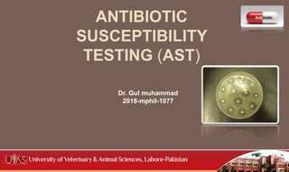
- 1. ANTIBIOTIC SUSCEPTIBILITY TESTING (AST) Dr. Gul muhammad 2018-mphil-1077
- 2. Terminology Antibiotic: A chemical compound derived either naturally or semi-synthetically from living micro-organisms (fungi or bacteria) which is capable, in small concentration,of inhibiting the life process of other micro-organisms. Bacteriostatic: prevents the growth of bacteria. Bactericidal: it kills bacteria.
- 3. Cont.. Antimicrobial: A compound that is derived from natural, semi- synthetic or synthetic sources and is effective against any sort of micro-organisms (e.g., bacteria, virus, fungi, protozoa). Antibacterial: A compound that is derived from natural, semi- synthetic or synthetic sources and is capable to inhibit bacterial growth or cause bacterial destruction.
- 4. Cont... Minimum inhibitory concentration: MIC is the smallest amount of an agent needed to inhibit growth of a microorganism. Minimum bactericidal concentration: MBC is the smallest amount of an agent needed to kill the Bacteria.
- 5. Bactericidal and Bacteriostatic Bactericidal Bacteriostatic Penicillins Cholramphenicol Cephalosporins Tetracyclines Aminoglycosides Macrolides Quinolones Sulphonamides Polymixins Bacitracin Rifampicin Bactericidal+ bactericidal= synergistic effect Bacteriostatic + bacteriostatic = additive effect bactericidal + bacteriostatic = antagonistic
- 6. Action of antimicrobial agents
- 7. Introduction Antibiotic sensitivity testing (AST) The in vitro testing of bacterial cultures with antibiotics to determine the Susceptibility or Resistance of Bacteria. AST is performed only for pathogenic bacteria isolated from the specimens and not for the commensals.
- 8. Types of AST Diffusion method Strokes disc diffusion method Kirby-Bauer disc diffusion method Dilution method Agar dilution method Broth dilution method Types of AST
- 9. Kirby-Bauer disc diffusion method Placing of discs impregnated with antimicrobial agents onto an agar plate seeded with the bacterium to be tested The antimicrobial agents diffuse into the agar creating a zone saturated with the agent, in which an organism susceptible to that agent will not grow.
- 10. Procedure Select a pure culture plate of one of the organisms to be tested. Aseptically emulsify a colony from the plate in the sterile saline solution. Mix it thoroughly to ensure that no solid material from the colony is visible in the saline solution. Repeat until the turbidity of the saline solution visually match that of the standard turbidity. Take a sterile swab and dip it into the broth culture of organism. Gently squeeze the swab against the inside of the tube in order to remove excess fluid in the swab. Reference :by Amrita virtual lab
- 11. Cont.. Take a sterile Mueller-Hinton agar (MHA) plate or a nutrient agar (NA) plate. Use the swab with the test organism to streak a MHA plate or a NA plate for a lawn of growth. After the streaking is complete, allow the plate to dry for 5 minutes. Antibiotic discs can be placed on the surface of the agar using sterilized forceps. Gently press the discs onto the surface of the agar using flame sterilized forceps or inoculation loop. Carefully invert the inoculated plates and incubate for 24 hours at 37° C.
- 12. Result
- 13. Result
- 14. Measuring Zone of inhibition After incubation, use a metric ruler to measure the diameter of the zone of inhibition for each antibiotic used. Compare the measurement obtained from the individual antibiotics with the standard table to determine the sensitivity zone. Compare the measurement obtained from the individual antibiotics to the standard table to determine whether the tested bacterial species is sensitive or resistant to the tested antibiotic Animation link : http://vlab.amrita.edu/?sub=3&brch=73&sim=1628&cnt=3419
- 15. Measuring zones of inhibition. Gray shading represents a confluent lawn of bacterial growth. The white circle represents no growth of the test organism.
- 16. Zone diameter interpretative standard Zone diameter interpretative standards for E. coli and other enteric gram-negative rods ASM microbelibrary
- 17. Factors affecting Antibiotic Susceptibility Testing pH A more acidic pH decreases the activity of aminoglycosides and macrolide antibiotics such as erythromycin. A more alkaline pH favours the action of tetracycline, novobiocin and fusidic acid. Moisture The presence of moisture content on the medium can counteract with accuracy of the susceptibility testing. Reference : Amrita virtual lab
- 18. Cont.. Effect of medium component If the media selected for the antibiotic susceptibility contains excessive amounts of thymine or thymidine compounds, they will reversibly inhibit the action of certain antimicrobial agents such as trimethoprim. Amount of organism Too light inoculum Inhibition zones will be larger even though the sensitivity of the organism is unchanged Relatively resistant strains may be falsely reported as susceptible. Reference : Amrita virtual lab
- 19. Cont… Too heavy inoculum Inhibition zones will be smaller Relatively susceptible strains may then be falsely reported as resistant. Timing of disk application: Plate seeded with inoculum, left at room tempreture for long period. Multiplication of inoculum starts before disks applied. Reduction in zone will lead to false result, susceptible strain will reported as resistant.
- 20. Prepartion of turbidity Standard A 0.5 McFarland standard is prepared by Mixing 0.05 mL of 1.175% barium chloride dihydrate (BaCl2•2H2O), with 9.95 mL of 1% sulfuric acid (H2SO4).
- 21. McFarland standards 0.5 1 2 McFarland standards# A 0.5 McFarland Latex Standard is comparable to a bacterial suspension of 1.5 X 10⁸ CFU/ml.
- 22. Mueller Hinton Agar (MHA) Howard Mueller and Jane Hinton developed Mueller Hinton Agar (MHA) in 1941 for the isolation of pathogenic Neisseria and Moraxella species. Nowadays, it is more commonly used for the routine susceptibility testing of non-fastidious microorganism by the Kirby-Bauer disk diffusion technique.
- 23. Preparation of MHA Suspend 38 gm of the medium in one liter of distilled water. Heat with frequent agitation and boil for one minute to completely dissolve the medium. Autoclave at 121°C for 15 minutes. Cool to room temperature. Pour cooled Mueller Hinton Agar into sterile petri dishes (25 ml) on a level, horizontal surface to give uniform depth. Allow to cool to room temperature. Check for the final pH 7.3 ± 0.1 at 25ºC.
- 24. Why MHA It is a non-selective, non-differential medium. This means that almost all organisms plated on here will grow. It contains starch. Starch is known to absorb toxins released from bacteria, so that they cannot interfere with the antibiotics. Allows better diffusion of the antibiotics than most other plates. A better diffusion leads to a truer zone of inhibition.
- 25. Dilution method Used to determine the minimal concentration of antibiotic to inhibit or kill the microorganism. Achieved by dilution of antibiotic in either agar or broth media.
- 26. Procedure The MIC (Minimal Inhibitory Concentration) of a bacterium to a certain antimicrobial agent gives a quantitative estimate of the susceptibility. MIC is defined as the lowest concentration of antimicrobial agent required to inhibit growth of the organism. The principle is simple: Agar plates, tubes or microtitre trays with two-fold dilutions of antibiotics are inoculated with a standardised inoculum of the bacteria and incubated under standardised conditions following NCCLS guidelines. The next day, the MIC is recorded as the lowest concentration of antimicrobial agent with no visible growth. National Committee for Clinical Laboratory Standards
- 28. MBC The minimum bactericidal concentration (MBC) is determined by subculturing from each tube showing no growth on nutrient agar plate without any antimicrobial agent. Incubate the plates and examine them for growth. The tube containing the lowest concentration of the drug that fails to show growth, on sub culture, is the MBC of the drug for that test strain
- 29. You
- 30. References https://microbiologyinfo.com/mueller-hinton-agar-mha- composition-principle-uses-and-preparation/ https://www.asm.org/getattachment/2594ce26-bd44-47f6-8287- 0657aa9185ad/Kirby-Bauer-Disk-Diffusion-Susceptibility-Test- Protocol-pdf.pdf https://en.wikipedia.org/wiki/McFarland_standards https://www.baytril.com/en/farm animals/cattle/microbiology/