IRJET- Subcellular Localization of Transmembrane E-cadherin-GFP Fusion Pr...
GFP Stability Lab
1. Results
Plasmid Recovery and Gel Electrophoresis: Plasmids sent
to us from the scientific community were extracted from
the filter paper pieces they were transported on and
propagated in DH5alpha cells. Plasmids were isolated
using Qiagen plasmid miniprep kit and verified by
agarose gel electrophoresis.(Figure 5).
GFP Exploration
Ethan Barach and Boriana Marintcheva
Department of Biology, Bridgewater State University, Bridgewater, MA 02324
Abstract
Green fluorescent protein (GFP) in its natural form is derived from the jellyfish Aequorea
Victoria. GFP emits a green light when exposed to UV radiation. Scientists have created
various colors of GFP by introducing mutations in natural GFP. GFP variants could be
expressed in many organisms and used to study sub-cellular localization of proteins and
regulation of gene expression. GFP is also widely used as a teaching tool due to the fact
that it is easily traceable allowing straightforward visualization of processes and
procedures.
We have requested plasmids of various GFPs from the scientific community and have
propagated them. Currently, we are purifying the GFP proteins and collecting data on their
pH and temperature sensitivity. Our long-term goal is to generate a library of GFP proteins
fluorescing with different colors and characterize their properties for the purpose of
developing an inquiry-based teaching lab.
Acknowledgments
This project was supported by funds from the BSU Adrian Tinsley Program’s Fall
2011 and Spring 2012 Semester Grants. I would also like to thank my mentor Dr.
Boriana Marintcheva for her guidance and support. Special thanks to the rest of the
BSU Biology Department.
Materials and
Methods
Materials: Plasmids encoding GFP were obtained from
Biorad (pGLO encoding green GFP). pRSET-Honeydew,
pRSET-Cherry and pRSET- Tangerine were generously
provided by Tsien’s lab, University of California, San
Diego. pRSET –GFP-de1 and de-4 were generously
provided by Remington’s lab, University of Oregon,
Eugene.
Methods
Transformation: GFP plasmids were added to
competent cells (DH5aplpha for plasmid propagation and
BL21 for protein expression) for the purpose of
propagation and protein expression. Respectively the
transformation mixes were incubated on ice for ten
minutes and then heat shocked in a 42oC water bath for
40 seconds. This allowed the GFP plasmid to enter the
competent cells, which were incubated over night on LB
Ampiciline plates to select transformed cells carrying the
GFP plasmid. Single colonies were inoculated to grow in
overnight cultures for the purpose of a starter culture for
GFP expression and isolation.
Plasmid Preparation: Plasmids encoding GFP proteins
were purified from overnight bacteria cultures using
Qiagen plasmid miniprep kit following the
manufacturer’s protocol (Qiagen).
GFP Expression and Purification: pGlo plasmid
encoding green GFP was transformed in DH5a cells and
plated on LB agar plate supplemented with ampiciline
and arabinose. A single bacterial colony was selected and
inoculated to generate an overnight starter culture for
GFP expression. The starter culture was diluted 1:100 and
the new culture grown for 48 hours at 30 o C. Cells that
expressed the GFP were lysed with lysozyme and three
freezing/thawing cycles. Soluble proteins were separated
from cellular debris via centrifugation. A HIC
(Hydrophobic Interaction Chromatography) column was
prepared following the manufacturer’s protocol (Biorad).
The soluble proteins were loaded into the column and
washed with a wash buffer. GFP was eluted they were
eluted with the elution buffer.
Testing GFP Temperature Stability: The purified GFP
was put on ice and tested at several different temperatures
in 50 micro liter aliquots. The high temperatures that the
GFP was tested at were 25 o C (Room Temperature), 50 o
C, 75 o C, and 95 o C. These aliquots were tested at seven
different time intervals. The intervals were thirty minutes,
twenty five minutes, twenty minutes, fifteen minutes, ten
minutes, five minutes, and one minute. Two separate
aliquots of 50 micro liters were also tested at cold
temperatures, which were -29 o C and -80 o C; both of
which were done in an overnight incubation.
Conclusions
1. GFP is stable at 25 o C and 50 o C because it fluoresces green at all of the tested
time intervals.GFP starts to destabilize at 75 o C and it is completely unstable at
95 o C as judged by the loss of green fluorescence.
2. GFP is also stable at extremely cold temperatures such as -29 o C and – 80 o C.
3. GFP is stable in ethanol level ratios that range from 1:1 to 1:5.
Background
Green fluorescent protein (GFP) was discovered in the jelly fish species
Aequorea Victoria (Figure 1). The protein becomes bioluminescent and emits a green color
when it is illuminated with ultra violet light. It has been hypothesized that Aequorea
Victoria becomes bioluminescent to warn their predators, that they may become visible
themselves and thus easily hunted . The bioluminescence has been referenced as the
“burglar alarm” since the jelly fish has only been documented illuminating when
stimulated (1.)
Most of the discoveries on GFP came during the 1990’s. In 2008 Martin Chalfie, Osamu
Shimomura and Roger Tsien were awarded the Nobel Prize for their work on GFP.
Shinomura was the first to realize that the fluorescent entity in Aequorea Victoria was a
protein that fluoresced under ultra violet light. Chalfie expressed the coding sequence of
GFP and Tsien mutated GFP to increase its fluorescence and photo stability as well as,
engineered different colors (2).
The three dimensional structure of GFP is a cylinder made of beta sheets (Figure 2), which
contains the protein. Chromophore is a series of amino acids, which is inside the cylinder.
The chromophore is extremely tolerant of chemical modifications which in turn affect the
spectral properties of the protein (1). Scientists have discovered GFP mutants glowing with
different colors. These include, green, yellow, red, and cyan (Figure 3a). The difference in
colors is due to the engineered differences in the chromophore structures (Figure 3b).
Today GFP allows scientists to do things which they would not be able to without GFP.
One of the most important roles of GFP is to serve as a tool in cellular targeting. Since,
fluorescent proteins
are not harmful to the cells that they are expressed they can be used to visualize specific
proteins and cell structures (Figure 3). Examples of the organisms that have been studied
with the help of GFP include; fish, reptiles, bacteria, and mammals. Several colors of GFP
can be used so different proteins or cellular structures can be tracked. This approach allows
scientists to understand many biological processes that were difficult to observe before the
use of GFP which is why GFP is of great significance to the scientific community.
In addition to being a great research tool, GFP is also a great teaching tool since it allows
straightforward visualization of protein purification procedure, as well as low tech means
for following the integrity of the protein. The goal of this project is to examine GFP
stability in the context of a teaching lab in order to create an inquiry based learning
module. On the long term we hope of generate a library of GFP colors.
Figure 1: Aequorea Victoria - the jelly fish
species that was first discovered to produce the
naturally occurring GFP. Picture courtesy of
http://gfp.conncoll.edu/
Figure 2A: GFP and fluorescent
variants
(A) GFP has a 3D cylinder shape
composed of Beta sheets (green) and
chromophore (yellow) in the cavity of the
cylinder. Image originally from
www.pnas.org ; (B) Different colors of
GFP available for research purposes
ranging from cyan to magenta. Picture
courtesy of www.michaeleisen.org
GFP Expression and Purification: GFP expression of donated GFP variants
was attempted in E.coli BL21 (DE3) cells and was unsuccessful. Green GFP
coded by pGLO (Biorad) was expressed in DH5a cells and purified using HIC
chromatography as described in materials and methods. Protein purification was
monitored by following up GFP fluorescence.
Thermostability Tests: Temperature is a key environmental factor influencing
protein stability that is straightforward and cost effective to explore in the settings
of a teaching lab. Fifty microliter aliquots of
purified protein were incubated at various temperatures ranging between room
temperature and 95oC for temperature intervals ranging between 1 and 30 minutes.
As expected GFP lost its fluorescence fast at 95oC and it was quite stable at the
temperatures below 50oC. The results of the performed time courses are
summarized in Figure 6. GFP appeared stable to freezing at - 29oC and -80oC (data
not shown).
Ethanol exposure and GFP stability: Ethanol stability tests were done by mixing
100 microliters of GFP with ethanol in ratios ranging from 1:1 to 1:5. In all
scenarios the GFP was stable and fluoresced green under ultra violet light (data not
shown).
Figure 5: Plasmid gel
electrophoresis. Plasmids were ran
on 0.8% TBE/agarose gel at 100 volts.
Lane 1-pRSET-GFP-DE1alpha)
Lane 2 - pRSET-GFP-DE4alpha)
Lane 3-pRSET-GFP-Honeydew
Lane 4- DNA ladder
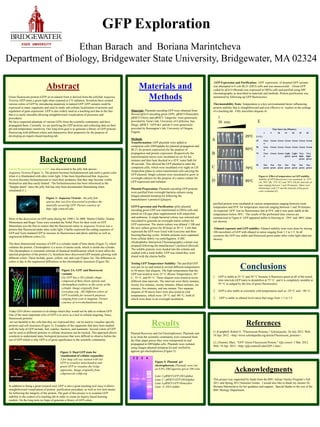
Time Intervals (Minutes)
1 5 10 15 20 25 30
T (o C)
25 Gree
n
Green Green Green Green Green Green
50 Gree
n
Green Green Green Green Green Green
75 Gree
n
Green Green Light
green
Light
green
Very
light
green
Very
light
green
95 Not
green
Not
green
Not
green
Not
green
Not
green
Not
green
Not
green
Figure 3: Dual GFP stain for
visualization of cellular organelles
A fox lung cell was stained with red
GFP to visualize mitochondria and
green GFP to visualize the Golgi
apparatus. Image originally from
cshprotocols.cshlp.org
1 2 3 4
References
(1.)Campbell, Robert E. "Fluorescent Proteins." Scholarpedia. 26 July 2012. Web.
10 Apr. 2012. <http://www.scholarpedia.org/article/Fluorescent_proteins>.
(2.) Zimmer, Marc. "GFP: Green Fluorescent Protein." Gfp.connol. 3 Mar. 2012.
Web. 18 Apr. 2012. <http://gfp.conncoll.edu/GFP-1.htm>.
A
B
C
D
25oC
50oC
75oC
95oC
1
30 min. E
Figure 6: Effect of temperature on GFP stability.
Stability of GFP fluorescence was examined at 25oC
(A), 50oC (B), 75oC (C) and 95oC (D) for intervals of
time ranging between 1 and 30 minutes. Tubes were
illuminated with UV and the intensity of the green
color evaluated (E)
A B