
Recommended
More Related Content
What's hot
What's hot (20)
Similar to Microbiology Culture Media & Methods Guide
Similar to Microbiology Culture Media & Methods Guide (20)
More from EDEMAWIILLIAM
Recently uploaded
Recently uploaded (20)
Microbiology Culture Media & Methods Guide
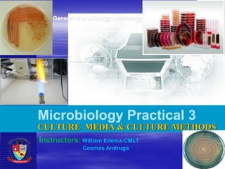
- 1. General Microbiology Laboratory Microbiology Practical 3 CULTURE MEDIA & CULTURE METHODS Instructors: William Edema-CMLT Cosmas Andruga
- 2. Aim of the practical Learn the different types of culture media Demonstrate preparation of culture media Demonstrate culturing methods
- 3. Introduction In natural environments, microorganisms usually exist as mixed populations. However, if we are to study, characterize, and identify microorganisms, we must have the organisms in the form of a pure culture. A pure culture is one in which all organisms are descendants of the same organism. Techniques for obtaining pure cultures from a mixed population will be studied in this Lab subsequently.
- 4. Bacteria have to be grown (cultured) for them to be identified. By appropriate procedures they have to be grown separately (isolated) on culture media and obtained as pure for study. History The original media used by Louis Pasteur – urine or meat broth Liquid medium – diffuse growth Solid medium – discrete colonies.
- 5. Colony – macroscopically visible collection of millions of bacteria originating from a single bacterial cell. Cooked cut potato by Robert Koch – earliest solid medium Gelatin – not satisfactory - liquefy at 24oC
- 6. Agar Frau Hesse Used for preparing solid medium Obtained from seaweeds. No nutritive value Not affected by the growth of the bacteria. Melts at 98oC & sets at 42oC 2% agar is employed in solid medium
- 7. Types of culture media I. Based on their consistency a) solid medium b) liquid medium c) semi solid medium II. Based on the constituents/ ingredients a) simple medium b) complex medium c) synthetic or defined medium d) Special media
- 8. Special media – Enriched media – Enrichment media – Selective media – Indicator media – Differential media – Sugar media – Transport media – Media for biochemical reactions III.Based on Oxygen requirement - Aerobic media - Anaerobic media
- 9. Solid media – contains 2% agar Distinctive colony morphology (size, shape, margins, pigmentation, hemolysis etc.) can be appreciated. Characteristics easy to identify Eg: Nutrient agar, Blood agar Liquid media – no agar. Diffuse growth, no x-tics for identification For inoculum preparation, Blood culture, for the isolation of pathogens from a mixture. Eg: Nutrient broth Semi solid medium – 0.5% agar. Eg: Motility medium Types of culture media: Based on consistency:
- 10. a) solid medium b) liquid medium c) semi solid medium Types of culture media: Based on consistency:
- 11. Simple media / basal media Most common in routine diagnostic labs: - Eg: Nutrient Broth, Nutrient Agar - NB consists of peptone, meat extract, NaCl, - NB+0.5 Glucose= Glucose Broth - NB + 2% agar = Nutrient agar - Agar conc. reduced to (0.2-0.5%)= Semi-solid media Types of culture media: Based on the constituents/ ingredients
- 12. Complex media Media other than basal media. They have added ingredients e.g. yeast extract and casein hydrolysate, which consist of a mixture of many chemical species of unknown proportions. Provide special nutrients Synthetic or defined media Media prepared from pure chemical substances and its exact composition is known; Eg: peptone water – 1% peptone + 0.5% NaCl in water
- 13. Enriched media Substances like blood, serum, egg are added to the basal medium. Used to grow bacteria that are exacting in their nutritional needs (fastidious organisms). E.g.: Blood agar, Chocolate agar Blood agar Chocolate agar
- 14. Enrichment media Liquid media used to isolate pathogens from a mixed culture. Media is incorporated with inhibitory substances to suppress the unwanted organism e.g.: – Selenite F Broth – for the isolation of Salmonella, Shigella – Alkaline Peptone Water – for Vibrio cholerae Selenite F Broth Alkaline Peptone Water Tetrathionate broth
- 15. Selective media Media which contains substances that prevent or slow the growth of microorganisms other than the bacteria for which the media is prepared for. The inhibitory substance is added to a solid media. E.g: – Mac Conkey’s medium for gram negative bacteria – TCBS – for V.cholerae – LJ medium – M.tuberculosis – Wilson and Blair medium – S.typhi – Potassium tellurite medium – Diphtheria bacilli
- 16. TCBS Mac Conkey’s medium Selective media examples
- 17. Potassium Tellurite media LJ media Selective media examples
- 18. Indicator media These media contain an indicator which changes its colour when a bacterium grows in them. Eg: – Blood agar – Mac Conkey’s medium – Christensen’s urease medium
- 19. Blood agar: shows three (3) types of hemolysis • Erythrocytes are incorporated into nutrient agar medium • Certain bacteria produce products that lyse Red Blood Cells Alpha-hemolytic- partial lysis Beta-hemolytic- complete lysis Gamma-hemolytic- no lysis
- 20. Urease medium
- 21. Differential media Contain indicators, dyes, etc, to differentiate microorganisms. E.g. MacConkey agar, which contains neutral red (pH indicator) and is used to differentiate lactose fermenter and non-lactose fermenter. (E.g. E. coli and Salmonella). – Lactose fermenters – Pink colonies – Non lactose fermenters – colourless colonies
- 22. Lactose fermenters – Pink colonies Non lactose fermenters – colourless colonies Chocolate agar MacConkey agar Examples of Lactose fermenters Escherichia coli, Enterobacter and Klebsiella Examples of Non-Lactose fermenters: Salmonella, Proteus species, Yersinia, Pseudomonas aeruginosa and Shigella
- 23. Transport media Media used for transporting the samples. Delicate organisms may not survive the time taken for transporting the specimen without a transport media. Eg: – Stuart’s medium – non nutrient soft agar gel containing a reducing agent – Buffered glycerol saline – enteric bacilli
- 24. Anaerobic media These media are used to grow anaerobic organisms. Eg: Robertson’s cooked meat medium, Thioglycolate medium.
- 25. Anaerobic media These media are used to grow anaerobic organisms. Eg: Robertson’s cooked meat medium, Thioglycolate medium.
- 26. Common media used in Microbiology Laboratory Chocolate Agar: Blood agar prepared by heating blood to 95C until medium becomes brown or chocolate in color. Heating the blood releases both X and V growth factors and also destroys the inhibitors of V factor. These factors are required for the growth of most species of Haemophilus and also Neisseria gonorrhea. MacConkey Agar: An inhibitory and differential medium used to distinguish lactose- fermenting Gram- negative organism from non fermentation. Crystal violet, bile salts and neutral red are inhibitor agent. neutral red is the PH
- 27. Common media used in Microbiology Laboratory Mannitol Salt Agar (both selective and differential) For selective isolation of coagulase positive, mannitol-fermenting staphylococcus. High salt concentration (7.5%) only permits Staphylococcous spp. growth. Mannitol is a sugar alcohol fermented by certain species o Pathogenic Staph will ferment mannitol o Non-pathogenic Staph will not ferment mannitol If mannitol is fermented, acidic products are formed. o Indicated by phenol red (yellow is acidic)
- 28. Common media used in Microbiology Laboratory Mannitol Salt Agar (both selective and differential)
- 29. Common media used in Microbiology Laboratory Mueller Hinton Agar: Rich medium that support the growth of most microorganism. It is commonly used for antibiotic susceptibility testing: o disk diffusion antibiotic susceptibility; o antibiotic serum level measurements; o MBC determination.
- 30. Common media used in Microbiology Laboratory Salmonella Shigella ( SS ) Agar: Isolation and differential medium for pathogenic Gram-negative bacilli in particular, Salmonella and Shigella. It is an Inhibitor for Coliforms. Triple Sugar Iron Agar (TSI): this is a key medium used in the beginning of the identification of Gram- negative bacilli of the enteric group. It contains glucose (0.1% ), Lactose (1%), sucrose(1%). And peptone (2%) as nutritional sources. Sodium Thiosulfate serves as the electron receptor for reduction of sulfur and production of H2S. Detects fermentation of sucrose, lactose, glucose, as well as production of hydrogen sulfide and /or gas . Indicators: Phenol red is the PH indicator; ferric ammonium citrate is H2S indicator.
- 31. Preparation of Culture Media When lab personnel make media, they measure out a quantity of dry powdered nutrient media, add water and check the pH. – Measurements are done according to manufacturer’s recommendation The media is then dispensed into bottles (flask, tube), capped and autoclaved at 121°C (15 psi) for 20 minutes. Once the media is autoclaved it is sterile (all microoranism forms killed)
- 35. CULTURE METHODS Culture methods employed depend on the purpose for which they are intended. The indications for culture are: – To isolate bacteria in pure cultures. – To demonstrate their properties. – To obtain sufficient growth for the preparation of antigens and for other tests. – For bacteriophage & bacteriocin susceptibility. – To determine sensitivity to antibiotics. – To estimate viable counts. – Maintain stock cultures.
- 36. Culture methods include: Streak culture Lawn culture Stroke culture Stab culture Pour plate method Liquid culture Anaerobic culture methods
- 37. STREAK CULTURE Used for the isolation of bacteria in pure culture from clinical specimens. Platinum wire or Nichrome wire is used. One loopful of the specimen is transferred onto the surface of a well dried plate. Spread over a small area at the periphery. The inoculum is then distributed thinly over the plate by streaking it with a loop in a series of parallel lines in different segments of the plate. On incubation, separated colonies are obtained over the last series of streaks.
- 38. STREAK CULTURE: Procedure Necessary Equipment
- 40. Identification of bacteria - Once a pure colony is obtained, the next step is to identify the bacterial species. This will be done in the next practical
- 43. LAWN CULTURE Provides a uniform surface growth of the bacterium. Uses – For bacteriophage typing. – Antibiotic sensitivity testing. – In the preparation of bacterial antigens and vaccines. Lawn cultures are prepared by flooding the surface of the plate with a liquid suspension of the bacterium.
- 45. STROKE CULTURE Stroke culture is made in tubes containing agar slope / slant. Uses –Provide a pure growth of bacterium for slide agglutination and other diagnostic tests.
- 46. STAB CULTURE Prepared by puncturing a suitable medium – gelatin or glucose agar with a long, straight, charged wire. Uses –Demonstration of gelatin liquefaction. –Oxygen requirements of the bacterium under study. –Maintenance of stoke cultures.
- 47. Gelatin liquefaction Oxidation – Fermentation medium
- 48. POUR PLATE CULTURE Agar medium is melted (15 ml) and cooled to 45oC. 1 ml of the inoculum is added to the molten agar. Mix well and pour to a sterile petri dish. Allow it to set. Incubate at 37oC, colonies will be distributed throughout the depth of the medium. Uses – Gives an estimate of the viable bacterial count in a suspension. – For the quantitative urine cultures.
- 49. LIQUID CULTURES Liquid cultures are inoculated by touching with a charged loop or by adding the inoculum with pipettes or syringes. Uses – Blood culture – Sterility tests – Continuous culture methods Disadvantage – It does not provide a pure culture from mixed inocula.
Editor's Notes
- TCBS
- C.Diphtheriae on Potassium tellurite media
- Mac Conkey’s medium
- Antibiotic sensitivity testing
- Motility medium