
Empfohlen
Weitere ähnliche Inhalte
Was ist angesagt?
Was ist angesagt? (20)
Ähnlich wie Maternal Anatomy by _ Dr. tejaswini [Autosaved].pptx
Ähnlich wie Maternal Anatomy by _ Dr. tejaswini [Autosaved].pptx (20)
Mehr von DrTejaswini7
Kürzlich hochgeladen
Kürzlich hochgeladen (20)
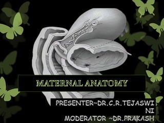
Maternal Anatomy by _ Dr. tejaswini [Autosaved].pptx
- 2. Contents Anterior Abdominal wall External Generative Organs Internal Generative Organs Pelvic Anatomy
- 3. ANTERIOR ABDOMINAL WALL Anterior Abdominal wall stretches to accommodate the expanding uterus & provide surgical access to the internal reproductive organs, thus comprehensive knowledge of it’s layered structure is required to surgically enter the peritoneal cavity. The anterior abdominal wall extends from the xiphoid process of the sternum and costal margins cranially to the iliac crest and pubic bones caudally
- 4. • Skin – Langer lines • Subcutaneous Layer – Camper’s fascia – Scarpa’s fascia • Primary fascia (aponeurosis)/rectus sheath • Abdominal wall muscles – Rectus abdominis – Pyramidalis – Obliques (Ext. & Int.) – Tranversus abdominis • Tranversalis fascia • Extra peritoneal tissue • Parietal layer of peritonuem LAYERS OF THE ANTERIOR ABDOMINAL WALL
- 6. Skin Langer lines : These describes the orientation of dermal fibers within the skin. In Ant. Abdominal wall they are arranged transversely. As a result of this, vertical skin incisions sustain greater and thus in general it develops wider scars. In contrast, Low Transverse incisions such as Pfannenstiel incision follows larger lines and lead to superior cosmetic result. LAYERS OF THE ANTERIOR ABDOMINAL WALL
- 7. Subcutaneous Layer – Camper’s fascia (Superficial fatty layer) – Scarpa’s fascia (Deeper membranous layer) Camper’s Fascia : Continues on to the perineum to provide fatty substance to Mons pubis, Labia majora and then to blend with the fat of Ischio – anal fossa. Scarpa’s Fascia : Continues inferiorly on to perineum as Colles fascia. LAYERS OF THE ANTERIOR ABDOMINAL WALL
- 8. Primary fascia (aponeurosis)/Rectus sheath – It is formed by Fibrous aponeurosis of External Obliques, Internal Obliques, Transverse abdominis, these fuse in the midline at the Linea alba [Normal 10 – 15mm] These three aponeuroses also invest the Rectus abdominis muscle . Rectus sheath construction is different above and below the arcuate line LAYERS OF THE ANTERIOR ABDOMINAL WALL
- 10. Abdominal wall muscles – Rectus abdominis – Pyramidalis – External Oblique – Internal Oblique – Tranversus abdominis LAYERS OF THE ANTERIOR ABDOMINAL WALL
- 13. Blood Supply of the ANTERIOR ABDOMINAL WALL BRANCHES OF FEMORAL ARTERY Superficial Epigastric Artery Superficial Circumflex Iliac Artery Superficial External Pudendal Artery supplies Skin, Subcutaneous layer & Mons Pubis. Inferior Deep Epigastric Vessels : Branch of External Iliac vessels – supplies Anterior Abdominal wall muscles & Fascia Lies above the Arcuate line between Posterior Rectus sheath & Rectus Abdominis.
- 14. Near the umbilicus : Inferior epigastric vessels anastomoses with Superior epigastric artery and vein which are the branches of internal thoracic vessels Clinically when a Maylard incision is used for cesarean delivery ,the inferior epigastric vessels may be lacerated lateral to the rectus belly during muscle transection .preventively ,identification &surgical occlusion are preferrable
- 17. Nerve Supply of the ANTERIOR ABDOMINAL WALL Inter coastal Nerve [ T7 – 11] Sub coastal Nerve [T12] Ilio hypogastric Nerve[L1] Ilio inguinal Nerve[L1] Derived from Anterior Rami of thoracic spinal nerves Intercoastal & Subcoastal nerves are the anterior rami spinal nerves, an Intercoastal nerve extends ventrally between the Trasversus abdominis and Internal oblique muscles. During this path, the nerve ives rise to Lateral & Anterior cutaneus branches – that innervates the Ant. Abdominal wall. The space between Transversus abdominis & Internal obliques is known as – TRANS ABDOMINIS PLANE. [Most widely used for Post C – section analgesia blockade.]
- 18. Ilio hypogastric and Ilio inguinal nerves supply the suprapubic area ,lower abdomen,and mons pubis. These nerve fibres run between the layers of rectus sheath lateral to rectus muscle and may be entrapped during closure, especially if incisions beyond the lateral borders of the rectus abdominis muscle. These nerves carry sensory information only, and injury leads to loss of sensations within the areas supplied. Chronic pain may develop in rare cases.
- 19. The T10 dermatome approximates the level of the umbilicus. Analgesia to this level is suitabe for labor & Vaginal Birth. Regional analgesia for Cesarean delivery or for puerperal sterilization ideally extends to T4.
- 20. EXTERNAL GENERATIVE ORGANS Vulva (Pudenda) Includes : Mons pubis Labia majora Labia minora Clitoris Vestibule Urethral opening Vaginal orifice Hymen Para urethral glands (skene’s gland) Bartholin glands
- 21. MONS PUBIS :This is a triangular area anterior to the pubic bones; It is continuous with the abdominal wall above and with labia below. It is filled with adipose tissue and covered by hairy skin :LABIA MAJORA:These are folds of fatty tissue covered by skin that extend from the mons pubis to the perineum to meet in front of the anus, forming the posterior fourchette. The skin on the lateral aspects of the labia majora is pigmented and covered by hair . The inner aspect is smooth and shiny and contains apocrine, sweat & sebaceous glands .
- 23. LABIA MINORA:Labia minora are folds of skin that lie medial to the labia majora,encircling the urethral and vaginal orificies. Posteriorly they fuse with the posterior fourchette but anteriorly they divide to form hood /prepuce & a frenulum for the clitoris
- 24. HART’S LINE:The outer side of L.Minora is lined by keratinized squamous epithelium. The medial (inner)side of L.minora is lined by non keratinized squamous epithelium. The line which divides outer and inner side of L.minora is called HART”S LINE
- 25. • Vulva (pudenda): includes all structures visible externally from the symphysis pubis to the perineal body. EXTERNAL GENERATIVE ORGANS
- 26. CLITORIS It is the main female erectile structure & is located anterior to the urethral orifice between the anterior folds of the labia minora. It is the homologous of the penis in men It is about 1.5-2cm in length. • Parts : – Glans – richly innervated – Corpus – (2) crura EXTERNAL GENERATIVE ORGANS
- 27. VESTIBULE It is an almond shaped area enclosed by labia minora laterally & extends from clitoris to fourchette Perforated By 6 Openings – Urethra – Vagina – Bartholin gland ducts – Skene glands EXTERNAL GENERATIVE ORGANS
- 28. VESTIBULE Boundaries – P: fourchette – A: clitoral frenulum anteriorly – L: Hart line laterally – M: external surface of hymen medially Fossa navicularis – posterior portion of the vestibule between the fourchette and the EXTERNAL GENERATIVE ORGANS
- 31. VESTIBULE • Bartholin glands – Greater vestibular glands – Bartholin cyst (obstructed) – Bartholin abscess (if infected) • Paraurethral glands • Skene glands – largest • Inflammation, duct obstruction –> urethral diverticulum
- 32. BARTHOLIN GLAND 1)pea shaped gland 2)2 in number 3)located in superficial perineal pouch between L.majora & L.minora at 4’o & 8’o clock postion . 4)These are homologous to cowper glands in male . 5)Duct of the Bartholin gland open in the vestibule outside the hymen i.e at the junction of anterior 2/3rd & posterior 1/3rd . 6)If duct gets blocked ,it leads to Bartholin cyst .
- 33. BARTHOLIN’S CYST POSTERIO- LATERAL WALL OF VAGINA GARTNERS’ CYST ANTERIO- LATERAL OF VAGINA
- 35. BARTHOLIN’S CYST :Intermittent painless mass on vulva which is aggravated by intercourse MANAGEMENT:
- 38. HYMEN It is a septum of mucous membrane which usually gets ruptured during first intercourse or during sternous exercise. The hymen gets badly torn at parturition to form different sized cicatrized nodules known as hymenal tags
- 39. HYMEN
- 44. INTERNAL GENERATIVE ORGANS • Vagina • Cervix • Uterus • Ovaries • Fallopian tubes
- 45. VAGINA
- 46. Anterior vaginal wall : 6 to 8 cm Posterior vaginal wall : 7 to 10 cm Lining : NKSS VAGINA There are NO VAGINAL GLANDS.
- 48. VAGINA BLOOD SUPPLY : Cervical branch of Uterine artery} upper 1/3rd Vaginal artery}middle 1/3rd Middle rectal artery Internal pudendal artery Lower 1/3rd
- 49. LYMPHATICS : Inguinal lymph nodes lower third, along with those of the vulva Internal iliac nodes middle third External, internal, and common iliac nodes upper third NERVE SUPPLY Sympathetic from hypogastric plexus Parasympathetic from S2,3,4 VAGINA
- 50. Cervix Small opening (nulli) Slit-like (parous) Ectocervix: NKSS epithelium Endocervix: Simple Columnar ep. SCJ is the mc site of malignant transformation Eversion – during pregnancy Composition: collagen, elastin, proteoglycans, very little SM
- 51. Uterus NULLIGRAVID MULTIPAROUS Weight 60kg Weighs more Length 6-8cm 9-10cm Fundus and cervix Equal in length Cervix is only a third of entire Length
- 52. ANGLE OF UTERUS also called as CORNUA OF UTERUS 3structures are attached on either side
- 53. FALLOPIAN TUBE ROUND LIGAMENT OVARIAN LIGAMENT FALLOPIAN TUBE ROUND LIGAMENT FALLOPIAN TUBE OVARIAN LIGAMENT ANTERIOR POSTERIOR LATERAL ANTERIOR POSTERIOR LATERAL ANTERIOR POSTERIOR LATERAL
- 54. WHY IS IT IMPORTANT TO KNOW RELATIONSHIPOF THESE STRUCTURES MC CAUSE of failure of female sterilization is identificationof wrong structure Therefore; to prevent this wrong identification ,tube should be identified by distal end
- 55. PERINEUM
- 60. ANATOMY OF THE ANTERIOR TRIANGLE
- 62. ANATOMY OF THE POSTERIOR TRIANGLE ISCHIO RECTAL FOSSAE Two fat-filled wedge-shaped spaces found on either side of the anal canal (comprise the bulk of the posterior triangle) ANAL CANAL distal continuation of the rectum: begins at the level of levator ani attachment to the rectum and ends at the anal skin (4 to 5-cm long) Anal cushion – aids in the complete closure of canal and fecal continence when apposed Lining (mucosa): columnar epithelium (upper) stratified squamous epithelium (dentate line) HEMORRHOIDS : External CC: pain (inferior rectal nerve) Internal CC: bleeding
- 63. The ANAL SPHINCTER Complex Description Function Symptoms EAS Striated muscle attaching to PB anteriorly Provides squeeze pressure responsible or maintaining fecal continence when continence is threatened 25% resting pressure Provides emergency control for liquid stool and flatus Fecal urgency Urge incontinence: liquid & flatus IAS Continuation of the rectal circular smooth Keeps anal canal Fecal soiling muscle. (70-85% resting pressure) closed at rest, Incontinence maintenance of fecal continence at rest continence of liquid of liquid Receives parasympathetic nerve fibers stool & flatus stool and flatus
- 64. PERINEAL BODY Central point of the perineum it is a fibromuscular node
- 65. MNEMONIC BELTS for support BULBOSPONGIOSUS EXTERNAL ANAL SPHINCTER LEVATOR ANI TRANSVERSE PERENEI-SUPERFICIAL &DEEP SPINCTER URETHRAE
- 66. PELVIC DIAPHRAGM • Broad muscular sling that provides substantial support to the pelvic viscera • composed of : – Levator Ani* Pubococcygeus Puborectalis Iliococcygeus – Coccygeus Muscle
- 69. PUDENDAL NERVE • Anterior rami of S2-4 • Lies within the Alcock canal • 3 terminal branches: – Dorsal nerve of the clitoris – Perineal nerve • Posterior labial branches • Muscular branches – Inferior rectal
- 71. Where does the ROUNDligament terminate? Labia Majora
- 72. Which ARTERY courses through the ROUND ligament ? Sampson Artery
- 73. Ligaments
- 74. The broad ligaments Two wing like structures that extend from the lateral uterine margins to the pelvic sidewalls. Peritoneum that folds over the fallopian tube mesosalpinx, round ligament is the mesoteres Ovarian ligament is the mesovarium. Ligaments
- 75. Ligaments
- 77. • CARDINAL LIGAMENT – Transverse Cervical Ligament – Mackenrodt ligament • anchors medially to the uterus and upper vagina. • PARAMETRIUM is the connective tissues adjacent and lateral to the uterus within the broad ligament. • PARACERVICAL tissues are those adjacent to the cervix • PARACOLPIUM is that tissue lateral to the vaginal walls. Ligaments
- 82. Venous drainage • The right ovarian vein empties into the vena cava • Left ovarian vein empties into the left renal vein
- 83. Lymphatics • Cervix : Internal iliac nodes • Uterine corpus : internal iliac nodes, para – aortic lymph nodes
- 85. Fallopian tubes (oviducts) • 8–14 cm from the uterine cornua
- 86. Ovaries Rests at the ovarian fossa of Waldeyer. Sympathetic nerves : ovarian plexus (originates in the renal plexus) Parasympathetic input : Vagus nerve Sensory afferents follow the ovarian artery and enter at T10
- 87. FEMALE REPRODUCTIVE TRACT Lining Epithelium Vulva Vagina Cervix Uterus FT Ovary Stratified Stratified Ectocervix: Simple Simple Cuboidal Squamous Squamous Stratified squamous Columnar Columnar Keratinizing Non- Non-keratinizing Ciliated Keratinizing Endocervix: Simple columnar Mucin-producing ?? Transformation zone: Squamo-columnar Junction