

Empfohlen
Weitere ähnliche Inhalte
Was ist angesagt?
Was ist angesagt? (20)
Ähnlich wie Triangles of the neck
Ähnlich wie Triangles of the neck (20)
Mehr von Dr. Mohammad Mahmoud
Mehr von Dr. Mohammad Mahmoud (20)
Kürzlich hochgeladen
Kürzlich hochgeladen (20)
Triangles of the neck
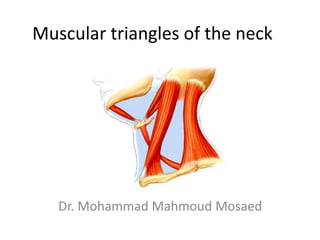
- 1. Muscular triangles of the neck Dr. Mohammad Mahmoud Mosaed
- 2. Lateral side of the neck • The lateral side of the neck is bounded by; • Anteriorly: the midline of the neck • Posteriorly: the anterior border of the trapizuis muscle • Superiorly: the mandible • Inferiorly: the clavicle
- 3. • The lateral side of the neck is divided by sternocleidomastoid muscle into 2 triangles: • Anterior triangle • Posterior triangle
- 4. The posterior trangle • Boundaries: • Posteriorly: by the trapezius muscle • Anteriorly: by the sternocleidomastoid muscle • Inferiorly: by the clavicle. • Floor: prevertebral muscles "semispinalis capitis, splenius capitis, levator scapulae and scalenus medius” • Roof: skin, superficial fascia and platysma • The posterior triangle of the neck is further subdivided by the inferior belly of the omohyoid muscle into a large occipital triangle above and a small supraclavicular triangle below.
- 7. Structures in Posterior Triangle • The brachial plexus and subclavian artery pass between the scalenus anterior and medius muscles. • The subclavian vein passes anterior to the scalenus anterior muscle.
- 9. Anterior triangle of the neck: Boundaries: • Superior: the lower border of the mandible. • Lateral: the posterior border of the sternomastoid muscle. • Medially: the midline. • Roof: The skin, platysma muscle, superficial veins and nerves, deep fascia. Subdivisions of anterior triangle • Digastric triangle • Submental triangle • Carotid triangle • Muscular triangle
- 11. The muscular triangle: • Boundaries: • Laterally the posterior border of the sternomastoid, • Superiorly the superior belly of the omohyoid muscle • Anteriorly the midline. • Contents: • Superior belly of omohyoid • Sternohyoid • Sternothyroid • Thyrohyoid • Thyroid gland • Parathyroid gland • Cervical part of trachea and esophagus
- 13. The digastric triangle • Boundaries: the lower border of the mandible above and the two bellies of the digastric muscles on each side. • Floor: the myelohyoid muscle in front and the hyoglossus behind. • Contents: • The submandibular salivary gland with its duct, • The submandibular lymph nodes, • The facial artery. • The lingual nerve and submandibular ganglion • The hypoglossal nerve
- 14. Boundaries mastoid & mandible above anterior belly of digastric posterior belly of digastric
- 15. The submental triangle: • Boundaries: • Anteriorly: the midline. • Laterally: the anterior belly of digastric • Inferiorly: the hyoid bone • Floor: The myelohyoid muscle • Contents: • Submental vessels, nerve and lymph nodes.
- 17. The carotid triangle Boundaries: • Laterally the posterior border of sternomastoid • Above and medially the posterior belly of digastric muscle inferiorly and medially the superior belly of omohyoid muscle • Floor: the middle constrictor of the pharynx. Contents: • Vessels; the external carotid artery with its superior thyroid, ascending pharyngeal, lingual and facial branches, the carotid sheath containing the internal carotid artery, internal jugular vein and vagus nerve, • Nerves; the ansa cervicalis overlying the carotid sheath, the sympathetic trunk deep to the sheath and the hypoglossal nerve crosses both the internal and external carotids. The accessory nerve runs on the lateral side of the vessels for a short distanc • Lymph Nodes; deep cervical lymph nodes
- 18. The boundaries of the carotid triangle • posterior belly of digastric muscle (pbd) • superior belly of the omohyoid muscle (so) • anterior border of sternomastoid muscle (st)