Cranial nerve nuclei
•Download as PPT, PDF•
43 likes•20,098 views

Neuroanatomy lecture by dr. Mohammed Mahmoud Mosaed Northern borderd faculty of medicine Al-azhar faculty of medicine

Recommended
More Related Content
What's hot
What's hot (20)
Similar to Cranial nerve nuclei
Similar to Cranial nerve nuclei (20)
More from Dr. Mohammad Mahmoud
More from Dr. Mohammad Mahmoud (20)
Recently uploaded
Recently uploaded (20)
Cranial nerve nuclei
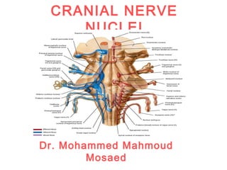
- 1. CRANIAL NERVE NUCLEI Dr. Mohammed Mahmoud Mosaed
- 2. Neural tube • The wall of a recently closed neural tube consists of neuroepithelial cells, they divide rapidly, producing more and more neuroepithelial cells which constitute the neuroepithelial layer. • Once the neural tube closes, neuroepithelial cells begin to give rise to another cell type, the primitive nerve cells or neuroblasts which form the mantle layer. • The outermost layer of the neural tube is the Marginal layer which contains nerve fibers emerging from neuroblasts in the mantle layer
- 3. Basal, Alar, Roof, and Floor Plates As a result of continuous addition of neuroblasts to the mantle layer, each side of the neural tube shows a ventral and a dorsal thickening. The ventral thickenings, The Basal plates, which form the motor areas in the neural tube; The dorsal thickenings, The Alar plates, form the sensory areas. The dorsal and ventral midline portions of the neural tube, known as the roof and floor plates, respectively, do not contain neuroblasts
- 5. Cranial nerve nuclei in the brain stem The basal plate contains motor nuclei which divided into 3 groups: (a) General Somatic Efferent group (medial in position) (b) Special Visceral Efferent group (intermediate) (c) General Visceral Efferent group (lateral in position) The alar plate contains 4 groups of sensory relay nuclei (a) Special Somatic Afferent group (lateral in position), receives impulses from the ear by way of the vestibulocochlear nerve. (b) General Somatic Afferent receives impulses from the head and face (c) Special Visceral Afferent group receives impulses from taste buds of the tongue and from the palate, oropharynx, and epiglottis. (d) General Visceral Afferent, group (medial in position) receives interoceptive information from the gastrointestinal tract and heart
- 6. • Somatic refers to head, body wall, and extremities; • Visceral refers to viscera; • Afferent refers to sensory (input); • Efferent refers to motor (output); • General refers to wide areas of the head and body; • Special refers to the specialized functions of olfaction (smell), gustation (taste), vision, audition, equilibrium (vestibular system), and branchiomeric muscles. •All cranial nerve nuclei present in the brain stem except The olfactory nerve (SVA) and the optic nerve (SSA) are telencephalic derivatives, not brainstem cranial nerves Sensory nuclei where afferent fibers terminate (called terminal nuclei) •The nuclei of origin of motor fibers of the cranial nerves are organized in discontinuous nuclear “columns” within the brainstem.
- 7. Cranial nerves • The 12 Cranial Nerves • There are 12 pairs of cranial nerves, which leave the brain and pass through foramina and fissures in the skull. All the nerves are distributed in the head and neck, except cranial nerve X, which also supplies structures in the thorax and abdomen. The cranial nerves are named as follows: • 1. Olfactory 2. Optic • 3. Oculomotor 4. Trochlear • 5. Trigeminal 6. Abducent • 7. Facial 8. Vestibulocochlear • 9. Glossopharyngeal 10. Vagus • 11. Accessory 12. Hypoglossal
- 9. MOTOR NUCLEI
- 10. The General Somatic Efferent Column • It includes • Nucleus of the oculomotor nerve (midbrain), • Nucleus of trochlear nerve (lower midbrain), • Nucleus of abducent nerve (lower pons) • Nucleus of hypoglossal nerve (medulla). • These nuclei are composed of lower motor neurons innervating the voluntary muscles of the eye and tongue.
- 11. The Special Visceral (Branchial) Efferent Column • It includes • Motor nucleus (masticator nucleus) of the trigeminal nerve (midpons), • Motor nucleus of the facial nerve (the facial nucleus) (lower pons), • Nucleus ambiguus (medulla) of glossopharyngeal, vagus and accessory nerves
- 12. The General Visceral Efferent Column (parasympathetic nuclei) It includes: The nucleus of Edinger–Westphal (midbrain). The superior salivatory nucleus (posterior tegmentum of lower pons). The inferior salivatory nucleus (posterior tegmentum of upper medulla). The dorsal motor nucleus of the vagus nerve (posterior tegmentum of medulla). These nuclei are composed of cell bodies of preganglionic parasympathetic neurons of the autonomic nervous system.
- 14. SENSORY NUCLEI
- 15. The Special Somatic Afferent Column It includes: • The vestibular and cochlear nuclei of the vestibulocochlear nerve, which are located in the posterolateral tegmentum of the upper medulla and lower pons. • Optic nerve which is a telencephalic derivatievs
- 16. The General Somatic Afferent Column It includes: • The mesencephalic nucleus of trigeminal nerve (proprioception), lies in the posteromedial midbrain tegmentum, • The main sensory nucleus of trigeminal nerve (crude touch), located in the lateral midpontine tegmentum. • The nucleus of spinal tract of trigeminal nerve (pain and temperature), located in the lateral tegmentum of the lower pons, medulla, and the upper two cervical spinal cord segments (fibers from nerves V, VII, IX, and X terminate in these nuclei).
- 17. The Visceral Afferent column It consists of: Special Visceral Afferent (smell and taste): The nucleus solitarius located in the mid posterior tegmentum of the medulla; General Visceral Afferent: The nucleus solitarius • The nucleus solitarius components include taste (SVA) and other sensations from the viscera (GVA) that enter the brainstem via facial, glossopharyngeal and vagus nerves.
- 18. CRANIAL NERVES
- 19. Olfactory Nerve (n. I) • The olfactory nerve (SVA) is composed of unmyelinated axons that extend from the nasal mucosa to the olfactory bulb.
- 20. Optic Nerve Optic nerve (SSA) • The optic nerve is actually a tract of the brain composed of axons of retinal ganglion cells,
- 21. Oculomotor, Trochlear and Abducent Nerves • These cranial nerves have lower motor neurons (GSE) that innervate the extraocular voluntary muscles and the levator palpebrae muscle (eyelid). • The integrated actions of these nerves are responsible for conjugate movements of the eye (called gaze; simultaneous movement of the two eyes in the same direction).
- 22. Oculomotor nerve (III( • 1. GSE • Oculomotor nerve has motor fibers (GSE) that innervate the extraocular muscles (superior rectus, inferior rectus, medial rectus, and inferior oblique muscles) and the levator palpebrae muscle (eyelid) • 2. GVE • From the nucleus of Edinger– Westphal that innervates the constrictor pupillae muscle and ciliaris muscle
- 23. • Trochlear nerve (n. IV) • The trochlear nerve has motor fibers (GSE) innervates the superior oblique muscle • Abducent nerve (n.VI) • Abducent nerve has motor fibers (GSE) innervates the lateral rectus muscle
- 24. Trigeminal Nerve Nuclei of trigeminal nerve 1. Mesencephalic nucleus (proprioception) 2. Main sensory nucleus (touch) 3. Nucleus of the spinal tract of the trigeminal nerve (pain and temperature) 4. Motor nucleus of trigeminal nerve
- 25. Type of fibers in trigeminal nerve 1. General Somatic Afferent (GSA) The innervated region comprises the face, orbit, mucous membranes of the nasal cavity, nasal sinuses and oral cavities, teeth, and most of the dura mater. The fibers terminate in the main sensory nucleus and the spinal nucleus of trigeminal nerve. The mesencephalic nucleus receives proprioceptive input via the mandibular nerve from the muscles of mastication and from pressure receptors in the periodontal ligaments of the teeth. 2. Special Visceral Efferent (SVE) innervates the muscles of mastication (masseter, pterygoids, and temporalis muscles), tensor tympani and some other muscles.
- 26. Facial Nerve (n. VII) The (SVE) Fibers from the motor nucleus of facial nerve innervate the muscles of the 2nd branchial arch; muscle of facial expression and stapedius (moves stapes bone) muscles. The (GVE) Parasympathetic preganglionic fibers from the superior salivatory nucleus make synaptic connections with postganglionic neurons in the pterygopalatine and submandibular ganglia; these fibers stimulate the lacrimal, nasal, oral, submandibular and sublingual glands, and blood vessels. The GSA input from the external acaustic meatus and a small area in the back of the auricle these fibers pass to the sensory nucleus of vagus The SVA (taste) input from the anterior two- thirds of the tongue terminate in the nucleus solitarius
- 28. Vestibulocochlear Nerve (n.VIII) It contains special somatic afferent fibers (SSA) • The cochlear nerve concerned with hearing, consists of fibers of bipolar neurons with cell bodies in the spiral ganglion. • The vestibular nerve concerned with equilibrium and orientation of the head in space, consists of fibers of bipolar neurons with cell bodies in the vestibular ganglion.
- 29. Glossopharyngeal Nerve (IX) The GVA input from the palatine, tonsillar, and pharyngeal regions and from the carotid sinus and carotid body is conveyed to the nucleus solitarius. The SVA input (taste) from the posterior third of the tongue is relayed via to the gustatory portion of the nucleus solitarius, which is located at its rostral end. The GSA afferents from the tympanic cavity and external auditory meatus terminate in the spinal trigeminal nucleus. The SVE (branchiomotor) from the nucleus ambiguus innervate the stylopharyngeal muscle of the 3rd pharyngeal arch (elevates upper pharynx). The GVE (preganglionic parasympathetic) component from the inferior salivatory nucleus is relayed via the otic ganglion to the parotid gland.
- 31. Vagus Nerve (X( The GVA input from the respiratory system (larynx, trachea, and lungs), cardiovascular system (carotid sinus and body, heart, and various blood vessels), gastrointestinal tract, and dura mater in the posterior fossa. The peripheral processes extend from the organs to the cell bodies located in the inferior ganglion of the vagus; the central processes terminate in the nucleus solitarius. The SVA (taste). Their peripheral processes originate in the most posterior part of the tongue and epiglottis and extend to cell bodies in the inferior ganglion adjacent to the medulla; central processes terminate in the gustatory portion of the nucleus solitarius. The GSA component consists of fibers from the tympanic cavity (middle ear) and the external auditory meatus (with cell bodies in the superior ganglion) that terminate in the spinal trigeminal nucleus.
- 32. The GVE component consists of preganglionic parasympathetic fibers from the dorsal vagal nucleus that project to terminal ganglia close to their target structures. From there, postganglionic neurons go to the cardiovascular, respiratory, and gastrointestinal systems of the thorax and abdomen. The SVE output takes origin from lower motor neurons in the nucleus ambiguus that innervate the voluntary muscles of the soft palate, pharynx, and intrinsic laryngeal muscles
- 35. Accessory nerve The accessory nerve consists of two roots, spinal and cranial. The spinal root innervates the ipsilateral sternomastoid and the upper half of the trapezius muscles. • SVE • The fibers of the cranial root originate from the nucleus ambiguous (SVE(, and joining the branches of the vagus nerve which innervates the intrinsic laryngeal muscles
- 37. Hypoglossal Nerve (XII( 1. GSE Lower motor neurons originating in the hypoglossal nucleus (GSE) innervate the ipsilateral tongue musculature, including the intrinsic muscles and the genioglossus, styloglossus, and hyoglossus muscles.