SlideShare a Scribd company logo
1 of 45
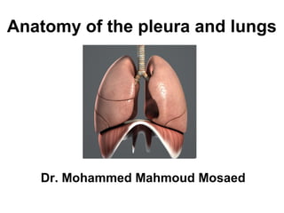
Anatomy of the pleura and lungs
Dr. Mohammed Mahmoud Mosaed
Chest Cavity
• The chest cavity is bounded by the chest
wall and below by the diaphragm.
• It extends upward into the root of the neck
about one fingerbreadth above the
clavicle on each side
• The chest cavity can be divided into a
median partition, called the mediastinum,
and the laterally placed pleurae and lungs
Pleura
• It is a serous membrane arranged as a closed invaginated
sac that covers the lung and lines the chest wall
• Each pleura has two parts:
• 1. Parietal layer (outer layer), which lines the thoracic
wall,
• 2. Visceral layer (inner layer), which completely covers
the outer surfaces of the lungs
• The two layers become continuous with one another by
means of a cuff of pleura at the hilum of each lung. the
pleural cuff hangs down as a loose fold called the
pulmonary ligament
• The parietal and visceral layers of pleura are separated
from one another by the pleural cavity (pleural space) that
contains a small amount of the pleural fluid
Division of the parietal pleura
• parietal pleura divided according to the region
in which it lies or the surface that it covers.
• The cervical pleura extends up into the neck.
It reaches a level 1 to 1.5 in. (2.5 to 4 cm)
above the medial third of the clavicle.
• The costal pleura lines the inner surfaces of
the ribs, the costal cartilages, the intercostal
spaces, the sides of the vertebral bodies, and
the back of the sternum
• The diaphragmatic pleura covers the
thoracic surface (upper surface) of the
diaphragm.
• The mediastinal pleura covers and
forms the lateral boundary of the
mediastinum. At the hilum of the lung, it is
reflected as a cuff around the vessels and
bronchi that constitute the lung root and
here becomes continuous with the visceral
pleura.
The pleural Recesses
• The costodiaphragmatic recesses are slitlike
spaces between the costal and diaphragmatic parietal
pleurae that are separated only by a layer of pleural
fluid. During inspiration, the lower margins of the lungs
descend into the recesses. During expiration, the
lower margins of the lungs ascend again.
• The costomediastinal recesses are situated along
the anterior margins of the pleura. They are slitlike
spaces between the costal and the mediastinal
parietal pleurae. During inspiration and expiration, the
anterior borders of the lungs slide in and out of the
recesses
Lines of pleural reflection
• The lines, which indicate the limits of the parietal pleura where it lies
close to the body surface, are referred to as the lines of pleural
reflection.
• The cervical pleura (dome of the pleura) bulges upward
into the neck A curved line may be drawn, convex upward, from the
sternoclavicular joint to a point 1 in. (2.5 cm) above the junction of the
medial and intermediate thirds of the clavicle
• The anterior border of the right pleura runs down behind
the sternoclavicular joint, almost reaching the midline behind the
sternal angle. It then continues downward until it reaches the
xiphisternal joint.
• The anterior border of the left pleura has a similar course,
but at the level of the fourth costal cartilage it deviates laterally and
extends to the lateral margin of the sternum to form the cardiac
notch. It then turns sharply downward to the xiphisternal joint.
• The lower border of the pleura on both sides follows a
curved line, which crosses the 8th rib in the midclavicular line and
the 10th rib in the midaxillary line, and reaches the 12th rib
adjacent to the vertebral column that is at the lateral border of the
erector spinae muscle
Dome of the pleura
2nd
costal cartilage
4th
costal cartilage
6th
costal cartilage
8th
rib in
midclavicular line
10th
rib in
midaxillary line
12th
rib
Nerve Supply of the Pleura
• The parietal pleura is sensitive to pain,
temperature, touch, and pressure and is supplied as
follows:
The costal pleura is segmentally supplied by the
intercostal nerves.
The mediastinal pleura is supplied by the phrenic nerve.
The diaphragmatic pleura is supplied over the domes by
the phrenic nerve and around the periphery by the
lower six intercostal nerves.
• The visceral pleura covering the lungs is sensitive
to stretch but is insensitive to common sensations
such as pain and touch. It receives an autonomic
nerve supply from the pulmonary plexus
lungs Anatomy of
Lungs
• The lungs are the essential organs of
respiration.
• They are situated on either side of the
heart and other mediastinal contents
• the lungs are soft, spongy and very
elastic.
• Each lung is conical in shape, covered
with visceral pleura, being attached to the
mediastinum only by its root
• In the child, they are pink, but with age,
they become dark and mottled
Anatomical features of the lungs
• Each lung has an apex, base, three borders and two surfaces
• Apex each lung has a blunt apex, which projects upward into the neck
for about 1 in. (2.5 cm) above the clavicle;
• Base is concave, and rests upon the upper surface of the diaphragm ;
• The costal surface is smooth and convex which corresponds to the
concave chest wall;
• The medial surface has a posterior vertebral part and anterior
mediastinal part.
The vertebral part lies in contact with the sides of the thoracic vertebrae
and intervertebral discs
The mediastinal part is deeply concave, and related to the mediastinal
content which causes impressions on this surface. The hilum, where
various structures enter or leave the lung lies on this surface
• The anterior border is thin and overlaps the heart; it is here on the left
lung that the cardiac notch is found.
• The posterior border is thick and lies beside the vertebral column.
• Inferior border
The mediastinal part of the medial
surface
• The mediastinal part is deeply
concave, and related to the
mediastinal content which causes
impressions on this surface.
• Also it contains the hilum, where
various structures enter or leave the
lung lies on this surface
Impressions on medial surface
of the right lung
1.The cardiac impression is related to the right atrium.
2. The impression for the superior vena cava and the
end of the right brachiocephalic vein ascends anterior
to the hilum.
3. The impression for azygos vein arches forwards
above the hilum forming deep sulcus,.
4. Oesophageal impression: The right side of the
oesophagus makes a shallow vertical groove behind
the hilum and the pulmonary ligament.
5. Groove for the inferior vena cava: lies
posteroinferior to the cardiac impression.
6. Groove for the trachea and right vagus nerve:
Between the apex and the groove for the azygos,.
Impressions on medial surface
of the left lung
• 1. The cardiac impression is related to left ventricle
• 2. Impression ascends in front of the hilum to
accommodate the pulmonary trunk.
• 3. Groove for the aortic arch and descending aorta: A
large groove arches over the hilum, and descends behind
it and the pulmonary ligament,
• 4. groove for the left subclavian artery: From the
summit of the aortic arch a narrower groove ascends to
the apex
• 5. The groove for thoracic duct and oesophagus lies
behind the groove for the left subclavian artery above the
aortic groove. Inferiorly, the oesophagus may mould the
surface in front of the lower end of the pulmonary ligament
Hilum of the lung
• The hilum is the part of the lung on its medial surface which
gives passage to the structures enters or leaves the lung (root
of the lung)
• The pulmonary root is formed by a group of structures
that enter or leave the hilum.
• These structures are:
• the principal bronchus, pulmonary artery, two pulmonary
veins, bronchial vessels, a pulmonary autonomic plexus,
lymph vessels, bronchopulmonary lymph nodes and loose
connective tissue.
• which are all enveloped by pleura. The pulmonary roots lie
opposite the bodies of the fifth to seventh thoracic vertebrae.
• The major structures in both roots are similarly arranged, so
that the upper of the two pulmonary veins is in front, the
pulmonary artery and principal bronchus are behind, and the
bronchial vessels are most posterior.
Lobes and fissures of right lung
Right Lung
• The right lung is divided by the oblique and
horizontal fissures into three lobes: the
upper, middle and lower lobes.
The oblique fissure runs from the inferior
border upward and backward across the
medial and costal surfaces until it cuts the
posterior border about 2.5 in. (6.25 cm)
below the apex.
The horizontal fissure runs horizontally
across the costal surface at the level of the
4th
costal cartilage to meet the oblique
fissure in the midaxillary line. The middle
lobe is thus a small triangular lobe
bounded by the horizontal and oblique
fissures.
Lobes and fissures of left lung
• Left Lung
• The left lung is divided by
oblique fissure into two
lobes: the upper and lower
lobes
• There is no horizontal fissure
in the left lung.
Bronchopulmonary Segments
• The bronchopulmonary segments are the
anatomic, functional, and surgical units of the
lungs.
• Each lobar (secondary) bronchus, which
passes to a lobe of the lung, gives off branches
called segmental (tertiary) bronchi.
• Each segmental bronchus passes to a
structurally and functionally independent unit of
a lung lobe called a bronchopulmonary
segment, which is surrounded by connective
tissue.
Structure of bronchopulmonary segment
• Main bronchus lobar bronchus
segmental bronchus
• Each segmental bronchus divides repeatedly.The
smallest bronchi divide and give rise to bronchioles,
The bronchioles then divide and give rise to terminal
bronchioles, which show delicate outpouchings from
their walls (respiratory bronchiole). The respiratory
bronchioles end by branching into alveolar ducts,
which lead into alveolar sacs.
• Each alveolus is surrounded by a rich network of
blood capillaries.
The main characteristics of a
bronchopulmonary segment
• It is a subdivision of a lung lobe.
• It is pyramid shaped, with its apex toward the lung
root.
• It is surrounded by connective tissue.
• It has a segmental bronchus, a segmental artery,
lymph vessels, and autonomic nerves.
• The segmental vein lies in the connective tissue
between adjacent bronchopulmonary segments.
• Because it is a structural unit, a diseased segment can
be removed surgically.
The main bronchopulmonary segments
Right lung
• Superior lobe:
1. Apical,
2. posterior,
3. anterior
Middle lobe:
4. Lateral,
5. medial
• Inferior lobe:
6. Superior (apical),
7. medial basal,
8. anterior basal,
9. lateral basal,
10.posterior basal
Left lung
•Superior lobe:
1. Apical,
2. posterior,
3. anterior,
4. superior lingular,
5. inferior lingular
• Inferior lobe:
6. Superior (apical),
7.medial basal,
8. anterior basal,
9.lateral basal,
10. posterior basal
Blood Supply of the Lungs
• The bronchi, the connective tissue of the lung,
and the visceral pleura receive their blood
supply from the bronchial arteries, which are
branches of the descending aorta.
• The bronchial veins drain into the azygos and
hemiazygos veins.
• The alveoli receive deoxygenated blood from
the terminal branches of the pulmonary
arteries. The oxygenated blood leaving the
alveolar capillaries drains into the tributaries of
the pulmonary veins, to empty into the left
atrium of the heart.
Lymph Drainage of the Lungs
• The lymph vessels originate in superficial and deep
plexuses.
• The superficial (subpleural) plexus lies beneath the
visceral pleura and drains over the surface of the lung
toward the hilum, where the lymph vessels enter the
bronchopulmonary nodes.
• The deep plexus travels along the bronchi and
pulmonary vessels toward the hilum of the lung,
passing through pulmonary nodes located within the
lung substance; the lymph then enters the
bronchopulmonary nodes in the hilum of the lung.
• All the lymph from the lung leaves the hilum and
drains into the tracheobronchial nodes and then into
the bronchomediastinal lymph trunks
Nerve Supply of the Lungs
• At the root of each lung is a pulmonary plexus
composed of efferent and afferent autonomic nerve
fibers.
• The plexus is formed from branches of the
sympathetic trunk and receives parasympathetic fibers
from the vagus nerve.
• The sympathetic efferent fibers produce
bronchodilatation and vasoconstriction.
• The parasympathetic efferent fibers produce
bronchoconstriction, vasodilatation, and increased
glandular secretion.
• Afferent impulses derived from the bronchial mucous
membrane and from stretch receptors in the alveolar
walls pass to the central nervous system in both
sympathetic and parasympathetic nerves.
Surface anatomy of the lungs
• The apex bulges upward into the neck A curved line may be drawn,
convex upward, from the sternoclavicular joint to a point 1 in. (2.5 cm)
above the junction of the medial and intermediate thirds of the
clavicle
• The anterior border of the right lung runs down behind the
sternoclavicular joint, almost reaching the midline behind the sternal
angle. It then continues downward until it reaches the xiphisternal
joint ) 6th
costal cartilage)
• The anterior border of the left lung has a similar course, but
at the level of the fourth costal cartilage it deviates laterally and
extends to the lateral margin of the sternum to form the cardiac
notch. It then turns sharply downward to the xiphisternal joint.
• The lower border of the pleura on both sides follows a
curved line, which crosses the 6th
rib in the midclavicular line and the
8th
rib in the midaxillary line, and reaches the 10th
rib adjacent to the
vertebral column that is, at the lateral border of the erector spinae
muscle

More Related Content

What's hot (20)

The pulmonary trunk
The pulmonary trunkThe pulmonary trunk
The pulmonary trunk
 
thoracic duct
thoracic ductthoracic duct
thoracic duct
 
Mediastinum
MediastinumMediastinum
Mediastinum
 
Anatomy of The Diaphragm
 Anatomy of The Diaphragm Anatomy of The Diaphragm
Anatomy of The Diaphragm
 
5. Trachea
5. Trachea5. Trachea
5. Trachea
 
Diaphragm Anatomy
Diaphragm AnatomyDiaphragm Anatomy
Diaphragm Anatomy
 
Thoracic cavity & mediastinum
Thoracic cavity & mediastinumThoracic cavity & mediastinum
Thoracic cavity & mediastinum
 
Mediastinum
MediastinumMediastinum
Mediastinum
 
Arch of aorta
Arch of aortaArch of aorta
Arch of aorta
 
Lung anatomy.
Lung anatomy.Lung anatomy.
Lung anatomy.
 
Blood vessels and lymphatic of the thoracic wall
Blood vessels and lymphatic of the thoracic wallBlood vessels and lymphatic of the thoracic wall
Blood vessels and lymphatic of the thoracic wall
 
Anatomy of the Cardiovascular system
Anatomy of the Cardiovascular systemAnatomy of the Cardiovascular system
Anatomy of the Cardiovascular system
 
Pleura and its recesses
Pleura and its recessesPleura and its recesses
Pleura and its recesses
 
Blood supply to heart
Blood supply to heartBlood supply to heart
Blood supply to heart
 
Nerves, vessels of thoracic wall
Nerves, vessels of thoracic wallNerves, vessels of thoracic wall
Nerves, vessels of thoracic wall
 
lungs bp segments
lungs bp segmentslungs bp segments
lungs bp segments
 
1. pericardium
1. pericardium1. pericardium
1. pericardium
 
Thoracic wall
Thoracic wallThoracic wall
Thoracic wall
 
Anatomy of trachea & lungs
Anatomy of  trachea & lungsAnatomy of  trachea & lungs
Anatomy of trachea & lungs
 
Thorax anatomy presentation
Thorax anatomy presentationThorax anatomy presentation
Thorax anatomy presentation
 

Similar to 6. Pleura and lung

respiratory system.pptx
respiratory system.pptxrespiratory system.pptx
respiratory system.pptxssuser65b0c5
 
lungs-131018180202-phpapp02.pdf
lungs-131018180202-phpapp02.pdflungs-131018180202-phpapp02.pdf
lungs-131018180202-phpapp02.pdfSonuSharma887555
 
Gross anatomy and development of respiratory system
Gross anatomy and development of respiratory systemGross anatomy and development of respiratory system
Gross anatomy and development of respiratory systemDr Laxman Khanal
 
Gross anatomy of lungs
Gross anatomy of lungsGross anatomy of lungs
Gross anatomy of lungsSumayyaFarooq1
 
Lecture 3- Anatomy of the Pleura and Lung
Lecture 3- Anatomy of the Pleura and LungLecture 3- Anatomy of the Pleura and Lung
Lecture 3- Anatomy of the Pleura and LungIslam Ahmady
 
Anatomy of trachea, bronchus and lung
Anatomy of trachea, bronchus and lungAnatomy of trachea, bronchus and lung
Anatomy of trachea, bronchus and lungabrahammezgebu1
 
Lungs anatomy, organs in thorax
Lungs anatomy, organs in thoraxLungs anatomy, organs in thorax
Lungs anatomy, organs in thoraxFaarah Yusuf
 
LOWER RESPIRATORY SYSTEM.pptx upper respiratory tract, Respiratory system,Ana...
LOWER RESPIRATORY SYSTEM.pptx upper respiratory tract, Respiratory system,Ana...LOWER RESPIRATORY SYSTEM.pptx upper respiratory tract, Respiratory system,Ana...
LOWER RESPIRATORY SYSTEM.pptx upper respiratory tract, Respiratory system,Ana...roy456393
 
Anatomy of lungs, pleura and diaphragm
Anatomy  of lungs, pleura and diaphragmAnatomy  of lungs, pleura and diaphragm
Anatomy of lungs, pleura and diaphragmDr. Ashish kumar
 
Lower respiratory tract (2).pdf
Lower respiratory tract (2).pdfLower respiratory tract (2).pdf
Lower respiratory tract (2).pdfTeshaleTekle1
 
ANATOMY OF TRACHEA, BRONCHI & PLEURA.pptx
ANATOMY OF TRACHEA, BRONCHI & PLEURA.pptxANATOMY OF TRACHEA, BRONCHI & PLEURA.pptx
ANATOMY OF TRACHEA, BRONCHI & PLEURA.pptxMohitJagga2
 
Physiology of speech
Physiology of speechPhysiology of speech
Physiology of speechAmit kumar
 
Anatomy .pptx
Anatomy .pptxAnatomy .pptx
Anatomy .pptxSika30
 
Anatomy of Lower respiratory tract.pptx
Anatomy of Lower respiratory tract.pptxAnatomy of Lower respiratory tract.pptx
Anatomy of Lower respiratory tract.pptxSyedYusufSyed
 

Similar to 6. Pleura and lung (20)

Lecture 3 lungs & pleura
Lecture 3 lungs & pleuraLecture 3 lungs & pleura
Lecture 3 lungs & pleura
 
LUNGS ANATOMY
LUNGS ANATOMYLUNGS ANATOMY
LUNGS ANATOMY
 
respiratory system.pptx
respiratory system.pptxrespiratory system.pptx
respiratory system.pptx
 
Lungs
LungsLungs
Lungs
 
lungs-131018180202-phpapp02.pdf
lungs-131018180202-phpapp02.pdflungs-131018180202-phpapp02.pdf
lungs-131018180202-phpapp02.pdf
 
Gross anatomy and development of respiratory system
Gross anatomy and development of respiratory systemGross anatomy and development of respiratory system
Gross anatomy and development of respiratory system
 
Gross anatomy of lungs
Gross anatomy of lungsGross anatomy of lungs
Gross anatomy of lungs
 
Lecture 3- Anatomy of the Pleura and Lung
Lecture 3- Anatomy of the Pleura and LungLecture 3- Anatomy of the Pleura and Lung
Lecture 3- Anatomy of the Pleura and Lung
 
Anatomy of trachea, bronchus and lung
Anatomy of trachea, bronchus and lungAnatomy of trachea, bronchus and lung
Anatomy of trachea, bronchus and lung
 
Lungs anatomy, organs in thorax
Lungs anatomy, organs in thoraxLungs anatomy, organs in thorax
Lungs anatomy, organs in thorax
 
LOWER RESPIRATORY SYSTEM.pptx upper respiratory tract, Respiratory system,Ana...
LOWER RESPIRATORY SYSTEM.pptx upper respiratory tract, Respiratory system,Ana...LOWER RESPIRATORY SYSTEM.pptx upper respiratory tract, Respiratory system,Ana...
LOWER RESPIRATORY SYSTEM.pptx upper respiratory tract, Respiratory system,Ana...
 
usg Lung.pptx
usg Lung.pptxusg Lung.pptx
usg Lung.pptx
 
Anatomy of lungs, pleura and diaphragm
Anatomy  of lungs, pleura and diaphragmAnatomy  of lungs, pleura and diaphragm
Anatomy of lungs, pleura and diaphragm
 
Lower respiratory tract (2).pdf
Lower respiratory tract (2).pdfLower respiratory tract (2).pdf
Lower respiratory tract (2).pdf
 
ANATOMY OF TRACHEA, BRONCHI & PLEURA.pptx
ANATOMY OF TRACHEA, BRONCHI & PLEURA.pptxANATOMY OF TRACHEA, BRONCHI & PLEURA.pptx
ANATOMY OF TRACHEA, BRONCHI & PLEURA.pptx
 
Lungs.pptx
Lungs.pptxLungs.pptx
Lungs.pptx
 
Physiology of speech
Physiology of speechPhysiology of speech
Physiology of speech
 
Anatomy .pptx
Anatomy .pptxAnatomy .pptx
Anatomy .pptx
 
Thorax second part anatomy
Thorax second part anatomyThorax second part anatomy
Thorax second part anatomy
 
Anatomy of Lower respiratory tract.pptx
Anatomy of Lower respiratory tract.pptxAnatomy of Lower respiratory tract.pptx
Anatomy of Lower respiratory tract.pptx
 

More from Dr. Mohammad Mahmoud

Embryology Lecture 4 Ovulation and fertilization
 Embryology Lecture 4  Ovulation and fertilization Embryology Lecture 4  Ovulation and fertilization
Embryology Lecture 4 Ovulation and fertilizationDr. Mohammad Mahmoud
 
Embryology Lecture 3 spermatogenesis
 Embryology Lecture 3 spermatogenesis Embryology Lecture 3 spermatogenesis
Embryology Lecture 3 spermatogenesisDr. Mohammad Mahmoud
 
Arteries of head and neck; subclavian vessels
Arteries of head and neck; subclavian vesselsArteries of head and neck; subclavian vessels
Arteries of head and neck; subclavian vesselsDr. Mohammad Mahmoud
 
Anatomy of the neck; skin, fascia and muscles
Anatomy of the neck;  skin, fascia and musclesAnatomy of the neck;  skin, fascia and muscles
Anatomy of the neck; skin, fascia and musclesDr. Mohammad Mahmoud
 
Anatomy Lecture 4: Skeletal system
Anatomy  Lecture 4: Skeletal systemAnatomy  Lecture 4: Skeletal system
Anatomy Lecture 4: Skeletal systemDr. Mohammad Mahmoud
 
Anatomy of the liver and gallbladder
Anatomy of the liver and gallbladderAnatomy of the liver and gallbladder
Anatomy of the liver and gallbladderDr. Mohammad Mahmoud
 
Anatomy of small and large intestine
Anatomy of  small and large intestineAnatomy of  small and large intestine
Anatomy of small and large intestineDr. Mohammad Mahmoud
 

More from Dr. Mohammad Mahmoud (20)

Lymphatic system.pptx
Lymphatic system.pptxLymphatic system.pptx
Lymphatic system.pptx
 
Anatomy of spleen.pptx
Anatomy of spleen.pptxAnatomy of spleen.pptx
Anatomy of spleen.pptx
 
Anatomy of medulla oblongata
Anatomy of medulla oblongataAnatomy of medulla oblongata
Anatomy of medulla oblongata
 
Anatomy of pons
Anatomy of ponsAnatomy of pons
Anatomy of pons
 
Anatomy of midbrain
Anatomy of midbrainAnatomy of midbrain
Anatomy of midbrain
 
Embryology Lecture 4 Ovulation and fertilization
 Embryology Lecture 4  Ovulation and fertilization Embryology Lecture 4  Ovulation and fertilization
Embryology Lecture 4 Ovulation and fertilization
 
Embryology Lecture 3 spermatogenesis
 Embryology Lecture 3 spermatogenesis Embryology Lecture 3 spermatogenesis
Embryology Lecture 3 spermatogenesis
 
Embryology Lecture 2 oogenesis
 Embryology Lecture 2 oogenesis  Embryology Lecture 2 oogenesis
Embryology Lecture 2 oogenesis
 
Embryology Lecture 1
Embryology Lecture 1Embryology Lecture 1
Embryology Lecture 1
 
Triangles of the neck
Triangles of the neckTriangles of the neck
Triangles of the neck
 
Veins of the neck
Veins of the neckVeins of the neck
Veins of the neck
 
Arteries of head and neck; subclavian vessels
Arteries of head and neck; subclavian vesselsArteries of head and neck; subclavian vessels
Arteries of head and neck; subclavian vessels
 
Arteries of the head and neck
Arteries of the head and neckArteries of the head and neck
Arteries of the head and neck
 
Anatomy of the neck; skin, fascia and muscles
Anatomy of the neck;  skin, fascia and musclesAnatomy of the neck;  skin, fascia and muscles
Anatomy of the neck; skin, fascia and muscles
 
Anatomy Lecture 4: Skeletal system
Anatomy  Lecture 4: Skeletal systemAnatomy  Lecture 4: Skeletal system
Anatomy Lecture 4: Skeletal system
 
Anatomy lecture 3
Anatomy lecture 3Anatomy lecture 3
Anatomy lecture 3
 
Anatomy lecture 2
Anatomy lecture 2 Anatomy lecture 2
Anatomy lecture 2
 
Anatomy 1st lecture
Anatomy   1st lecture Anatomy   1st lecture
Anatomy 1st lecture
 
Anatomy of the liver and gallbladder
Anatomy of the liver and gallbladderAnatomy of the liver and gallbladder
Anatomy of the liver and gallbladder
 
Anatomy of small and large intestine
Anatomy of  small and large intestineAnatomy of  small and large intestine
Anatomy of small and large intestine
 

Recently uploaded

Inclusivity Essentials_ Creating Accessible Websites for Nonprofits .pdf
Inclusivity Essentials_ Creating Accessible Websites for Nonprofits .pdfInclusivity Essentials_ Creating Accessible Websites for Nonprofits .pdf
Inclusivity Essentials_ Creating Accessible Websites for Nonprofits .pdfTechSoup
 
ANG SEKTOR NG agrikultura.pptx QUARTER 4
ANG SEKTOR NG agrikultura.pptx QUARTER 4ANG SEKTOR NG agrikultura.pptx QUARTER 4
ANG SEKTOR NG agrikultura.pptx QUARTER 4MiaBumagat1
 
How to Add Barcode on PDF Report in Odoo 17
How to Add Barcode on PDF Report in Odoo 17How to Add Barcode on PDF Report in Odoo 17
How to Add Barcode on PDF Report in Odoo 17Celine George
 
USPS® Forced Meter Migration - How to Know if Your Postage Meter Will Soon be...
USPS® Forced Meter Migration - How to Know if Your Postage Meter Will Soon be...USPS® Forced Meter Migration - How to Know if Your Postage Meter Will Soon be...
USPS® Forced Meter Migration - How to Know if Your Postage Meter Will Soon be...Postal Advocate Inc.
 
Student Profile Sample - We help schools to connect the data they have, with ...
Student Profile Sample - We help schools to connect the data they have, with ...Student Profile Sample - We help schools to connect the data they have, with ...
Student Profile Sample - We help schools to connect the data they have, with ...Seán Kennedy
 
Virtual-Orientation-on-the-Administration-of-NATG12-NATG6-and-ELLNA.pdf
Virtual-Orientation-on-the-Administration-of-NATG12-NATG6-and-ELLNA.pdfVirtual-Orientation-on-the-Administration-of-NATG12-NATG6-and-ELLNA.pdf
Virtual-Orientation-on-the-Administration-of-NATG12-NATG6-and-ELLNA.pdfErwinPantujan2
 
AMERICAN LANGUAGE HUB_Level2_Student'sBook_Answerkey.pdf
AMERICAN LANGUAGE HUB_Level2_Student'sBook_Answerkey.pdfAMERICAN LANGUAGE HUB_Level2_Student'sBook_Answerkey.pdf
AMERICAN LANGUAGE HUB_Level2_Student'sBook_Answerkey.pdfphamnguyenenglishnb
 
Science 7 Quarter 4 Module 2: Natural Resources.pptx
Science 7 Quarter 4 Module 2: Natural Resources.pptxScience 7 Quarter 4 Module 2: Natural Resources.pptx
Science 7 Quarter 4 Module 2: Natural Resources.pptxMaryGraceBautista27
 
Transaction Management in Database Management System
Transaction Management in Database Management SystemTransaction Management in Database Management System
Transaction Management in Database Management SystemChristalin Nelson
 
ISYU TUNGKOL SA SEKSWLADIDA (ISSUE ABOUT SEXUALITY
ISYU TUNGKOL SA SEKSWLADIDA (ISSUE ABOUT SEXUALITYISYU TUNGKOL SA SEKSWLADIDA (ISSUE ABOUT SEXUALITY
ISYU TUNGKOL SA SEKSWLADIDA (ISSUE ABOUT SEXUALITYKayeClaireEstoconing
 
Earth Day Presentation wow hello nice great
Earth Day Presentation wow hello nice greatEarth Day Presentation wow hello nice great
Earth Day Presentation wow hello nice greatYousafMalik24
 
4.16.24 21st Century Movements for Black Lives.pptx
4.16.24 21st Century Movements for Black Lives.pptx4.16.24 21st Century Movements for Black Lives.pptx
4.16.24 21st Century Movements for Black Lives.pptxmary850239
 
4.18.24 Movement Legacies, Reflection, and Review.pptx
4.18.24 Movement Legacies, Reflection, and Review.pptx4.18.24 Movement Legacies, Reflection, and Review.pptx
4.18.24 Movement Legacies, Reflection, and Review.pptxmary850239
 
Barangay Council for the Protection of Children (BCPC) Orientation.pptx
Barangay Council for the Protection of Children (BCPC) Orientation.pptxBarangay Council for the Protection of Children (BCPC) Orientation.pptx
Barangay Council for the Protection of Children (BCPC) Orientation.pptxCarlos105
 
MULTIDISCIPLINRY NATURE OF THE ENVIRONMENTAL STUDIES.pptx
MULTIDISCIPLINRY NATURE OF THE ENVIRONMENTAL STUDIES.pptxMULTIDISCIPLINRY NATURE OF THE ENVIRONMENTAL STUDIES.pptx
MULTIDISCIPLINRY NATURE OF THE ENVIRONMENTAL STUDIES.pptxAnupkumar Sharma
 
Karra SKD Conference Presentation Revised.pptx
Karra SKD Conference Presentation Revised.pptxKarra SKD Conference Presentation Revised.pptx
Karra SKD Conference Presentation Revised.pptxAshokKarra1
 
FILIPINO PSYCHology sikolohiyang pilipino
FILIPINO PSYCHology sikolohiyang pilipinoFILIPINO PSYCHology sikolohiyang pilipino
FILIPINO PSYCHology sikolohiyang pilipinojohnmickonozaleda
 

Recently uploaded (20)

Inclusivity Essentials_ Creating Accessible Websites for Nonprofits .pdf
Inclusivity Essentials_ Creating Accessible Websites for Nonprofits .pdfInclusivity Essentials_ Creating Accessible Websites for Nonprofits .pdf
Inclusivity Essentials_ Creating Accessible Websites for Nonprofits .pdf
 
ANG SEKTOR NG agrikultura.pptx QUARTER 4
ANG SEKTOR NG agrikultura.pptx QUARTER 4ANG SEKTOR NG agrikultura.pptx QUARTER 4
ANG SEKTOR NG agrikultura.pptx QUARTER 4
 
How to Add Barcode on PDF Report in Odoo 17
How to Add Barcode on PDF Report in Odoo 17How to Add Barcode on PDF Report in Odoo 17
How to Add Barcode on PDF Report in Odoo 17
 
USPS® Forced Meter Migration - How to Know if Your Postage Meter Will Soon be...
USPS® Forced Meter Migration - How to Know if Your Postage Meter Will Soon be...USPS® Forced Meter Migration - How to Know if Your Postage Meter Will Soon be...
USPS® Forced Meter Migration - How to Know if Your Postage Meter Will Soon be...
 
Student Profile Sample - We help schools to connect the data they have, with ...
Student Profile Sample - We help schools to connect the data they have, with ...Student Profile Sample - We help schools to connect the data they have, with ...
Student Profile Sample - We help schools to connect the data they have, with ...
 
Virtual-Orientation-on-the-Administration-of-NATG12-NATG6-and-ELLNA.pdf
Virtual-Orientation-on-the-Administration-of-NATG12-NATG6-and-ELLNA.pdfVirtual-Orientation-on-the-Administration-of-NATG12-NATG6-and-ELLNA.pdf
Virtual-Orientation-on-the-Administration-of-NATG12-NATG6-and-ELLNA.pdf
 
AMERICAN LANGUAGE HUB_Level2_Student'sBook_Answerkey.pdf
AMERICAN LANGUAGE HUB_Level2_Student'sBook_Answerkey.pdfAMERICAN LANGUAGE HUB_Level2_Student'sBook_Answerkey.pdf
AMERICAN LANGUAGE HUB_Level2_Student'sBook_Answerkey.pdf
 
Model Call Girl in Tilak Nagar Delhi reach out to us at 🔝9953056974🔝
Model Call Girl in Tilak Nagar Delhi reach out to us at 🔝9953056974🔝Model Call Girl in Tilak Nagar Delhi reach out to us at 🔝9953056974🔝
Model Call Girl in Tilak Nagar Delhi reach out to us at 🔝9953056974🔝
 
Science 7 Quarter 4 Module 2: Natural Resources.pptx
Science 7 Quarter 4 Module 2: Natural Resources.pptxScience 7 Quarter 4 Module 2: Natural Resources.pptx
Science 7 Quarter 4 Module 2: Natural Resources.pptx
 
FINALS_OF_LEFT_ON_C'N_EL_DORADO_2024.pptx
FINALS_OF_LEFT_ON_C'N_EL_DORADO_2024.pptxFINALS_OF_LEFT_ON_C'N_EL_DORADO_2024.pptx
FINALS_OF_LEFT_ON_C'N_EL_DORADO_2024.pptx
 
Transaction Management in Database Management System
Transaction Management in Database Management SystemTransaction Management in Database Management System
Transaction Management in Database Management System
 
ISYU TUNGKOL SA SEKSWLADIDA (ISSUE ABOUT SEXUALITY
ISYU TUNGKOL SA SEKSWLADIDA (ISSUE ABOUT SEXUALITYISYU TUNGKOL SA SEKSWLADIDA (ISSUE ABOUT SEXUALITY
ISYU TUNGKOL SA SEKSWLADIDA (ISSUE ABOUT SEXUALITY
 
Earth Day Presentation wow hello nice great
Earth Day Presentation wow hello nice greatEarth Day Presentation wow hello nice great
Earth Day Presentation wow hello nice great
 
4.16.24 21st Century Movements for Black Lives.pptx
4.16.24 21st Century Movements for Black Lives.pptx4.16.24 21st Century Movements for Black Lives.pptx
4.16.24 21st Century Movements for Black Lives.pptx
 
4.18.24 Movement Legacies, Reflection, and Review.pptx
4.18.24 Movement Legacies, Reflection, and Review.pptx4.18.24 Movement Legacies, Reflection, and Review.pptx
4.18.24 Movement Legacies, Reflection, and Review.pptx
 
LEFT_ON_C'N_ PRELIMS_EL_DORADO_2024.pptx
LEFT_ON_C'N_ PRELIMS_EL_DORADO_2024.pptxLEFT_ON_C'N_ PRELIMS_EL_DORADO_2024.pptx
LEFT_ON_C'N_ PRELIMS_EL_DORADO_2024.pptx
 
Barangay Council for the Protection of Children (BCPC) Orientation.pptx
Barangay Council for the Protection of Children (BCPC) Orientation.pptxBarangay Council for the Protection of Children (BCPC) Orientation.pptx
Barangay Council for the Protection of Children (BCPC) Orientation.pptx
 
MULTIDISCIPLINRY NATURE OF THE ENVIRONMENTAL STUDIES.pptx
MULTIDISCIPLINRY NATURE OF THE ENVIRONMENTAL STUDIES.pptxMULTIDISCIPLINRY NATURE OF THE ENVIRONMENTAL STUDIES.pptx
MULTIDISCIPLINRY NATURE OF THE ENVIRONMENTAL STUDIES.pptx
 
Karra SKD Conference Presentation Revised.pptx
Karra SKD Conference Presentation Revised.pptxKarra SKD Conference Presentation Revised.pptx
Karra SKD Conference Presentation Revised.pptx
 
FILIPINO PSYCHology sikolohiyang pilipino
FILIPINO PSYCHology sikolohiyang pilipinoFILIPINO PSYCHology sikolohiyang pilipino
FILIPINO PSYCHology sikolohiyang pilipino
 

6. Pleura and lung

  • 1. Anatomy of the pleura and lungs Dr. Mohammed Mahmoud Mosaed
  • 2. Chest Cavity • The chest cavity is bounded by the chest wall and below by the diaphragm. • It extends upward into the root of the neck about one fingerbreadth above the clavicle on each side • The chest cavity can be divided into a median partition, called the mediastinum, and the laterally placed pleurae and lungs
  • 3.
  • 4. Pleura • It is a serous membrane arranged as a closed invaginated sac that covers the lung and lines the chest wall • Each pleura has two parts: • 1. Parietal layer (outer layer), which lines the thoracic wall, • 2. Visceral layer (inner layer), which completely covers the outer surfaces of the lungs • The two layers become continuous with one another by means of a cuff of pleura at the hilum of each lung. the pleural cuff hangs down as a loose fold called the pulmonary ligament • The parietal and visceral layers of pleura are separated from one another by the pleural cavity (pleural space) that contains a small amount of the pleural fluid
  • 5.
  • 6. Division of the parietal pleura • parietal pleura divided according to the region in which it lies or the surface that it covers. • The cervical pleura extends up into the neck. It reaches a level 1 to 1.5 in. (2.5 to 4 cm) above the medial third of the clavicle. • The costal pleura lines the inner surfaces of the ribs, the costal cartilages, the intercostal spaces, the sides of the vertebral bodies, and the back of the sternum
  • 7. • The diaphragmatic pleura covers the thoracic surface (upper surface) of the diaphragm. • The mediastinal pleura covers and forms the lateral boundary of the mediastinum. At the hilum of the lung, it is reflected as a cuff around the vessels and bronchi that constitute the lung root and here becomes continuous with the visceral pleura.
  • 8.
  • 9. The pleural Recesses • The costodiaphragmatic recesses are slitlike spaces between the costal and diaphragmatic parietal pleurae that are separated only by a layer of pleural fluid. During inspiration, the lower margins of the lungs descend into the recesses. During expiration, the lower margins of the lungs ascend again. • The costomediastinal recesses are situated along the anterior margins of the pleura. They are slitlike spaces between the costal and the mediastinal parietal pleurae. During inspiration and expiration, the anterior borders of the lungs slide in and out of the recesses
  • 10.
  • 11. Lines of pleural reflection • The lines, which indicate the limits of the parietal pleura where it lies close to the body surface, are referred to as the lines of pleural reflection. • The cervical pleura (dome of the pleura) bulges upward into the neck A curved line may be drawn, convex upward, from the sternoclavicular joint to a point 1 in. (2.5 cm) above the junction of the medial and intermediate thirds of the clavicle • The anterior border of the right pleura runs down behind the sternoclavicular joint, almost reaching the midline behind the sternal angle. It then continues downward until it reaches the xiphisternal joint. • The anterior border of the left pleura has a similar course, but at the level of the fourth costal cartilage it deviates laterally and extends to the lateral margin of the sternum to form the cardiac notch. It then turns sharply downward to the xiphisternal joint. • The lower border of the pleura on both sides follows a curved line, which crosses the 8th rib in the midclavicular line and the 10th rib in the midaxillary line, and reaches the 12th rib adjacent to the vertebral column that is at the lateral border of the erector spinae muscle
  • 12. Dome of the pleura 2nd costal cartilage 4th costal cartilage 6th costal cartilage 8th rib in midclavicular line 10th rib in midaxillary line 12th rib
  • 13.
  • 14. Nerve Supply of the Pleura • The parietal pleura is sensitive to pain, temperature, touch, and pressure and is supplied as follows: The costal pleura is segmentally supplied by the intercostal nerves. The mediastinal pleura is supplied by the phrenic nerve. The diaphragmatic pleura is supplied over the domes by the phrenic nerve and around the periphery by the lower six intercostal nerves. • The visceral pleura covering the lungs is sensitive to stretch but is insensitive to common sensations such as pain and touch. It receives an autonomic nerve supply from the pulmonary plexus
  • 16. Lungs • The lungs are the essential organs of respiration. • They are situated on either side of the heart and other mediastinal contents • the lungs are soft, spongy and very elastic. • Each lung is conical in shape, covered with visceral pleura, being attached to the mediastinum only by its root • In the child, they are pink, but with age, they become dark and mottled
  • 17.
  • 18. Anatomical features of the lungs • Each lung has an apex, base, three borders and two surfaces • Apex each lung has a blunt apex, which projects upward into the neck for about 1 in. (2.5 cm) above the clavicle; • Base is concave, and rests upon the upper surface of the diaphragm ; • The costal surface is smooth and convex which corresponds to the concave chest wall; • The medial surface has a posterior vertebral part and anterior mediastinal part. The vertebral part lies in contact with the sides of the thoracic vertebrae and intervertebral discs The mediastinal part is deeply concave, and related to the mediastinal content which causes impressions on this surface. The hilum, where various structures enter or leave the lung lies on this surface • The anterior border is thin and overlaps the heart; it is here on the left lung that the cardiac notch is found. • The posterior border is thick and lies beside the vertebral column. • Inferior border
  • 19.
  • 20.
  • 21. The mediastinal part of the medial surface • The mediastinal part is deeply concave, and related to the mediastinal content which causes impressions on this surface. • Also it contains the hilum, where various structures enter or leave the lung lies on this surface
  • 22.
  • 23. Impressions on medial surface of the right lung 1.The cardiac impression is related to the right atrium. 2. The impression for the superior vena cava and the end of the right brachiocephalic vein ascends anterior to the hilum. 3. The impression for azygos vein arches forwards above the hilum forming deep sulcus,. 4. Oesophageal impression: The right side of the oesophagus makes a shallow vertical groove behind the hilum and the pulmonary ligament. 5. Groove for the inferior vena cava: lies posteroinferior to the cardiac impression. 6. Groove for the trachea and right vagus nerve: Between the apex and the groove for the azygos,.
  • 24.
  • 25. Impressions on medial surface of the left lung • 1. The cardiac impression is related to left ventricle • 2. Impression ascends in front of the hilum to accommodate the pulmonary trunk. • 3. Groove for the aortic arch and descending aorta: A large groove arches over the hilum, and descends behind it and the pulmonary ligament, • 4. groove for the left subclavian artery: From the summit of the aortic arch a narrower groove ascends to the apex • 5. The groove for thoracic duct and oesophagus lies behind the groove for the left subclavian artery above the aortic groove. Inferiorly, the oesophagus may mould the surface in front of the lower end of the pulmonary ligament
  • 26.
  • 27. Hilum of the lung • The hilum is the part of the lung on its medial surface which gives passage to the structures enters or leaves the lung (root of the lung) • The pulmonary root is formed by a group of structures that enter or leave the hilum. • These structures are: • the principal bronchus, pulmonary artery, two pulmonary veins, bronchial vessels, a pulmonary autonomic plexus, lymph vessels, bronchopulmonary lymph nodes and loose connective tissue. • which are all enveloped by pleura. The pulmonary roots lie opposite the bodies of the fifth to seventh thoracic vertebrae. • The major structures in both roots are similarly arranged, so that the upper of the two pulmonary veins is in front, the pulmonary artery and principal bronchus are behind, and the bronchial vessels are most posterior.
  • 28.
  • 29. Lobes and fissures of right lung Right Lung • The right lung is divided by the oblique and horizontal fissures into three lobes: the upper, middle and lower lobes. The oblique fissure runs from the inferior border upward and backward across the medial and costal surfaces until it cuts the posterior border about 2.5 in. (6.25 cm) below the apex. The horizontal fissure runs horizontally across the costal surface at the level of the 4th costal cartilage to meet the oblique fissure in the midaxillary line. The middle lobe is thus a small triangular lobe bounded by the horizontal and oblique fissures.
  • 30. Lobes and fissures of left lung • Left Lung • The left lung is divided by oblique fissure into two lobes: the upper and lower lobes • There is no horizontal fissure in the left lung.
  • 31. Bronchopulmonary Segments • The bronchopulmonary segments are the anatomic, functional, and surgical units of the lungs. • Each lobar (secondary) bronchus, which passes to a lobe of the lung, gives off branches called segmental (tertiary) bronchi. • Each segmental bronchus passes to a structurally and functionally independent unit of a lung lobe called a bronchopulmonary segment, which is surrounded by connective tissue.
  • 32.
  • 33.
  • 34. Structure of bronchopulmonary segment • Main bronchus lobar bronchus segmental bronchus • Each segmental bronchus divides repeatedly.The smallest bronchi divide and give rise to bronchioles, The bronchioles then divide and give rise to terminal bronchioles, which show delicate outpouchings from their walls (respiratory bronchiole). The respiratory bronchioles end by branching into alveolar ducts, which lead into alveolar sacs. • Each alveolus is surrounded by a rich network of blood capillaries.
  • 35.
  • 36. The main characteristics of a bronchopulmonary segment • It is a subdivision of a lung lobe. • It is pyramid shaped, with its apex toward the lung root. • It is surrounded by connective tissue. • It has a segmental bronchus, a segmental artery, lymph vessels, and autonomic nerves. • The segmental vein lies in the connective tissue between adjacent bronchopulmonary segments. • Because it is a structural unit, a diseased segment can be removed surgically.
  • 37. The main bronchopulmonary segments Right lung • Superior lobe: 1. Apical, 2. posterior, 3. anterior Middle lobe: 4. Lateral, 5. medial • Inferior lobe: 6. Superior (apical), 7. medial basal, 8. anterior basal, 9. lateral basal, 10.posterior basal Left lung •Superior lobe: 1. Apical, 2. posterior, 3. anterior, 4. superior lingular, 5. inferior lingular • Inferior lobe: 6. Superior (apical), 7.medial basal, 8. anterior basal, 9.lateral basal, 10. posterior basal
  • 38.
  • 39.
  • 40.
  • 41. Blood Supply of the Lungs • The bronchi, the connective tissue of the lung, and the visceral pleura receive their blood supply from the bronchial arteries, which are branches of the descending aorta. • The bronchial veins drain into the azygos and hemiazygos veins. • The alveoli receive deoxygenated blood from the terminal branches of the pulmonary arteries. The oxygenated blood leaving the alveolar capillaries drains into the tributaries of the pulmonary veins, to empty into the left atrium of the heart.
  • 42. Lymph Drainage of the Lungs • The lymph vessels originate in superficial and deep plexuses. • The superficial (subpleural) plexus lies beneath the visceral pleura and drains over the surface of the lung toward the hilum, where the lymph vessels enter the bronchopulmonary nodes. • The deep plexus travels along the bronchi and pulmonary vessels toward the hilum of the lung, passing through pulmonary nodes located within the lung substance; the lymph then enters the bronchopulmonary nodes in the hilum of the lung. • All the lymph from the lung leaves the hilum and drains into the tracheobronchial nodes and then into the bronchomediastinal lymph trunks
  • 43.
  • 44. Nerve Supply of the Lungs • At the root of each lung is a pulmonary plexus composed of efferent and afferent autonomic nerve fibers. • The plexus is formed from branches of the sympathetic trunk and receives parasympathetic fibers from the vagus nerve. • The sympathetic efferent fibers produce bronchodilatation and vasoconstriction. • The parasympathetic efferent fibers produce bronchoconstriction, vasodilatation, and increased glandular secretion. • Afferent impulses derived from the bronchial mucous membrane and from stretch receptors in the alveolar walls pass to the central nervous system in both sympathetic and parasympathetic nerves.
  • 45. Surface anatomy of the lungs • The apex bulges upward into the neck A curved line may be drawn, convex upward, from the sternoclavicular joint to a point 1 in. (2.5 cm) above the junction of the medial and intermediate thirds of the clavicle • The anterior border of the right lung runs down behind the sternoclavicular joint, almost reaching the midline behind the sternal angle. It then continues downward until it reaches the xiphisternal joint ) 6th costal cartilage) • The anterior border of the left lung has a similar course, but at the level of the fourth costal cartilage it deviates laterally and extends to the lateral margin of the sternum to form the cardiac notch. It then turns sharply downward to the xiphisternal joint. • The lower border of the pleura on both sides follows a curved line, which crosses the 6th rib in the midclavicular line and the 8th rib in the midaxillary line, and reaches the 10th rib adjacent to the vertebral column that is, at the lateral border of the erector spinae muscle