
Empfohlen
Empfohlen
Weitere ähnliche Inhalte
Was ist angesagt?
Was ist angesagt? (20)
Ähnlich wie Ipertensione polmonare secondaria clina e terapia
Ähnlich wie Ipertensione polmonare secondaria clina e terapia (20)
Mehr von Dr Salvatore Mazzuca
Mehr von Dr Salvatore Mazzuca (9)
Ipertensione polmonare secondaria clina e terapia
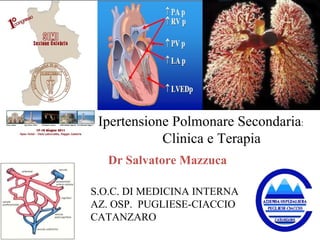
- 1. Ipertensione Polmonare Secondaria : Clinica e Terapia S.O.C. DI MEDICINA INTERNA AZ. OSP. PUGLIESE-CIACCIO CATANZARO Dr Salvatore Mazzuca
- 4. Diagnosi sempre tardiva 24% 63% 12% 1% 100 90 80 70 60 50 40 30 20 10 0 WHO FC I WHO FC II WHO FC III WHO FC IV n = 674 Humbert M, et al. Am J Respir Crit Care Med 2006. %
- 6. Lo screening è efficace per diagnosticare la malattia 1% 24% 75% 12% Patients (%) 63% 100 80 60 40 20 0 I II III IV 44% 28% 11% II III IV Patients (%) 39% No screening 1 With screening 2 WHO FC WHO FC 1 Hachulla et al. Arthritis Rheum 2005; 2 Humbert et al. Am J Respir Crit Care Med 2006 100 80 60 40 20 0
- 9. Sospetto Clinico di PH in caso di insorgenza di dispnea, faticabilità, ripetute sincopi senza motivi... in tutte le condizioni patologiche che possono complicarsi con l’ipertensione polmonare in caso di rilievo incidentale di segni all’ECG o al radiogramma del torace
- 10. Quali sono i più comuni segni/sintomi iniziali in PH?
- 11. Miyamoto et al Am J Respir Crit Care Med 2000;161:487-492 Distanza percorsa < 332 m (n = 22) Distanza percorsa 332 m (n = 21) Mesi 0 20 40 60 80 100 % di sopravvivenza 0 10 30 40 20 50 60 p < 0.001 (logrank test)
- 13. Guidelines for the diagnosis and treatment of pulmonary hypertension 2009 Algoritmo diagnostico Linee Guida ESC 2009
- 16. DLCO IN PH La riduzione della DLCO può essere dovuta alla fibrosi sia per ispessimento di membrana, sia per perdita di volume polmonare, ma anche per le lesioni vasculitiche. Una riduzione della DLCO isolata, senza anomalie spirometriche può essere segno di sviluppo di ipertensione polmonare.
- 18. Ecocardiografia doppler Ipertensione arteriosa polmonare di grado lieve presente in caso di rilievo di una pressione sistolica in arteria polmonare pari a 36 - 50 mmHg, che corrisponde ad una velocità di rigurgito tricuspidalico pari a 2,8 – 3,4 m/sec JACC, CHEST 2004
- 23. Secondary ( Non Category- 1) Pulmonary Hypertension Pulmonary Hypertension Due to left heart disease (group 2)
- 25. Secondary ( Non Category- 1) Pulmonary Hypertension Pulmonary Hypertension Due to left heart disease (group 2) POST –CAPYLLARY PASSIVE PH Transpulmonary pressure gradient ( mean PAP – mean PWP) and PVR are within the normal range POSTCAPYLLARY REACTIVE PH Transpulmonary pressure gradient is increased: PAP is greater than that PWP PVR is increasd
- 26. Fibrous intimal thickening Marked Lynphatic dilation Congested alveolar capillaries An increase in pulmonary arteries vasomotor tone Vasoconstrictive reflexes arising from stretch receptors localized in the left trium and pulmonary veins Endothelial dysfunction of pulmonary arteries that may favour vasoconstriction and proliferation of vessel wall cells PULMONARY HYPERTENSION DUE TO LEFT HEART DISEASE ( group 2)
- 28. Secondary ( Non Category- 1) Pulmonary Hypertension Pulmonary Hypertension Due to left heart disease (group 2)
- 31. Secondary ( Non Category- 1) Pulmonary Hypertension Pulmonary Hypertension Due to lung disease and/or Hypoxaemia (group 3)
- 33. Secondary ( Non Category- 1) Pulmonary Hypertension Pulmonary Hypertension Due to lung disease and/or Hypoxaemia (group 3) I sintomi clinici ed i segni fisici dell’ ipertensione polmonare sono difficilmente identificabili nei paz con patologia respiratoria. Inoltre, nella BPCO, l’edema periferico può non essere il segno di insuff del ventricolo Dx, poiché può essere l’effetto dell’ipossiemia ed ipercapnia sul sistema renina – angiotensina- aldosterone. Una concomitante patologia del cuore Sx, che è comunemente associata con la patologia cronica respiratoria, può anche contribuire ad aumentare la PAPs.
- 34. Secondary ( Non Category- 1) Pulmonary Hypertension Pulmonary Hypertension Due to lung disease and/or Hypoxaemia (group 3) Pulmonary muscolar artery: Aboundant amount of elastina And collagen in the intimal layer
- 35. PULMONARY HYPERTENSION DUE TO LUNG DISEASE (group 3) Pulmonary muscolar artery: Aboundant amount of elastina And collagen in the intimal layer Hypoxic vasoconstriction Mechanical stress of hyperinflated lungs Loss of capillaries Inflammation Toxic effects of cigarette smoke
- 37. Normal Endothelin-1 Angiotensin II serotonin NO PGI 2 Vasoconstrictors > vasodilators Pulmonary hypertension ANP Adrenomedullin Minimal resting tone Increased tone Vascular remodelling LO SQUILIBRIO TRA VASOCOSTRITTORI E VASODILATATORI CONTRIBUISCE ALLA PH Endothelin-1 Angiotensin II serotonin NO PGI 2 ANP Adrenomedullin Vasoconstrictors = vasodilators
- 38. PULMONARY HYPERTENSION DUE TO LUNG DISEASE (group 3)
- 43. Recommendations for PH due to Lung Disease
- 44. Secondary ( Non Category – 1) Pulmonary Hypertension Chronic Thromboembolic Pulmonary Hypertension ( Group 4)
- 45. IPERTENSIONE POLMONARE CRONICA TROMBOEMBOLICA Causata da ostruzione delle principali diramazioni dell’arteria polmonare da ripetuti episodi di embolia polmonare e dalla particolare organizzazione/progressione di questi emboli
- 46. IPERTENSIONE POLMONARE CRONICA TROMBOEMBOLICA EMBOLIA POLMONARE Risoluzione completa (minoranza dei casi) Risoluzione parziale (maggioranza dei casi) Progressione verso ipertensione polmonare ? 0,1%
- 48. Secondary ( Non Category – 1) Pulmonary Hypertension Ostruzione delle piccole arterie elastiche subsegmentali Ostruzione nelle piccole arterie Muscolari: ispessimento della media, proliferazione intimale, lesione plessiforme Piccole arterie muscolari e Arteriole distali ai grossi vasi elastici ostruiti Chronic Thromboembolic Pulmonary Hypertension ( Group 4)
- 50. Secondary ( Non Category – 1) Pulmonary Hypertension Chronic Thromboembolic Pulmonary Hypertension ( Group 4) Obstructive lesions identical to those observed in PAH Shear stress Pressure Inflammation Release of cytochines and Vasculotrophic mediators
- 53. CC, 18 yo, f PPH CE, 63 yo, f CTEPH Esami per diagnosi differenziale: Scintigrafia polmonare perfusionale
- 56. Secondary ( Non Category – 1) Pulmonary Hypertension Pulmonary Hypertension With unclear and / or Multifactorial mechanism ( Group 5)
- 57. Pulmonary Hypertension With unclear and / or Multifactorial mechanism ( Group 5)
- 59. CONCLUSIONI
- 60. CONCLUSIONI
Hinweis der Redaktion
- Because the symptoms of PAH can be mild and unspecific, PAH patients often present late, and a definitive diagnosis of PAH is made only when symptoms become marked and the underlying disease is more advanced. In fact, three qua rters of patients are diagnosed when they are in WHO FC III/IV – that is, when they are already in the advanced stages of the disease. However, around one quarter of PAH patients are diagnosed when they are in WHO FC II and are still mildly symptomatic. Humbert et al. Am J Respir Crit Care Med 2006;173:1023–1030 .
- The reason that PAH is difficult to diagnose and patients present so late are two-fold: • T he symptoms of PAH can be mild and are non-specific • The symptoms can be difficult to differentiate from those of other pulmonary or cardiovascular diseases As a result, PAH is often not diagnosed until the patient has advanced disease. The average interval from onset of symptoms to diagnosis is approximately 2 years. During this time, patients are likely to see a number of different physicians before a definitive diagnosis is made. Despite these difficulties, the number of patients diagnosed with and treated for PAH is growing, due to increased disease awareness, improved diagnostic techniques and the development of management guidelines.
- Annual screening using Doppler echocardiography for the detection of PAH is recommended for high-risk patient populations, including family members of a patient with FPAH, SSc and portopulmonary hypertension (PoPH) patients, even if they are asymptomatic. This enables detection of early disease that might otherwise remain undiagnosed until much later in the disease course. Other high risk populations include patients with other CTDs and HIV patients, both of whom should be screened if they present with dyspnea . The benefits of screening can be clearly seen from the findings of two studies shown on this slide. As seen previously, with no screening program in place, the majority of patients (~¾) are in WHO FC III/IV at the time of identification (with only a quarter in WHO FC II). By comparison, with screening in place, nearly half the patients are detected while they are still in WHO FC II. 1 Humbert et al. Am J Respir Crit Care Med 2006;173:1023–1030; 2 Hachulla et al. Arthrit Rheum 2005; 52:3792–3800.
- The importance of early diagnosis to long-term prognosis is highlighted in this slide. Studies have shown that there is a marked impact of WHO FC (i.e. degree of disease severity) on the prognosis of these patients – with patients in WHO FC I/II having a median survival time of ~5 years, compared with ~2.6 years for WHO FC III patients and only ~6 months for WHO FC IV patients. As such, if left untreated, the prognosis for patients with PAH is worse than some forms of cancer, as shown on the slide. D’Alonzo et al. Ann Internal Med 1991;115:343–349; Kato et al. Cancer 2001;92:2211–2219; Bjoraker et al. Am J Resp Crit Care Med 1998;157:199–203.
- Il sospetto clinico di IP polmonare emerge in tre circostanze particolari: in presenza di condizioni morbose che possono essere complicate da IP od associate ad IP (per esempio: malattie dell’apparato respiratorio, valvulopatie, cardiopatie congenite con shunt…); in ogni caso di dispnea da sforzo di non chiara origine; in caso di rilievo strumentale (radiologico o elettrocardiografico) occasionale di segni indicativi di dilatazione/ipertrofia del Vdx o dilatazione delle arterie polmonari.
- PAH: is a progressive disease characterized by an increase in PAP, decline in cardiac output, and increase in pulmonary vascular resistance, and eventually heart failure and death. is associated with a marked vasculopathy in the pulmonary vasculature and subsequent hypertrophy of the right ventricle. may be idiopathic (formerly called primary pulmonary hypertension [PPH], which is now divided into idiopathic and familial ) or related to a number of underlying diseases - the classification of PAH has been recently revised as shown in the slide. IPAH is uncommon (reported to occur in 2 cases per million). Simonneau G, Galiè N, Rubin LJ et al. Clinical classification of pulmonary hypertension. JACC 2004;43(Suppl):S5-S12
- PAH: is a progressive disease characterized by an increase in PAP, decline in cardiac output, and increase in pulmonary vascular resistance, and eventually heart failure and death. is associated with a marked vasculopathy in the pulmonary vasculature and subsequent hypertrophy of the right ventricle. may be idiopathic (formerly called primary pulmonary hypertension [PPH], which is now divided into idiopathic and familial ) or related to a number of underlying diseases - the classification of PAH has been recently revised as shown in the slide. IPAH is uncommon (reported to occur in 2 cases per million). Simonneau G, Galiè N, Rubin LJ et al. Clinical classification of pulmonary hypertension. JACC 2004;43(Suppl):S5-S12
- La scintigrafia polmonare perfusionale è sostanzialmente normale nei casi di IPP.