
Recommended
More Related Content
What's hot
What's hot (20)
Similar to Humidification & inhalation therapy
Similar to Humidification & inhalation therapy (20)
Recently uploaded
Recently uploaded (20)
Humidification & inhalation therapy
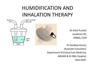
- 1. HUMIDIFICATION AND INHALATION THERAPY Dr Ankit Purohit Academic SR, DNBSS, CCM Dr Sandeep Kumar, Associate Consultant, Department of Critical Care Medicine, ABVIMS & Dr RML Hospital, New Delhi.
- 2. What is Humidification • Respiratory humidification is method of artificially warming and humidifying of respiratory gases for mechanically ventilated patients. • RESPIRATORY GAS CONDITIONING 1) Warming 2) Humidification 3) Purification • Naturally, the function is performed by upper airway and lungs.
- 3. Outlook of topic 1) Physical principles 2) Physiology 3) Guidelines 4) Type of humidifiers & Devices.
- 4. Physical principles Absolute humidity (AH) – mass of water vapour in volume of gas at a temperature, when saturated. Relative humidity (RH) – Percent saturation, it is percentage of amount of total water vapour, that can be contained by gas when saturated, at the temperature. REF – 1)Dorsch & Dorsch. 2) Plotnikow et al; Humidification and heating of inhaled gas in patients with artificial airway. A narrative review. Res Bras Ter Intensiva 2018;30(1): 86 -97.
- 5. Interrelationship between AH and RH • Gas saturated with water vapour, once heated, its capacity to retain water vapour increases. • So gas at room temperature( 27◦C), fully saturated, when enter the respiratory tract and attains 37◦C, would become desaturated. • The gas would absorb water from respiratory mucusa, till it becomes saturated. • So gradually desiccation of mucusa would occur. • Hence, optimal conditioning of air for mechanically breathing patient is required. REF – Dorsch & Dorsch
- 7. Physiology of conditioning • 75% of conditioning takes place in upper airway, rest 25% in lungs. • Upper airway cleans and humidifies air ranging from 1000 l to 21,000 litres, everyday, depending on body size and activity.
- 8. Warming • Network of capillaries & resistive vessels underlying nasal and oral mucosa, performs function of warming. • Inhaling cold air increase blood flow to mucosa. • Blood is regulated by autonomic nervous system. • Diameter of resistive vessels is mediated by SNS, while PSNS regulates glandular secretions. • Temperature of nasal mucosa is approx 32◦C. • Nasal turbinates and conchae generate current in inhaled air, moving them through sinuses, leading to warming.
- 9. Isothermal Saturation Limit • Isothermal Saturation Limit – The point at airway where inhaled gas acquires full water vapour saturation at 37◦C . At 4th or 5th generation bronchi. • Critical as at this limit air doesn't damage mucosa & ciliary epithelium. • Artificial respiration require the limit to be obtained at level of carina. REF -1) Plotnikow et al; Humidification and heating of inhaled gas in patients with artificial airway. A narrative review. Res Bras Ter Intensiva 2018;30(1): 86 -97. 2) Andrew D Bersten; Humdification and inhalational therapy, Oh’s intensive care manual; 6th edition.
- 10. Humidification The isothermal saturation boundary is reached at 4th to 5th generation bronchi and not at carina, as shown by red dashed line.
- 11. Humidification • Humidification is progressive as air moves down track. • Occurs till isothermal saturation point. • Under normal resting conditions, 250 ml of water is lost in 24 hours & 350 kcal of energy. • During expiration, upto 25% of moisture is retained by mucusa. Preserved via condensation. • Ciliary function is reduces at RH 75% at 37◦C ( AH of 32 gm/m3 ) & ceases at RH 50% at 37◦C. • It is recommended, AH> 33 gm/m3 to be maintained for normal ciliary function.
- 13. Mucociliary function; Cleaning • Clearance of surface particles depends on – 1) Beating cilia. 2) Airway mucus. 3) Transepithelial water flux. • mucus is derived from goblet cells, Submucosal glands & Clara cells & capillary transduate. • Conducting airways lined with pseudostratified, ciliated columnar epithelium. • On descending, airway epithelium gradually turns stratified and cuboidal having less ciliated cells. • Cilia beats at rate of 10 mm/min at 37◦C and RH 100%.
- 14. Factors Affecting mucociliary function 1) RH & temperature of inhaled air. 2) URTI 3) Chronic bronchitis. 4) Cystic fibrosis. 5) Bronchiectasis. 6) Immotile Cilia syndrome (Kartagener’s syndrome). 7) Dehydration. 8) Hyperventilation. 9) Anesthesia, Opioids, Atropine 10) Exposure to noxious gases 11) High FiO2 . 12) Beta agonist improve nasociliary function.
- 15. Adverse effects of decreased humidification 1) Increased mucus viscosity. 2) Depressed ciliary function. 3) Cytological damage to the tracheobronchial epithelium, including mucosal ulceration, tracheal inflammation and necrotising tracheobronchitis. 4) Microatelectasis from obstruction of small airways, and reduced surfactant leading to reduced lung compliance 5) Airway obstruction due to tenacious or inspissated sputum with increased airway resistance. 6) Metaplasia of the tracheal epithelium occurs over weeks to months in patients with a permanent tracheostomy.
- 16. Bioaerosol • Defined as collection of particles suspended on a column of air derived from or incorporating material of biological origin. • Particles < 100 microns are in inhalable range. • Airborne dissemination depends on air mass movement, turbulence and thermal convection. • Mass Median Aerodynamic Diameter(MMAD) decreases with increase distance of spread. • Particles 1 to 100 micron. Particles >8 micron get trapped to airways, thrown out by cilia. • Particle size 1 – 3micron reach alveoli and deposits there. • Aerodynamic size 0.5 – 5 micron have maximum tendency to reach alveoli.
- 18. REF – Thomas R; particle size and pathogenecity in the respiratory tract; virulence4:8, November 2013,847- 858. Landes Bioscience.
- 19. Ideal Humidification 1) The inspired gas is delivered into the trachea at 32–36°C with a water content of 30–43 g/m3. 2) The set temperature remains constant and does not fluctuate. 3) Humidification and temperature remain unaffected by high flows. 4) The device must be simple to use and to service. 5) The humidifier could be used with spontaneous or controlled ventilation. 6) There must be safety mechanisms, with alarms, against overheating, overhydration and electrocution. 7) The resistance, compliance and dead-space characteristics must not adversely affect ventilation. 8) The sterility of the inspired gas must not be compromised. REF- Andrew D Bersten; Humdification and inhalational therapy, Oh’s intensive care manual; 6th edition
- 20. Guidelines • DESCRIPTION- 1) Upper airway gives 75% humidification and warming. 2) Optimal required moisture at carina at 37◦C with optimal RH 44 mg/l. Temperature range (34◦C – 41◦C). 3) Inspired gas with temperature >37◦C & 100% RH, gives reduces viscosity and increased pericellular fluid depth.
- 21. Indications & Settings • Humidification – 1) Critical care, acute care, operating theatre, transport. 2) Humidification is inspired gas during mechanical ventilation is mandatory for ETT and tracheostomy. 3) Humidity level 33 mg/l to 44 mg/l at temperature between 34◦C to 41◦C. 4) Optional for NIV - Heated humidifier provides better CO2 clearance and lower work of breathing than does heat-and- moisture exchanger, because heated humidifier adds less dead space. 5) Inspired gas should never exceed 41◦C and alarm set at 43◦C.
- 22. Types of Humidifiers • PASSIVE – Heat and moisture exchangers with filter. • ACTIVE – 1) Unheated bubble type. 2) Heated ; pass over ( stream of gas over heated water)/ Blow by ( Stream of gas passing over wicks dipped in water)
- 23. HME( heat moist exchangers) • Efficient, simple & convenient passive humidifiers used in ICU. • Principle of heat and moisture conservation of expired air. • Simple condensers made of disposable foam, synthetic fibers or paper. • Have large surface area to create entrapping heat and moisture of exhaled gas. • Retained heat and moisture is delivered in next inspired breath. • Modern HME are light with dead space of 30 – 95 ml. • They have male and female connectors of 15 mm or Y piece connector of 22 mm (concentric).
- 24. REF - Plotnikow et al; Humidification and heating of inhaled gas in patients with artificial airway. A narrative review. Res Bras Ter Intensiva 2018;30(1): 86 -97.
- 25. Hygroscopic HME • Also known as Hygroscopic condenser humidifiers. • Made of synthetic fiber coated with hygroscopic material(Calcium chloride or Lithium chloride) • Synthetic fiber help to decrease accumulation of condensed water. • Tends to increase their efficiency ( AH around 30 g/m3) compared to hydrophobic HMEs (i.e. AH 20–25 g/m3). • DRAWBACK – 1) tend to loose filtration efficiency when wet. 2) resistance increases on being wet.
- 26. Hydrophobic HME • Contains hydrophobic membrane with small pores. • Moderate humidity (RH at 37◦C of 25 mg/l). • Efficient bacterial and viral filter. • Prevent transmission of Hepatitis C virus. • Better than hygroscopic filters. • Allow only small passage of water vapour. • Add less resistance to circuit, when wet. • DRAWBACK – high ambient temperatures decrease its performance.
- 27. HME with filter • Filter material is embedded with electrostatic charge. • The charge help to prevent flow of bacteria & viruses. • Types of available filters 1) HMEF – heat moisture exchanger with filter. 2) HCHF – Hygroscopic condenser humidified filter. 3) HHMEF – Hygroscopic heat and moisture exchange filters. • HMEF have not shown to reduce the incidences of nosocomial pneumonia, as they are mostly attributed to aspiration.
- 28. Combined HME • Contain both hygroscopic and hydrophobic elements. • Better performance than hygroscopic ones. • They have better retention of moisture and maintain humidity levels to 30 mg/l at 37◦C.
- 29. Comparison of hydrophobic & hygroscopic HME • Type 1) Heat moisture exchanging capacity 2) Effect of increased TV 3) Filtration efficiency (dry) 4) Filtration efficiency(wet) 5) Resistance (dry) 6) Resistance(wet) 7) Effect of nebulisation Hygroscopic Excellent Slight decrease Good Poor Low Slight increase Great increase in resistance Hydrophobic Good Significant decrease Excellent Excellent Low Slight increase Little effect.
- 31. HME for aerosol Therapy • Gibeck Humid –Flo • Remain in line with circuit during application of aersols. • Reduce opening & contamination of breathing circuit. • HME mode – conventional • AEROSOL mode – aerosol directly reach patient without contacting humidifier. • REF - Plotnikow et al; Humidification and heating of inhaled gas in patients with artificial airway. A narrative review. Res Bras Ter Intensiva 2018;30(1): 86 -97. Gibeck® Humid-Flo Heat & Moisture Exchanger (HME)
- 32. HME Booster • Hybrid active humidifier. • ‘T’ connector contain self regulated heater. • Small volume chamber for continuous infusion of distilled water. • Upper part of device has hydrophobic membrane to allow passage of water, only when it evaporates. • Dead space is only 9 ml. • Active humidification & passive retention during expiration. • Advantage – 1) Easy to use 2) Have bacterial filter. 3) Useful for hypothermic patient. 4) Eliminates excessive condensation in tubing • Disadvantage – 1) No temperature monitoring. 2) Increased resistance. 3) Require source of electricity
- 33. Dead Space of HME • Higher volume of condenser material will yield better performance. • Ideal dead space for humidifier is 50 ml, for standard adult patients. • Dead space is compensated by increasing Ventilatory minute volume. • Additional dead space leads to CO2 retention. • This increase work load of breathing in patients. • HME with smaller dead space volume have been developed. • Low humidification capacity and it worsens with increase in TV and supplementary O2.
- 34. Resistance • Normally unused HME have low resistance; 5 cmH2O/sec. • It increases with impaction of secretions, condensation of water, increase in flow and TV. • So it is important to change filters regularly.
- 35. Volume of HME & Relation to Humidification capacity • Internal volume of HME & AH are factors affecting delivery of humidity to patient. • Eckerbom et al, conducted study relating dead space of HME to AH. • They found poor correlation. • But they used only 6 devices.
- 36. Volume of HME & Relation to Humidification capacity • Branson et al conducted similar study with more devices. • Excluding bacterial filter, they found strong correlation (r = 0.91, p < 0.0001). • The ratio of delivered AH to Dead space in ml is efficacy measure for device. • Humidifiers reach level recommended of 30 mg/l at 60 ml dead space.
- 37. Performance of HME & Minute volume • Increase in MV decrease the humidification of HME. • Decrease in delivery of AH with increase in TV.(Lucato et al, Eckerbom et al). • The flow has variable effect on performance (Unal et al). • In 2014, Lellouche et al found no effect of minute ventilation and room temperature on performance of new generation devices. • MV = TV X Resp frequency. • Eckerbom et al & Branson et al, found TV to be affecting factor on performance of device. • Increasing TV decreased performance.
- 38. HME & Time of contact with air • Eckerbom et al , found no comparable differences between delivered humidity , when compared to time of contact of inspired air at 1st second & 2nd second. • Branson et al found, as expiratory flow increases, the time of contact between the gas and the HME decrease with decrease in the AH delivered to the patient.
- 39. Time of changing HME 1) Due to excessive condensation increasing resistance of circuit. 2) Visible impaction of blood or secretions. 3) In COPD patients, every 48 hours. 4) 96 hours to 1 week in remaining patients.
- 40. Indications 1) Preferred first device for humidification while initiating mechanical ventilation. 2) In OT, while performing elective surgeries. 3) Transporting intubated patient. 4) On tracheostomized patients, breathing spontaneously. 5) On supraglottic airway, to supplement oxygen,by connecting oxygen tubing to gas sampling port.
- 41. Contraindications for HME 1) In patients with bloody thick mucus secretions. 2) When expired TV is less than 70% of inspired (bronchopleurocutaneous fistula, ETT cuff malfunction). 3) In pediatric age group, increase dead space. 4) In acute respiratory failure , HME increase work of breathing. 5) When patient temperature is less than 32◦C. 6) When patient has high spontaneous minute ventilation.(relative) 7) When patient is being nebulised, HME to be removed. 8) NIV, large mask leak.
- 42. Active Humidifiers • Electric heater placed in plastic casing with metal base in which sterile distilled water is placed. • Parts – 1) Humidification chamber – clear chamber disposable, made f plastic. 2) Heat source – supplied by heating rods immersed in water or heating case at bottom of container. 3) Inspiratory tube- it carries heated humidified air. It contains heated element to maintain temperature. 4) Temperature monitor – temperature sensors placed at inspiratory end and at thermostat. 5) Thermostat (SERVO CONTROLLED) – it is equipped with temperature sensors and autoregulation .
- 43. Bubble type Humidifiers • The gas flow passes through the water level (bubbled through). • May be heated or non heated. • Rising air gains temperature and humidity. • Amount of water and Flow rate affects the humidification. • Higher water column, more air water interfacing. • Increased flow , decreased performance. • Inexpensive, but are inefficient. • Water content of around 9m g/l (i.e. about 50% RH at ambient temperatures). • They are also a potential source of microbiological contamination. • Leads to aerosol formation, source of infection.
- 44. Pass over humidifiers • Air flows over the heated water interface. • Attains humidity and gain the temperature. • The water bath temperature is thermostatically controlled (e.g. at 45– 60°C) to compensate for cooling along the inspiratory tubing, targeting an inspired RH of 100% at 37°C. • A heated wire is seated in the inspiratory tubing to maintain preset gas temperature and humidity. • They produce microdroplets (<5 microns), decreased probability of infection. REF – Haitham et al; Humidification during Mechanical Ventilation in the Adult Patient; Hindwai, May 2014.
- 45. Wick & Hydrophobic membrane Humidifiers • Porous membrane absorbs humidity as it is dipped in water. • The dry air enters chamber and comes in contact with water saturated wicks. • It increases gas liquid interface. • Hydrophobic membrane humidifiers, gas passes through membrane and attains water vapour.
- 46. Standard Requirement for humidifiers 1) Must produce an output of 10 mg/l of water. For supraglottic airways, it is 33mg/l. 2) Average temperature at delivery outlet would not exceed by 2◦C from set temperature. 3) Volume of liquid exiting humidifiers will not exceed 1 ml/min or 20 ml/hr. For neonates, it is 5 ml/min or 20 ml/hour. 4) Maximum temperature set at gas delivery outlet is 41◦C. 5) Accessible surface temperature of the delivery tube must not exceed more than 44◦C within 25 cm of connection port. 6) All calibrated controls must be within 5% of their range. 7) Flow direction should be marked on humidifiers.
- 48. Hazards & complications 1) Potential for electrical shock. 2) Hypothermia when Heated humidifier(HH) is inadequately set. 3) Hyperthermia due to overheating. 4) Burn injury or melting of circuit due to heated wires. 5) Underhydration ( <26 mg/l of AH). 6) Hypercapnia due to added dead space with inadequate air flow. 7) Condensation of water in circuits leads to rise in nosocomial infection.
- 50. Inhalational Therapy • Route of choice to deliver drugs for respiratory disorders. • Provides high concentration at pulmonary site and less systemic concentration. • This accounts for less side effects. • PHARMODYNAMIC AIRWAY SELECTIVITY – 1) ratio of pulmonary efficacy to airway selectivity. 2) It is favourable to this route. 3) Faster onset of action with less systemic side effects.
- 51. Inhalational therapy • Lungs should not be considered as single organ. • Various morphologic structures, network of intricate airways, bronchioles , capillaries and interstitial tissues. • Lungs have unique Pharmacokinetics(PK). • Multiple PK processes with each one having unique characteristics & have their interplays. • Depends on aspects of inhaled medications, their physiochemical characteristics of the drugs, drug formulation & Inhalational device.
- 53. PHARMACOKINETICS 1) Drug or droplet deposition 2) Drug dissolution in the lung fluids 3) Mucociliary clearance in the larger airways & macrophage clearance in the alveolar space. 4) Absorption of the drug in lung space 5) Pulmonary tissue retention and metabolism 6) Absorptive blood clearance.
- 55. Drug particle/ Drug deposition • Total dose in device, a fraction is deposited in device only. • LUNG DOSE – amount of drug deposited in lungs. • Central (larger airways) and peripheral (smaller airways) • Deposition depends on 1) Aerodynamic size 2) Inhalational flow 3) Device characteristics 4) Disease related factors • Independent of drug own physiochemical characteristics.
- 56. Aerodynamic particle diameter • It the diameter of particle aerosol generated from device. • 0.5 – 5 microns size easily deposited at the airways. • Smaller particles (1-3 microns), more peripheral distribution. • Larger (>5 microns) distributed at the larger airways, mouth throat areas. 1) Inertial impaction- large particle travel less, deposit at early. 2) Maximum airflow velocity – cause larger particle to impact on airways. Faster rate less peripheral deposition and vice versa. 3) Sedimentation – smaller particles. Due to gravity. Enhanced by breath holding once inspiration is over. 4) Diffusion or Brownian motion - submicron particles.
- 59. Pulmonary Drug Dissolution • Particles to diffuse through fluids of lung epithelium. • Depends on 1) Drug formulation 2) Physiochemical drug properties. 3) Physiologic factors. • Biphasic gel (mucus)– aqueous layer present at CONDUCTING AIRWAYS. Mucus hinders absorption. • Surfactant & alveoli fluid at alveolar level. Surfactant enhance absorption. • Properties of drug – free water soluble drugs(albuterol) diffuse easily into pulmonary lining. Fat soluble lesser diffusion.(steroid) • Slow dissolution is rate limiting factor.
- 61. Mucociliary & Phagocytic Clearance • Upward movement of mucus by underlying beating cilia of epithelium. • Drugs particles here are transported to pharynx and undergoes GI absorption. • Larger airway diameter , faster clearance. • Mucus thickness and higher viscosity decreases the mucociliary clearance. • Phagocytic - macrophages at alveoli phagocytise drug. • Slower than mucociliary.
- 63. Absorption to lung tissue • Lipophilic drugs are more rapidly absorbed once dissolution through diffusion occurs. • Hydrophillic drugs undergoes paracellular diffusion through aquaporin pores at gap junction. • Physiologic characteristics of pulmonary environment . 1) Phagocytosis increases absorption. 2) High perfusion at pulmonary level increase the absorption. 3) Large alveolar surface area. 4) Epithelial layer thickness.
- 64. Pulmonary tissue retention and tissue thickness • Physiochemical characteristic – 1) Tissue affinity. 2) Pulmonary tissue partition coefficient. • partition coefficient is the ratio of the concentration of a substance in one medium or phase (C1) to the concentration in a second phase (C2) when the two concentrations are at equilibrium. • Here, ratio of amount of drug at outside to inside. Defines convenience of diffusion. • Patient specific characteristic - Alveolar membrane interface. • LONG ACTING BETA AGONIST. – low permeability, longer retention. • Long permeability – enhanced efficacy. • Metabolising enzymes - CYP1A1 CYP2E1, more active in smokers.
- 65. Absorptive drug Clearance to systemic perfusion • Largely dependent on perfusion. • Larger airways have lower perfusion so less drug absorption. • Alveoli have fluent absorption of drugs. • Short half life of same drug. • Despite high tissue retention of drug in alveoli, rapid diffusion to circulation, makes drug short acting. In larger airways, long tissue retention means , longer half life as drug reached circulation at constant rate.
- 67. Spacer with mask
- 68. Spacer
- 75. PLANAR SCINTIGRAPHY IMAGE OF AEROSOL DEPOSITION IN MECHANICAL VENTILATED PATIENTS.
- 76. HIGHLIGHTS • MDI showed highest dose deposition, administering at Y piece of circuit.(38% of nominal dose). • Constant output jet nebulizers deposited only3 – 7% of nominal dose. • Inspiratory synchronized jet nebulizers deposited 1 – 16% of nominal doses. • Constant output nebulizer deposited 5% of nominal doses. • SPECT CT showed 33% & 17% deposition of drug to right and left lungs, when drug is directly instilled to ETT. • Post operative intubated patients SPECT CT is showed predominant proximal deposition of nebulized drugs. • Jet nebulizers showed 50% of ND retained in resevoir and T piece. • 15 – 30 % with ultrasonic nebulizers. • 3 – 10 % with vibrating mash nebulizers. • 10 – 44% was trapped in circuits. • No data on MDI
- 77. Administration technique • Nebulizer placed at inspiratory limb. • Vibrating mash type preferred. • TV – 8 ml/kg, respiratory rate 12 /min (to prevent expiratory phase loss). • Flow rate of 30 l/min with end inspiratory pause of 20%. • Heating humidifier system not to be used. • 63% of ND reached airways and only 37% deposited at circuits.
- 79. Highlights 1) ARDS- limited to patients with optimal ventilation, maintianing PEEP and on IRV. NO is introduced when they have PaO2 less than 90 mmHg at Fio2 100%. 2) RV failure due to PULMONARY HTN - defined as severe RVF with PUL HTN( mean PAP >24 mmHg), PVR > 400 dyne/sec/cm5. NO decreases PAP and PVR and help alleviate RVF. 20 – 40ppm. 3) If there is no response to 20 ppm dose for 30 minutes (more than 20% rise of PaO2), it is increased to 40 ppm. 4) Toxicity – methemoglobinemia, mucous membrane irritation.
- 85. Forest plot for mortality
- 86. Forest plot for microbiological cure
- 87. Highlights • Nebulised antibiotics were associated with higher rate of clinical cure. • No association was found with microbiological cure, improved mortality, duration of mechanical ventilation, ICU length of stay & renal toxicity. • Used as adjunct to main therapy. • Delivery of aerosolized particles to lungs 1) Ultrasonic or vibrating mesh type nebulizers are best. 2) Spontaneous ventilator modes are associated with high turbulent flows and more proximal distribution of drug 3) Ventilator circuits should have smooth surface and no acute angle connections. 4) HME removed.
- 88. Epinephrine Nebulisation in children • Numa et al ,in their article, the effect of nebulized epinephrine on respiratory mechanics and gas exchange, Resp critical care med journal, 2001. 1) Gave 0.5 mg/kg epinephrine (undiluted) with maximum dose of 5 mg to infants with bronchiolitis, asthma. 2) They measured respiratory mechanics and hemodynamics. 3) They found decrease in total resistance in airway pressure post nebulisation. 4) There was rise in heart rate and mean BP post nebulisation. 5) Oxygenation and ventilation didn't show much improvement.
- 89. 1) They found no difference in length of hospital stay in infants of bronchiolitis. 2) They recommended use of nebulised epinephrine in hospitalized infants only as rescue agents. 3) There is no good evidence to support treatment with nebulized epinephrine in infants.
- 90. THANK YOU