
Empfohlen
Empfohlen
Weitere ähnliche Inhalte
Was ist angesagt?
Was ist angesagt? (20)
Ähnlich wie Slitlampexamination dinesh
Ähnlich wie Slitlampexamination dinesh (20)
Mehr von Dinesh Madduri
Mehr von Dinesh Madduri (16)
Kürzlich hochgeladen
Kürzlich hochgeladen (20)
Slitlampexamination dinesh
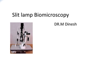
- 1. DR.M Dinesh
- 2. Introduction • It is a dynamic examination in which eye and ocular adnexa are examined anteroposteriorly and horizontally. • Slit Lamp term is a misnomer since slit is only one of the various other diaphragmatic opening present in the instrument. • Term “Biomicroscopy” introduced by Mawas in 1925 means “ Examination of living eye by means of microscope and slit lamp.”
- 3. • It is one of the important examination tools of ophthalmologists. • most important advantages is that one can examine the eye structure in three dimensions (3D). • The 3 basic requirements for appreciation of depth with a slit-lamp. – 1.the clinician possessing a third grade of binocular vision called stereopsis. – 2.the direction of the incoming light source,is dependent upon the fact that the light beam can be moved so it comes in from one side or the other. – 3.the shape of the slit.
- 4. History • 1806 - karl Himley used a convex lens for observation • 1823 - Purkinje, attempted to develop a slit lamp -used hand held magnifying lens in one hand -another hand held lens to focus strong oblique illumination • 1863 - Louis de Wecker, -devised portable ophthalmomicroscope -combination of small monocular microscope with condensed lens -lacks stereoscopic view
- 5. • 1891 - Albert and Greenough developed binocular microscope with stereoscopic view • 1897 - Siegfried Czapski –modified binocular corneal microscope. -However, none of the units had sufficient and adjustable illumination. • 1911 - Allvar Gullstrand,an ophthalmologist and Nobel laureate -introduced the illumination system which had a slit diaphragm ,for the first time -therefore credited with the invention of the slit lamp.
- 7. • 1916 - Henker and Vogt - improved upon Gullstrand’s device by creating an adjustable slit-lamp -combined Czapski’s binocular corneal microscope with Gullstrand’s slit-lamp illumination. • 1933 - Hans Goldmann - improvise by placing all vertical and horizontal adjustments of slit lamp & slit beam on a single mechanical stage - it is marketed as Haag-Streit model 360 slit lamp
- 8. • 1950 - Littmann - introduced new optical principle for the biomicroscope - incorporated rotatory magnification changer based on principle of Galilean telescope ,which is fore runner of current ziess slit lamp series
- 9. Basic design of slit lamp • The three main components of the modern slit-lamp are: 1)Observation system 2) Illumination system 3) Mechanical system
- 10. Observation system (microscope) Should have following Characteristics : • Optimum stereoscopic observation. • Selectable magnification. • Large field of view. • Large depth of field. • Enough space in front of the microscope for manipulations on the eye.
- 11. Observation system is composed of • Compound microscope : composed of -An objective lens: Two planoconvex lenses with their convexities put together (Composite power +22D) -Eyepiece: has lens of +10 D • Pair of prisms: To reinvert the image B/W objective & eye piece • Converging tubes: They are converged at an angle of 10-15 degree, to provide good stereopsis. • telescope
- 13. Magnification systems Range of magnification in slit lamp -6x to 40x Moder slit lamps use one of the following 3 systems to produce range of magnification • Czapskiscope with rotating objective • The littmann-Galilean telescope principle • Zoom system
- 14. Czapskiscope with rotating objective -one of the oldest -mc used technique -different objectives are usually placed on a turrent type of arrangement that allows them to be fairly rapidly changed during examination eg: Haag-streit model Bausch&lomb model Thorpe model
- 15. The littmann-Galilean telescope principle • The Galilean Magnification changer(G) developed by litmann in 1950 • G is a completely separate optical system • Sits neatly b/w objective & eyepiece ,does not require either of them to change • It utilises Galilean telescopes to alter the magnification. • It has two optical components: • 1)Positive lens 2)Negative lens
- 16. • Lenses are arranged in turret arrangement. • It provides large range of magnifications, typically five. • It fits within the slit-lamp microscope along with relay lens (R) & prism ejector (P)
- 17. The Galilean Magnification changer Knob to change magnification (3 or 5step)
- 18. Zoom system • allows continuously various degree of magnification. • Range of magnififcation 7x-35x • E.g, Nikon photo slit lamp Zeiss-75 Sl
- 19. Magnification can also be changed by changing the eyepiece power
- 20. Illumination System Gullstrand s illumination system comprises of 1.Light source: -originally Nernst lamp was used -f/b nitra lamp f/b arc lamp f/b mercury vapour l. finally halogen lamps -provides illumination of 2-4x 105 lux 2.Condensor lens system: -couple of planoconvex lenses with their convex surfaces in apposition
- 21. 3.Slit & other diaphragms: • Height & width of slit can be varied • There are stenopaeic slits of 2 & 0.5 mm to provide conical beam of light • To assist in examination of fundus & angle of ant chamber -there is facility to rotate slit away from vertical meridian -also ability to tilt projection system about a horizontal axis
- 22. 4.Filters : -diff filters can be inserted eg.,cobalt blue & red free filters • Cobalt blue filter – Used in conjunction with fluorescein stain – Dye pods in area where the corneal epithelium is broken or absent. The dye absorbs blue light and emits green. Uses: • Ocular staining • RGP lenses fitting • Tear layer
- 23. • Red free(green)filter: – Obscure any thing that is red hence the red free light , thus blood vessels or haemorrhages appears black. – This increases contrast ,revealing the path and pattern of inflammed blood vessels. – Fleischer ring can also be viewed satisfactorily with the red free filter.
- 24. 5.Projection lens : -forms an image of slit at eye -its diameter is fairly small -it has 2 adv: a)keeps aberrations of lens down qlty image b) depth of focus of slit better optical section of eye
- 25. 6.Reflecting mirror or prism : -last component of illumination system -the illumination system of a slit lamp has to be able to pass easily from one side of microscope to another, to allow this projection system is arranged along a vertical axis With either mirror or prism final reflecting light along horizontal axis -use of narrow prism so that illumination axis can be made coincide with viewing axis ,without obstructing filed of view
- 27. • The illumination system of most slit-lamps consists of 2 designs. 1. The Haag-Streit type illumination -allows de-coupling in the vertical meridian. - -vertical de-coupling is useful in a)gonioscopy to minimize reflections b)indirect fundus examination to gain increased peripheral views. -illumination comes from above
- 28. • The Haag-Streit type illumination • illumination comes from above
- 29. 2. The Zeiss type illumination system : -the illumination comes from below -does not allow decoupling in the vertical meridian.
- 30. Optics of illumination system • Koeller illumination system has been used in slit lamp • Optically it is identical to that of a 35mm slide projection with exception that a variable aperture takes place of slit • Projection lens has much shorter focal length The interowen beam path in koeller illumination
- 31. •The light source L is imaged in the objective O by the collector system K. The objective in turn produces an image at S in the mechanical slit located next to the collector system. The image of the light source at O is the exit pupil of the system. •The Light source is imaged on to the objective lens but the mechanical slit is imaged on to the patient’s eye. MS
- 32. Mechanical system It is mainly concerned with: • Positioning of patient. • Adjustment for observer and patient. • Adjustment of illumination and observations system. • It generally contains following hard ware.
- 33. movable Fixation target Head rest Canthal alignment mark Chin rest Lock for slit lamp base Joy stick Power unit
- 35. • Mechanical coupling: Mechanical system provides coupling of microscope and illumination system along a common axis of rotation that coincides their focal planes. • This arrangement ensures that light falls on the point where microscope is focused.
- 36. Mechanical coupling disadvantage • In gonioscopy or using three-mirror fundus lens -suboptimal images are seen or -refocussing is necessary because slit µscope do not reach a common focal point in those conditions
- 37. Technique of biomicroscopy • Before using the slit-lamp, it is important to ensure that the instrument is correctly set up. • The eyepieces should be focused for the observer for his/her own refractive error. • Often a little more minus correction is required than the observer’s actual refractive error due to accommodation and proximal convergence. • The Pupillary distance (pd) is adjusted for the observer (perhaps the pd should be slightly less than that usually measured to account for proximal convergence).
- 38. • Check that the observation and illumination systems are coupled, and the slit-beam is of even illumination and has sharply demarcated edge (otherwise irregularity of the beam may be falsely interpreted as irregularity of tissues). • The slit-lamp examination is conducted in a semi dark room.
- 39. Patient adjustment • Patient is seated comfotably in front of slit-lamp on an adjustable stool in front of slit lamp with his/her chin on chin-rest and his forehead on the bar of head-rest. • Adjust the chin-rest so that the patient’s eyes are approximately level with the black marker on the side of the head rest. • Focus the slit-beam on the eye by moving the joystick either towards or away from the patient.
- 40. • The examination should be commenced using the X10 eyepieces and the lower powered objective to locate the pathology and higher magnification should then be used to examine it. • There should be min exposure of retina to light • Select the longest slit-length by means of the appropriate lever. • Medications like ointments &anaesthetic e/d produce corneal surface disturbances which can be mistaken for pathology
- 41. BASE LINE EXAM FLOW CHART lids conjunctiva limbus tears cornea AC iris pupil lens
- 42. MAGNIFICATIONS • Low magnification:7X - 10X : General eye – Lids. – Bulbar conjunctiva/sclera. – Cornea/limbus. – Tears. – Anterior chamber/iris/crystalline lens.
- 43. • Medium magnification: 20X - 25X : Structure of individual layers – Epithelium/epithelial breakdown. – Stroma. – Endothelium. – Contact lens fit/lens condition.
- 44. • High magnification: 30X - 40X : Details • Epithelium – vacuoles – microcysts – dystrophies. • Stroma – striae – folds. • Endothelium – Polymegathism – guttata – blebs – cell density.
- 45. METHODS OF ILLUMINATION • BERLINER Described 7 basic methods 1. Diffuse Illumination 2. Direct Focal Illumination 3. Indirect Focal Illumination 4. Retroillumination 5. Specular Reflection 6. Sclerotic Scatter 7. Oscillatory Illumination of Koeppe
- 46. Diffuse illumination with slit lamp The set is as given 1)Angle b/w microscope & illumination - 30-450 2)slit width - widest 3)Filter - diffusing filter. 4)Magnification - low to med 5)illumination- med to high Beam is only 8-14 mm diameter and therefore must be moved over the eyelids and ocular surface. It can reveal location and general pattern of eyelid, conjunctival, corneal lesions.
- 47. Applications: • General view of anterior of eye: lids,lashes,sclera,cornea ,iris, pupil, • Gross pathology and media opacities • Contact lens fitting. Diffuse illumination
- 48. Direct focal slit illumination • Terminology : Projection , of a narrow slit beam at an angle, to the corneal surface, producing an optical section that slices through the cornea and eye. • Principle: A direct narrow slit beam optically cuts through the cornea , providing a cross sectional view that reveals its contour and its internal structure. • It forms two parallel curved surface, one that follows anterior corneal surface and one that follows posterior corneal surface. • Two surfaces are joined by a block of light scattered in the stroma to create a geometric figure that resembles an elongated ice cube.
- 50. • Angle b/w microscope & illumination - 30-450 • slit width- narrow • Heterogenous tissues like cornea & lens disperse light & become visible as bright objects against dark background • Direct illumination is carried out utilising 3 slit beam effects i.e., -optical section -parallelepiped -conical beam effect
- 51. Optical section: is produced by a very narrow slit beam focused obliquely Corneal optical section:Consists of segment of arc with following concentric zones • Tear layer - bright anterior most zone • Epithelium - dark line immediately behind tear layer • BM - bright line • Stroma - wider granular & greyer zone • DM & endothelium – posterior most bright zone (not as bright as tear layer)
- 52. •
- 53. • Examination of the optical section of the cornea gives useful information about 1) Changes in the corneal curvature 2) Changes in the corneal thickness 3) Depth of the corneal pathologies 4) Anterior chamber angle grading by the Van Herrick method
- 54. OPTICAL SECTION OF THE LENS • On examination with the slit lamp the stratification of the lens into the following layers is seen 1) Anterior capsule (Ca) 2) Sub capsular clear zone (First cortical clear zone C1a) 3) A bright narrow scattering zone of discontinuity (First zone of disjunction C1b) 4) Second cortical clear zone (C2) 5) Light scattering zone of the deep cortex (C3) 6) Clear zone of the deep cortex (C4) 7) Nucleus
- 56. • The entire optical section of lens cannot be focused in one field & thus microscope is shifted forward to focus more posterior layer OPTICAL SECTION OF THE VITREOUS • Optical section of the anterior 1/3rd of the vitreous can be studied by the slit lamp beam
- 57. Parallelopiped of the cornea • Slit width - 2-3mm • Pathologies of the epithelium and stroma are better studied • Corneal scars and infiltrates appear brighter than surroundings because they have more densities • Water clefts have decreased optical density and so appear black in the optical block • Cells and flare in the anterior chamber can be graded by a parellelopiped 4mm height x 2mm in width
- 59. Conical beam • a small circular beam used to examine the presence of aqueous flare • Beam : Small and circular in pattern • Light source : 45 – 60 degrees temporally and directed towards the pupil • Bio microscope : Directly in front of the eye • Magnification : High ( 16-20X )
- 60. Focusing : • Beam is focused b/w cornea & ant. lens surface • The dark zone b/w cornea & lens is observed • This zone is normally optically empty and appears black • Flare appears as grey or milky and cells are seen as white dots • Locating the cells can be facilitated by gently oscillating the illuminator
- 62. Indirect illumination • The slit beam is focused on a position just beside the area to be examined • Angle between the slit lamp & microscope - 30-450 • Beam width - moderate • Illumination - low medium or high • Slit lamp can be offset
- 63. It is useful to observe • Corneal infiltrates • Corneal micro cysts • Corneal vacuoles • Epithelial cells
- 64. Retro illumination • Light is reflected off the iris or the fundus, while the microscope is focused on the cornea • Graves divided retro illumination as -direct -indirect depending upon the angle between the observer and the light
- 65. Principle: When light of direct focal illumination strikes a corneal opacity, it scatters and some of the light is reflected back towards the examiner. This form of illumination often washes out and obscures details of the lesion and provide little information about optical qualities and internal structure of small lesion. When retroillumination is used these details stands out more prominently because the lesion is less likely to scatter and more likely to obstruct and refract the reflected light.
- 66. Slit beam: narrow to medium slit height : reduced area of corneal pathology is positioned directly over the slit beam light reflected from the iris, either by moving the instrument or by altering the patient’s gaze.
- 67. Retroillumination from Iris Terminology: It is a technique of illuminating an area of cornea using light reflected from structure posterior to cornea such as iris. Direct type: • Observer is in the direct pathway of light reflected from structures • The pathology is seen against an Illuminated background
- 70. Indirect type : Cornea viewed adjacent to area of illuminated by the reflected light. • Observer is at right angles to the observed structure and therefore not in line with light. • Hence the pathology is seen against a dark, non illuminated background
- 73. • Based on the optical properties, Graves divided the pathologies as: 1) Obstructive 2) Respersive 3) Refractile Obstructive : • These are opaque to light and seen as dark against a bright back ground • Eg Pigment blood filled vessel
- 74. • A solid or an opaque object -seen as a dark area in direct retro illumination -In indirect retro illumination it will show reversed illumination i.e the side away from the illumination will be bright.
- 75. Respersive : • These scatter light but do not obstruct light completely • The pathology is seen brightly against a dark background • Eg Epithelial edema precipitates Infiltrates are -relucent in direct focal illumination but -respersive in direct retro illumination
- 76. Refractile : • These refractile pathologies distort the view of junction of illuminated and dark area because their refractive index is different from the surroundings • Eg Vacuole -seen as an illuminated area bordered by a dark line in direct retro illumination - In indirect retro illumination it appears as a black area with a bright surface towards the illuminated area i.e unreversed illumination
- 77. Retroillumination from the fundus: • Terminology : Light entering the pupil is reflected from RPE and choroid and emerges from the pupil with orange red glow, commonly called as red reflex. • When examiner views the cornea against this reflex he/she is able to detect lesions that are to subtle for visualization by other techniques.
- 78. • Technique: The pupil is dilated and the slit beam and microscope are made coaxial then decentered to the edge of the pupil. • Slit width: Medium and curved at one edge to fit in pupil. • Slit height: Reduced to 1/3 • This technique is used to observe cornea lens and vitreous pathologies
- 79. • The light is directed so that it strikes the fundus and creates a glow behind the opacity in the media • The media opacity creates a shadow in the glow • The microscope is then focused on the pathology directly and 10-16X magnification is used • Retro illumination of crystalline lens to classify and grade both cortical and posterior sub capsular cataracts using LOCS (lens opacity classifying system)
- 80. • Dense scars -Obstructs the reflected and they appear as dark silhouettes. • Translucent/transparent objects: Corneal guttae, DM Folds, Lattice corneal dystrophy, epithelial oedema stands out as brightly refractile contours
- 81. Schematic of retroillumination from the retina Light reflected of the retina
- 83. LOCS
- 84. SPECULAR REFLECTION • Reflection of light occurs when a beam of light is incidental on an optical surface which is called the zone of discontinuity , may be found in the cornea and the lens • When the observer is placed in the pathway of reflected light a dazzling reflex will be seen which is called specular reflection • Surface pathologies will scatter light the light irregularly and there fore create dark areas in the reflex
- 85. • To get specular reflection the patient is asked to look 30 degrees temporally • Light beam is directed from the opposite side • Optical block is focused under high magnification, 3- 4mm from the limbus • Towards the light source a shining reflex is seen on the cornea • When the light source is rotated still temporally the optical block will approach the reflex
- 86. • When the angle between the microscope and the slit beam is about 60 degrees i.e when the angle of incidence becomes equal to the angle of reflection, at this point a dazzling reflex which is coming from the tear meniscus will show meniscus irregularities • At the same time a deeper less luminous glow will be seen which when focused will show the endothelial mosaic • A parello piped beam with high illumination and high magnification is used in this technique
- 87. Schematic of specular reflection
- 88. • Similarly, specular reflection from the anterior and posterior lens capsule can be obtained Uses • Mostly used for examination of the endothelial layer of the cornea • Changes in the Corneal Endothelium like polymegathism ,guttae can be seen • Using an eyepiece reticule endothelial cells can be measured and counted • to study tear film details Lens surfaces Corneal epithelium
- 90. Sclerotic scatter • It is used to outline even the faintest corneal pathology • Light beam is focused at the limbus • Due to the phenomenon of total internal reflection, rays of light pass through the substance of the cornea and illuminate the opposite side of the limbus • If there is any pathology it becomes visible because it scatters the rays of light • Slit width - 0.5 mm • Illumination - max • Magnification - 6-10X microscope is directed straight ahead
- 91. Uses • Detect stromal haze, cellular or lipid infiltration • corneal scars • fine opacities • corneal edema Schematic of sclerotic scatter.
- 92. Oscillating illumination of koeppe • The slit beam is given oscillatory movements by which it is often possible to see minute objects or filaments • Especially when present in the aqueous as they would otherwise escape detection
- 93. Uses of slit lamp biomicroscopy • Diagnostic: – FFA – Anterior segment and posterior segment diseases – Dry eye
- 94. Specialized examination that can be accomplished with slit lamp biomicroscope using accessory devices are • Gonioscopy • Fundus examination • Pachymetry • Applanation tonometry • Ophthalmodynamometry • Slit lamp photography • Slit lamp as a delivery system for argon, diode and YAG laser • Laser interferometry • Potential acuity meter test
- 95. thank u