
Recommended
More Related Content
What's hot
What's hot (20)
Similar to Non-ST-Elevation Acute Coronary Sydromes
Similar to Non-ST-Elevation Acute Coronary Sydromes (20)
More from DilinaAarewatte
More from DilinaAarewatte (10)
Recently uploaded
Recently uploaded (20)
Non-ST-Elevation Acute Coronary Sydromes
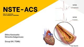
- 1. NSTE-ACS NON-ST-ELEVATION ACUTE CORONARY SYNDROMES Dilina Aarewatte Himesha Galgamuwa Group 9A | TSMU
- 2. What are NSTE-ACS? Non ST-elevation acute coronary syndrome is characterised by episodes of chest pain at rest or with minimal exertion, which increase in frequency or severity, often with dynamic ECG changes. NSTEMI Unstable Angina
- 4. Pathophysiology ● Most commonly caused by an imbalance between oxygen supply and oxygen demand resulting from a partially occluding thrombus ● This thrombus could be formed on a disrupted atherothrombotic coronary plaque or on an eroded coronary artery endothelium. ● The reduction of the blood flow caused by the thrombus (and by embolization / debris) can cause severe ischemia or myocardial necrosis.
- 5. ● Dynamic obstruction (e.g., coronary spasm,as in Prinzmetal’s variant angina) ● Severe mechanical obstruction due to progressive coronary atherosclerosis ● Increased myocardial oxygen demand produced by conditions such as fever, tachycardia, and thyrotoxicosis in the presence of fixed epicardial coronary obstruction. Other causes:
- 6. Diagnosis ● Diagnosis is based largely on the clinical presentation ● Typically, chest discomfort is severe and has at least one of three features: ○ it occurs at rest (or with minimal exertion), lasting >10 minutes; ○ it is of relatively recent onset (i.e., within the prior 2 weeks); and/or ○ it occurs with a crescendo pattern (i.e., distinctly more severe, prolonged, or frequent than previous episodes). ● NSTEMI diagnosis is established with evidence of abnormally elevated cardiac biomarker levels.
- 7. History and PE ● The chest discomfort, often severe, is typically located in the substernal region or sometimes in the epigastrium, and radiates to the left arm, left shoulder, and/or neck. ● Anginal “equivalents” such as dyspnea, epigastric discomfort, nausea, or weakness may occur instead of chest pain and appear to be more frequent in women, the elderly, and patients with diabetes mellitus. ● The physical examination resembles that in patients with stable angina and may be unremarkable. ● If the patient has a large area of myocardial ischemia or a large NSTEMI, the physical findings can include ○ diaphoresis; ○ pale, cool skin; ○ sinus tachycardia; ○ a third and/or fourth heart sound; ○ basilar rales; ○ Hypotension (sometimes).
- 8. Electrocardiogram ● ST-segment depression occurs in 20 to 25% of patients; it may be transient in patients without biomarker evidence of myocardial necrosis, but may be persistent for several days in NSTEMI. ● T-wave changes are common but are less specific signs of ischemia, unless they are new and deep T-wave inversions (≥0.3 mV).
- 10. Cardiac Biomarkers ● NSTEMI patients have elevated biomarkers of necrosis, such as cardiac troponin I or T, which are specific, sensitive, and the preferred markers of myocardial necrosis. ● The MB isoform of creatine kinase (CK-MB) is a less sensitive alternative. Elevated levels of these markers distinguish patients with NSTEMI from those with UA. ● There is a characteristic temporal rise and fall of the plasma concentration of these markers and a direct relationship between the degree of elevation and mortality.
- 12. Risk Stratification ● The diagnosis of a non-ST elevation ACS (NSTE-ACS) in the emergency department should be followed by risk stratification and treatment. ● Clinical guidelines recommend an early invasive strategy in higher risk NSTE-ACS. ● The Global Registry of Acute Coronary Events (GRACE) risk score is a validated risk stratification tool which has incremental prognostic value for risk stratification compared with clinical assessment or troponin testing alone.
- 13. Treatment and Complications Medical Treatment ● Patients should be placed at bed rest with continuous ECG monitoring for ST-segment deviation and cardiac arrhythmias. ● Ambulation is permitted if the patient shows no recurrence of ischemia (symptoms or ECG changes) and does not develop an elevation of a biomarker of necrosis for 12–24 h. ● Medical therapy involves simultaneous Anti-ischemic and Antithrombotic treatments and consideration of coronary revascularization
- 14. ANTI-ISCHEMIC TREATMENT To provide relief and prevention of recurrence of chest pain, initial treatment should include bed rest, nitrates, beta adrenergic blockers, and inhaled oxygen in the presence of hypoxemia 1. Nitrates i. Sublingual or buccal spray - if the patient is experiencing ischemic pain ii. Intravenous Nitroglycerin - If pain persists after three doses given 5 min apart a. The rate of the infusion may be increased by 10 μg/min every 3–5 min until symptoms are relieved, systolic arterial pressure falls to <100 mmHg, or the dose reaches 200 μg/min. iii. Topical or Oral Nitrates - can be used when the pain has resolved, or they may replace intravenous nitroglycerin when the patient has been pain free for 12–24h. ● Contraindications - The only absolute contraindications to the use of nitrates are hypotension or the use of sildenafil or other phosphodiesterase-5 inhibitors within the previous 24–48 h
- 15. 2. Beta Blockers ● Started by the intravenous route in patients with severe ischemia, but this is contraindicated in the presence of heart failure ● Oral beta blockade targeted to a heart rate of 50–60 beats/min is recommended. 3. Calcium Channel Blockers ● Heart rate–slowing calcium channel blockers, e.g. verapamil or diltiazem, are recommended for patients who have persistent symptoms or ECG signs of ischemia after treatment with full-dose nitrates and beta blockers and in patients with contraindications to either class of these agents ❖ Additional medical therapy includes angiotensin-converting enzyme (ACE) inhibitors or, if these are not tolerated, angiotensin receptor blockers. ❖ Early administration of intensive HMG-CoA reductase inhibitors (statins), such as atorvastatin 80 mg/d, prior to percutaneous coronary intervention (PCI), and continued thereafter, has been shown to reduce complications of the procedure and recurrences of ACS.
- 16. ANTITHROMBOTIC THERAPY 1. Antiplatelet Drugs Initial treatment should begin with the platelet cyclooxygenase inhibitor Aspirin. ● Contraindications are active bleeding or aspirin intolerance. Dual Antiplatelet Therapy (Asprin + Platelet P2Y12 Receptor Blocker) ● In the absence of a high risk for bleeding, patients with NSTE-ACS should receive a platelet P2Y12 receptor blocker to inhibit platelet activation. The Clopidogrel is an inactive prodrug that is converted into an active metabolite that causes irreversible blockade of the platelet P2Y12 receptor. When added to aspirin it has been shown to confer a 20% reduction in cardiovascular death, MI, or stroke, compared to aspirin alone, but to be associated with a moderate increase in major bleeding Triple Antiplatelet Therapy (Asprin + Platelet P2Y12 Receptor Blocker + Reversible platelet P2Y12 inhibitor)
- 17. 2. Anticoagulants (1) Unfractionated Heparin (UFH), long the mainstay of therapy (2) Low-Molecular-Weight Heparin (LMWH), enoxaparin, which has been shown to be superior to UFH in reducing recurrent cardiac events (3) Bivalirudin, a direct thrombin inhibitor that is similar in efficacy to either UFH or LMWH but causes less bleeding and is used just prior to and/or during PCI (4) Indirect factor Xa inhibitor, fondaparinux, which is equivalent in efficacy to enoxaparin but appears to have a lower risk of major bleeding
- 18. LONG-TERM MANAGEMENT ● Risk-factor modification ○ the caregiver should discuss with the patient the importance of smoking cessation, achieving optimal weight, daily exercise, blood-pressure control, following an appropriate diet, control of hyperglycemia (in diabetic patients), and lipid management ● long-term therapy with five classes of drugs are directed ○ Beta blockers, statins and ACE inhibitors or angiotensin receptor blockers are recommended for long-term plaque stabilization.
- 19. PRINZMETAL’S VARIANT ANGINA A syndrome of severe ischemic pain that usually occurs at rest and is associated with transient ST-segment elevation. PVA is caused by focal spasm of an epicardial coronary artery, leading to severe transient myocardial ischemia and occasionally infarction
- 20. ● Patients with PVA are generally younger and have fewer coronary risk factors (with the exception of cigarette smoking) than do patients with NSTE-ACS. ● Cardiac examination is usually unremarkable in the absence of ischemia. ● The clinical diagnosis of PVA is made by; ■ The detection of transient ST-segment elevation with rest pain. ■ Multiple episodes of asymptomatic ST-segment elevation (silent ischemia). ■ Small elevations of troponin ● Coronary angiography demonstrates transient coronary spasm as the diagnostic hallmark of PVA. ● Atherosclerotic plaques in at least one proximal coronary artery, and in these patients, spasm usually occurs within 1 cm of the plaque. ❖ Hyperventilation or intracoronary acetylcholine has been used to provoke focal coronary stenosis on angiography or to provoke rest angina with ST-segment elevation to establish the diagnosis Clinical and Angiographic Manifestations
- 21. TREATMENT ● Nitrates and calcium channel blockers are the main therapeutic agents. ● Aspirin may actually increase the severity of ischemic episodes, possibly as a result of the sensitivity of coronary tone to modest changes in the synthesis of prostacyclin. ● The response to beta blockers is variable. Coronary revascularization may be helpful in patients who also have discrete, flow-limiting, proximal fixed obstructive lesions.
- 22. ● Many patients with PVA pass through an acute, active phase, with frequent episodes of angina and cardiac events during the first 6 months after presentation. ● Patients with no or mild fixed coronary obstruction tend to experience a more benign course than do patients with associated severe obstructive lesions. ● Nonfatal MI occurs in up to 20% of patients by 5 years. ● Patients with PVA who develop serious arrhythmias during spontaneous episodes of pain are at a higher risk for sudden cardiac death. ● In most patients who survive an infarction or the initial 3- to 6-month period of frequent episodes, there is a tendency for symptoms and cardiac events to diminish over time. PROGNOSIS
- 23. References 1. Sarkees, M. L., & Bavry, A. A. (2010, November 15). Non St-elevation acute coronary syndrome. BMJ clinical evidence. Retrieved July 8, 2022, from https://www.ncbi.nlm.nih.gov/pmc/articles/PMC3217799/#:~:text=Introduction,often%20with%20dynamic%20ECG%20changes. 2. Harrison, T. R., & Kasper, D. L. (2015). Harrison's principles of Internal Medicine. McGraw-Hill Medical Publ. Division. 3. Kumar, P. J., & Clark, M. L. (2002). Kumar & Clark clinical medicine. Edinburgh: Saunders. 4. Corcoran, D., Grant, P., & Berry, C. (2015, September 1). Risk stratification in non-st elevation acute coronary syndromes: Risk scores, biomarkers and clinical judgment. International journal of cardiology. Heart & vasculature. Retrieved July 8, 2022, from https://www.ncbi.nlm.nih.gov/pmc/articles/PMC4691930/#:~:text=The%20optimal%20risk%20stratification%20of,ris k%20patients%20for%20invasive%20management. 5. Amsterdam EA, Wenger NK, Brindis RG, et al. 2014 AHA/ACC Guideline for the Management of Patients With Non-ST-Elevation Acute Coronary Syndromes: A Report of the American College of Cardiology/American Heart Association Task Force on Practice Guidelines. Circulation. 2014 Dec 23;130(25):e344-426full-text or in J Am Coll Cardiol. 2014 Dec 23;64(24):e139
- 24. Thank You!