
Empfohlen
Weitere ähnliche Inhalte
Kürzlich hochgeladen
Kürzlich hochgeladen (20)
Empfohlen
Empfohlen (20)
Venous Access Device Class Exercise
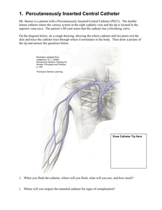
- 1. 1. Percutaneously Inserted Central Catheter Mr. Barnes is a patient with a Percutaneously Inserted Central Catheter (PICC). The double lumen catheter enters the venous system in the right cephalic vein and the tip is located in the superior vena cava. The patient’s ID card states that the catheter has a Groshong valve. On the diagram below, do a rough drawing, showing the where catheter and two ports exit the skin and trace the catheter tract through where it terminates in the body. Then draw a picture of the tip and answer the questions below. Illustration adapted from Josephson, D. L. (2004). Intravenous Infusion Therapy for Nurses: Principles and Practice. p. 159 Thompson Delmar Learning Draw Catheter Tip Here 1. When you flush the catheter, where will you flush, what will you use, and how much? Where will you inspect the tunneled catheter for signs of complication? 2.
- 2. 2. Implanted Vascular Access Device (Port-A-Cath) Mrs. Anderson is a patient with an Implanted Vascular Access Device (IVAD). The port pocket is in the right chest over the middle of the second rib. The IVAD catheter enters the venous system via the right internal jugular vein and the tip is located at the superior vena cava (SVC) – right atrial junction. The patient’s ID card states that the catheter tip has a Groshong valve. On the diagram below do a rough drawing that shows the location of the port and trace the catheter tract through where it terminates in the body. Then draw a picture of the tip and answer the questions below. Illustration adapted from Josephson, D. L. (2004). Intravenous Infusion Therapy for Nurses: Principles and Practice. p. 159 Thompson Delmar Learning Draw CatheterTip Here 1. When you flush the catheter, what will you use and how much? 2. Where will you inspect the IVAD for signs of complication?
- 3. 3. Subclavian Hickman Catheter Mrs. Jones is a patient with a right subclavian Hickman catheter. The dual lumen open-end catheter terminates in the superior vena cava – right atrial junction. On the diagram below, do a rough drawing, showing the where catheter and two ports exit the skin, and trace the catheter tract through where it terminates in the body. Then draw a picture of the tip and answer the questions below. Illustration adapted from Josephson, D. L. (2004). Intravenous Infusion Therapy for Nurses: Principles and Practice. p. 159 Thompson Delmar Learning Draw Catheter Tip Here 1. When you flush the catheter, where will you flush, what will you use, and how much? Where will you inspect the catheter for signs of complication? 2.
- 4. 4. Tunneled Dialysis Catheter Mr. Green is a patient with a tunneled dialysis catheter. The double lumen catheter exits the skin in the right chest above the third rib and enters the venous system via the right subclavian vein. The tip terminates in the superior vena cava. On the diagram below, do a rough drawing showing where the catheter and two ports exit the skin, and trace the catheter tract through where it terminates in the body. Then draw a picture of the tip and answer the questions below. Illustration adapted from Josephson, D. L. (2004). Intravenous Infusion Therapy for Nurses: Principles and Practice. p. 159 Thompson Delmar Learning Draw Catheter Tip Here 1. When you flush the catheter, where will you flush, what will you use, and how much? Where will you inspect the tunneled catheter for signs of complication? 2.
- 5. 5. Tunneled Catheter Mr. Johnson is a patient with a tunneled catheter. The double lumen catheter ports exit the skin in the right chest wall. Then the catheter enters the venous system in the right external jugular vein and the tip terminates at the superior vena cava – right atrial junction. The patient’s ID card states that the catheter has a Groshong tip. On the diagram below, do a rough drawing, showing where the catheter and two ports exit the skin, and trace the catheter tract through where it terminates in the body. Then draw a picture of the tip and answer the questions below. Illustration adapted from Josephson, D. L. (2004). Intravenous Infusion Therapy for Nurses: Principles and Practice. p. 159 Thompson Delmar Learning Draw Catheter Tip Here 1. When you flush the catheter, where will you flush, what will you use, and how much? Where will you inspect the tunneled catheter for signs of complication? 2.
- 6. 1. Percutaneously Inserted Central Catheter 1. 5 ml. NS flush to each lumen. 2. Inspect starting where the catheter exits the right basilic vein. Follow the catheter up the length of the arm. Look for signs of swelling, erythema, induration, pain, tenderness, and fluid leakage. Be alert for signs of tip migration forward (into right atrium) or backward (into subclavian vasculature). 2. Implanted Vascular Access Device (Port-A-Cath) 1. 5 ml. NS flush to each lumen. 2. Inspect starting where the port lies in the right chest. Follow the tract to the right internal jugular vein. Look for signs of swelling, erythema, induration, pain, tenderness, and fluid leakage. Be alert for signs of tip migration forward (into right atrium) or backward (into subclavian vasculature). 3. Subclavian Hickman Catheter 1. 3 ml. Heparin 100u/ml.flush to each lumen. 2. The venous entry and skin insertion site are very close to one another. Inspect around the skin entry site for signs of swelling, erythema, induration, pain, tenderness, and fluid leakage. 4. Tunneled Dialysis Catheter 1. Permission to use catheter, heparin concentration, flush amount, and frequency to be prescribed by MD. Heparin concentration usually 1,000u/ml. to 10,000u/ml. 2. Inspect starting at the skin exit site above the third rib. Follow the tract to the subclavian vein. Look for signs of swelling, erythema, induration, pain, tenderness, and fluid leakage. Be alert for signs of tip migration forward (into right atrium) or backward (into subclavian vasculature). 5. Tunneled Catheter 1. 5 ml. NS flush to each lumen. 2. Inspect starting at the skin in the right chest wall. Follow the tract to the right external jugular vein. Look for signs of swelling, erythema, induration, pain, tenderness, and fluid leakage. Be alert for signs of tip migration forward (into right atrium) or backward (into subclavian vasculature).