SlideShare ist ein Scribd-Unternehmen logo
1 von 21
What Is an Aneurysm?
An aneurysm (AN-u-rism) is a balloon-like bulge in an artery. Arteries are blood vessels that
carry oxygen-rich blood to your body.

Arteries have thick walls to withstand normal blood pressure. However, certain medical
problems, genetic conditions, and trauma can damage or injure artery walls. The force of
blood pushing against the weakened or injured walls can cause an aneurysm.

An aneurysm can grow large and rupture (burst) or dissect. A rupture causes dangerous
bleeding inside the body. A dissection is a split in one or more layers of the artery wall. The
split causes bleeding into and along the layers of the artery wall.

Both rupture and dissection often are fatal.

Overview
Most aneurysms occur in the aorta, the main artery that carries oxygen-rich blood from the
heart to the body. The aorta goes through the chest and abdomen.

An aneurysm that occurs in the chest portion of the aorta is called a thoracic (tho-RAS-ik)
aortic aneurysm. An aneurysm that occurs in the abdominal portion of the aorta is called an
abdominal aortic aneurysm.

Aneurysms also can occur in other arteries, but these types of aneurysm are less common.
This article focuses on aortic aneurysms.

About 13,000 Americans die each year from aortic aneurysms. Most of the deaths result from
rupture or dissection.

Early diagnosis and treatment can help prevent rupture and dissection. However, aneurysms
can develop and grow large before causing any symptoms. Thus, people who are at high risk
for aneurysms can benefit from early, routine screening.

Outlook
Doctors often can successfully treat aortic aneurysms with medicines or surgery if they’re
found in time. Medicines may be given to lower blood pressure, relax blood vessels, and
reduce the risk of rupture.

Large aortic aneurysms often can be repaired with surgery. During surgery, the weak or
damaged portion of the aorta is replaced or reinforced.


Types of Aneurysms
Aortic Aneurysms
The two types of aortic aneurysm are abdominal aortic aneurysm and thoracic aortic
aneurysm. Some people have both types.

                                     Aortic Aneurysms


Figure A shows a normal aorta. Figure B shows a thoracic aortic aneurysm, which is located
behind the heart. Figure C shows an abdominal aortic aneurysm, which is located below the
arteries that supply blood to the kidneys.

Abdominal Aortic Aneurysms

An aneurysm that occurs in the abdominal portion of the aorta is called an abdominal aortic
aneurysm (AAA). Most aortic aneurysms are AAAs.

These aneurysms are found more often now than in the past because of computed
tomography (to-MOG-rah-fee) scans, or CT scans , done for other medical problems.

Small AAAs rarely rupture. However, AAAs can grow very large without causing symptoms.
Routine checkups and treatment for an AAA can help prevent growth and rupture.
Thoracic Aortic Aneurysms

An aneurysm that occurs in the chest portion of the aorta (above the diaphragm, a muscle that
helps you breathe) is called a thoracic aortic aneurysm (TAA).

TAAs don't always cause symptoms, even when they're large. Only half of all people who
have TAAs notice any symptoms. TAAs are found more often now than in the past because
of chest CT scans done for other medical problems.

With a common type of TAA, the walls of the aorta weaken and a section close to the heart
enlarges. As a result, the valve between the heart and the aorta can't close properly. This
allows blood to leak back into the heart.

A less common type of TAA can develop in the upper back, away from the heart. A TAA in
this location may result from an injury to the chest, such as from a car crash.

Other Types of Aneurysms
Brain Aneurysms

Aneurysms in the arteries of the brain are called cerebral (seh-RE-bral)
aneurysms or brain aneurysms. Brain aneurysms also are called berry aneurysms because
they're often the size of a small berry.

                                       Brain Aneurysm


The illustration shows a typical location of a brain aneurysm in the arteries that supply blood
to the brain. The inset image shows a closeup view of the sac-like aneurysm.
Most brain aneurysms cause no symptoms until they become large, begin to leak blood, or
rupture (burst). A ruptured brain aneurysm can cause a stroke.

Peripheral Aneurysms

Aneurysms that occur in arteries other than the aorta and the brain arteries are called
peripheral (peh-RIF-eh-ral) aneurysms. Common locations for peripheral aneurysms include
the popliteal (pop-li-TE-al), femoral (FEM-o-ral), and carotid (ka-ROT-id) arteries.

The popliteal arteries run down the back of the thighs, behind the knees. The femoral arteries
are the main arteries in the groin. The carotid arteries are the two main arteries on each side
of your neck.

Peripheral aneurysms aren’t as likely to rupture or dissect as aortic aneurysms. However,
blood clots can form in peripheral aneurysms. If a blood clot breaks away from the aneurysm,
it can block blood flow through the artery.

If a peripheral aneurysm is large, it can press on a nearby nerve or vein and cause


Other Names for Aneurysm
       Abdominal aortic aneurysm
       Aortic aneurysm
       Berry aneurysm
       Brain aneurysm
       Cerebral aneurysm
       Peripheral aneurysm
       Thoracic aortic aneurysm


What Causes an Aneurysm?
The force of blood pushing against the walls of an artery combined with damage or injury to
the artery’s walls can cause an aneurysm.

Many conditions and factors can damage and weaken the walls of the aorta and cause aortic
aneurysms. Examples include aging, smoking, high blood pressure, and atherosclerosis (ath-
er-o-skler-O-sis). Atherosclerosis is the hardening and narrowing of the arteries due to the
buildup of a waxy substance called plaque (plak).

Rarely, infections—such as untreated syphilis (a sexually transmitted infection)—can cause
aortic aneurysms. Aortic aneurysms also can occur as a result of diseases that inflame the
blood vessels, such as vasculitis (vas-kyu-LI-tis).

A family history of aneurysms also may play a role in causing aortic aneurysms.

In addition to the factors above, certain genetic conditions may cause thoracic aortic
aneurysms (TAAs). Examples of these conditions include Marfan syndrome, Loeys-Dietz
syndrome, Ehlers-Danlos syndrome (the vascular type), and Turner syndrome.
These genetic conditions can weaken the body’s connective tissues and damage the aorta.
People who have these conditions tend to develop aneurysms at a younger age than other
people. They’re also at higher risk for rupture and dissection.

Trauma, such as a car accident, also can damage the walls of the aorta and lead to TAAs.

Researchers continue to look for other causes of aortic aneurysms. For example, they’re
looking for genetic mutations (changes in the genes) that may contribute to or cause
aneurysms.


Who Is at Risk for an Aneurysm?
Certain factors put you at higher risk for an aortic aneurysm. These factors include:

       Male gender. Men are more likely than women to have aortic aneurysms.
       Age. The risk for abdominal aortic aneurysms increases as you get older. These
       aneurysms are more likely to occur in people who are aged 65 or older.
       Smoking. Smoking can damage and weaken the walls of the aorta.
       A family history of aortic aneurysms. People who have family histories of aortic
       aneurysms are at higher risk for the condition, and they may have aneurysms before
       the age of 65.
       A history of aneurysms in the arteries of the legs.
       Certain diseases and conditions that weaken the walls of the aorta. Examples include
       high blood pressure and atherosclerosis.

Having a bicuspid aortic valve can raise the risk of having a thoracic aortic aneurysm. A
bicuspid aortic valve has two leaflets instead of the typical three.

Car accidents or trauma also can injure the arteries and increase the risk for aneurysms.

If you have any of these risk factors, talk with your doctor about whether you need screening
for aneurysms.


What Are the Signs and Symptoms of an
Aneurysm?
The signs and symptoms of an aortic aneurysm depend on the type and location of the
aneurysm. Signs and symptoms also depend on whether the aneurysm has ruptured (burst) or
is affecting other parts of the body.

Aneurysms can develop and grow for years without causing any signs or symptoms. They
often don't cause signs or symptoms until they rupture, grow large enough to press on nearby
body parts, or block blood flow.

Abdominal Aortic Aneurysms
Most abdominal aortic aneurysms (AAAs) develop slowly over years. They often don't cause
signs or symptoms unless they rupture. If you have an AAA, your doctor may feel a
throbbing mass while checking your abdomen.

When symptoms are present, they can include:

       A throbbing feeling in the abdomen
       Deep pain in your back or the side of your abdomen
       Steady, gnawing pain in your abdomen that lasts for hours or days

If an AAA ruptures, symptoms may include sudden, severe pain in your lower abdomen and
back; nausea (feeling sick to your stomach) and vomiting; constipation and problems with
urination; clammy, sweaty skin; light-headedness; and a rapid heart rate when standing up.

Internal bleeding from a ruptured AAA can send you into shock . Shock is a life-
threatening condition in which blood pressure drops so low that the brain, kidneys, and other
vital organs can't get enough blood to work well. Shock can be fatal if it’s not treated right
away.

Thoracic Aortic Aneurysms
A thoracic aortic aneurysm (TAA) may not cause symptoms until it dissects or grows large. If
you have symptoms, they may include:

       Pain in your jaw, neck, back, or chest
       Coughing and/or hoarseness
       Shortness of breath and/or trouble breathing or swallowing

A dissection is a split in one or more layers of the artery wall. The split causes bleeding into
and along the layers of the artery wall.

If a TAA ruptures or dissects, you may feel sudden, severe, sharp or stabbing pain starting in
your upper back and moving down into your abdomen. You may have pain in your chest and
arms, and you can quickly go into shock.

If you have any symptoms of TAA or aortic dissection, call 9–1–1. If left untreated, these
conditions may lead to organ damage or death.


How Is an Aneurysm Diagnosed?
If you have an aortic aneurysm but no symptoms, your doctor may find it by chance during a
routine physical exam. More often, doctors find aneurysms during tests done for other
reasons, such as chest or abdominal pain.

If you have an abdominal aortic aneurysm (AAA), your doctor may feel a throbbing mass in
your abdomen. A rapidly growing aneurysm about to rupture (burst) can be tender and very
painful when pressed. If you're overweight or obese, it may be hard for your doctor to feel
even a large AAA.
If you have an AAA, your doctor may hear rushing blood flow instead of the normal
whooshing sound when listening to your abdomen with a stethoscope.

Specialists Involved
Your primary care doctor may refer you to a cardiothoracic or vascular surgeon for diagnosis
and treatment of an aortic aneurysm.

A cardiothoracic surgeon does surgery on the heart, lungs, and other organs and structures in
the chest, including the aorta. A vascular surgeon does surgery on the aorta and other blood
vessels, except those of the heart and brain.

Diagnostic Tests and Procedures
To diagnose and study an aneurysm, your doctor may recommend one or more of the
following tests.

Ultrasound and Echocardiography

Ultrasound and echocardiography (echo) are simple, painless tests that use sound waves to
create pictures of the structures inside your body. These tests can show the size of an aortic
aneurysm, if one is found.

Computed Tomography Scan

A computed tomography scan, or CT scan        , is a painless test that uses x rays to take clear,
detailed pictures of your organs.

During the test, your doctor will inject dye into a vein in your arm. The dye makes your
arteries, including your aorta, visible on the CT scan pictures.

Your doctor may recommend this test if he or she thinks you have an AAA or a thoracic
aortic aneurysm (TAA). A CT scan can show the size and shape of an aneurysm. This test
provides more detailed pictures than an ultrasound or echo.

Magnetic Resonance Imaging

Magnetic resonance imaging (MRI) uses magnets and radio waves to create pictures of the
organs and structures in your body. This test works well for detecting aneurysms and
pinpointing their size and exact location.

Angiography

Angiography (an-jee-OG-ra-fee) is a test that uses dye and special x rays to show the insides
of your arteries. This test shows the amount of damage and blockage in blood vessels.

Aortic angiography shows the inside of your aorta. The test may show the location and size
of an aortic aneurysm.
How Is an Aneurysm Treated?
Aortic aneurysms are treated with medicines and surgery. Small aneurysms that are found
early and aren’t causing symptoms may not need treatment. Other aneurysms need to be
treated.

The goals of treatment may include:

       Preventing the aneurysm from growing
       Preventing or reversing damage to other body structures
       Preventing or treating a rupture or dissection
       Allowing you to continue doing your normal daily activities

Treatment for an aortic aneurysm is based on its size. Your doctor may recommend routine
testing to make sure an aneurysm isn't getting bigger. This method usually is used for
aneurysms that are smaller than 5 centimeters (about 2 inches) across.

How often you need testing (for example, every few months or every year) is based on the
size of the aneurysm and how fast it's growing. The larger it is and the faster it's growing, the
more often you may need to be checked.

Medicines
If you have an aortic aneurysm, your doctor may prescribe medicines before surgery or
instead of surgery. Medicines are used to lower blood pressure, relax blood vessels, and
lower the risk that the aneurysm will rupture (burst). Beta blockers and calcium channel
blockers are the medicines most commonly used.

Surgery
Your doctor may recommend surgery if your aneurysm is growing quickly or is at risk of
rupture or dissection.

The two main types of surgery to repair aortic aneurysms are open abdominal or open chest
repair and endovascular repair.

Open Abdominal or Open Chest Repair

The standard and most common type of surgery for aortic aneurysms is open abdominal or
open chest repair. This surgery involves a major incision (cut) in the abdomen or chest.

General anesthesia (AN-es-THE-ze-ah) is used during this procedure. The term ―anesthesia‖
refers to a loss of feeling and awareness. General anesthesia temporarily puts you to sleep.

During the surgery, the aneurysm is removed. Then, the section of aorta is replaced with a
graft made of material such as Dacron® or Teflon.® The surgery takes 3 to 6 hours; you’ll
remain in the hospital for 5 to 8 days.
If needed, repair of the aortic heart valve also may be done during open abdominal or open
chest surgery.

It often takes a month to recover from open abdominal or open chest surgery and return to
full activity. Most patients make a full recovery.

Endovascular Repair

In endovascular repair, the aneurysm isn't removed. Instead, a graft is inserted into the aorta
to strengthen it. Surgeons do this type of surgery using catheters (tubes) inserted into the
arteries; it doesn't require surgically opening the chest or abdomen. General anesthesia is
used during this procedure.

The surgeon first inserts a catheter into an artery in the groin (upper thigh) and threads it to
the aneurysm. Then, using an x ray to see the artery, the surgeon threads the graft (also called
a stent graft) into the aorta to the aneurysm.

The graft is then expanded inside the aorta and fastened in place to form a stable channel for
blood flow. The graft reinforces the weakened section of the aorta. This helps prevent the
aneurysm from rupturing.

                                     Endovascular Repair


The illustration shows the placement of a stent graft in an aortic aneurysm. In figure A, a
catheter is inserted into an artery in the groin (upper thigh). The catheter is threaded to the
abdominal aorta, and the stent graft is released from the catheter. In figure B, the stent graft
allows blood to flow through the aneurysm.
The recovery time for endovascular repair is less than the recovery time for open abdominal
or open chest repair. However, doctors can’t repair all aortic aneurysms with endovascular
repair. The location or size of an aneurysm may prevent the use of a stent graft.


How Can an Aneurysm Be Prevented?
The best way to prevent an aortic aneurysm is to avoid the factors that put you at higher risk
for one. You can’t control all aortic aneurysm risk factors, but lifestyle changes can help you
lower some risks.

For example, if you smoke, try to quit. Talk with your doctor about programs and products
that can help you quit smoking. Also, try to avoid secondhand smoke. For more information
about how to quit smoking, go to the Health Topics Smoking and Your Heart article.

Another important lifestyle change is following a healthy diet. A healthy diet includes a
variety of fruits, vegetables, and whole grains. It also includes lean meats, poultry, fish,
beans, and fat-free or low-fat milk or milk products. A healthy diet is low in saturated fat,
trans fat, cholesterol, sodium (salt), and added sugar.

For more information about following a healthy diet, go to the National Heart, Lung, and
Blood Institute’s (NHLBI’s) Aim for a Healthy Weight Web site, "Your Guide to a Healthy
Heart," and "Your Guide to Lowering Your Blood Pressure With DASH." All of these
resources include general information about healthy eating.

Be as physically active as you can. Talk with your doctor about the amounts and types of
physical activity that are safe for you. For more information about physical activity, go to the
Health Topics Physical Activity and Your Heart article and the NHLBI’s "Your Guide to
Physical Activity and Your Heart."

Work with your doctor to control medical conditions such as high blood pressure and high
blood cholesterol. Follow your treatment plans and take all of your medicines as your doctor
prescribes.

Screening for Aneurysms
Although you may not be able to prevent an aneurysm, early diagnosis and treatment can help
prevent rupture and dissection.

Aneurysms can develop and grow large before causing any signs or symptoms. Thus, people
who are at high risk for aneurysms may benefit from early, routine screening.

Your doctor may recommend routine screening if you’re:

       A man between the ages of 65 and 75 who has ever smoked
       A man or woman between the ages of 65 and 75 who has a family history of
       aneurysms
If you’re at risk, but not in one of these high-risk groups, ask your doctor whether screening
will benefit you.


What Is Atherosclerosis?
Atherosclerosis (ath-er-o-skler-O-sis) is a disease in which plaque (plak) builds up inside
your arteries. Arteries are blood vessels that carry oxygen-rich blood to your heart and other
parts of your body.

Plaque is made up of fat, cholesterol, calcium, and other substances found in the blood. Over
time, plaque hardens and narrows your arteries. This limits the flow of oxygen-rich blood to
your organs and other parts of your body.

Atherosclerosis can lead to serious problems, including heart attack, stroke, or even death.

                                       Atherosclerosis


Figure A shows a normal artery with normal blood flow. Figure B shows an artery with
plaque buildup.

Atherosclerosis-Related Diseases
Atherosclerosis can affect any artery in the body, including arteries in the heart, brain, arms,
legs, pelvis, and kidneys. As a result, different diseases may develop based on which arteries
are affected.

Coronary Heart Disease

Coronary heart disease (CHD), also called coronary artery disease, is the #1 killer of both
men and women in the United States. CHD occurs if plaque builds up in the coronary
arteries. These arteries supply oxygen-rich blood to your heart.

Plaque narrows the coronary arteries and reduces blood flow to your heart muscle. Plaque
buildup also makes it more likely that blood clots will form in your arteries. Blood clots can
partially or completely block blood flow.

If blood flow to your heart muscle is reduced or blocked, you may have angina (chest pain or
discomfort) or a heart attack.

Plaque also can form in the heart's smallest arteries. This disease is called coronary
microvascular disease (MVD). In coronary MVD, plaque doesn't cause blockages in the
arteries as it does in CHD.

Carotid Artery Disease

Carotid (ka-ROT-id) artery disease occurs if plaque builds up in the arteries on each side of
your neck (the carotid arteries). These arteries supply oxygen-rich blood to your brain. If
blood flow to your brain is reduced or blocked, you may have a stroke.

Peripheral Arterial Disease

Peripheral arterial disease (P.A.D.) occurs if plaque builds up in the major arteries that supply
oxygen-rich blood to your legs, arms, and pelvis.

If blood flow to these parts of your body is reduced or blocked, you may have numbness,
pain, and, sometimes, dangerous infections.

Chronic Kidney Disease

Chronic kidney disease can occur if plaque builds up in the renal arteries. These arteries
supply oxygen-rich blood to your kidneys.

Over time, chronic kidney disease causes a slow loss of kidney function. The main function
of the kidneys is to remove waste and extra water from the body.

Overview
The cause of atherosclerosis isn't known. However, certain traits, conditions, or habits may
raise your risk for the disease. These conditions are known as risk factors.

You can control some risk factors, such as lack of physical activity, smoking, and an
unhealthy diet. Others you can't control, such as age and a family history of heart disease.
Some people who have atherosclerosis have no signs or symptoms. They may not be
diagnosed until after a heart attack or stroke.

The main treatment for atherosclerosis is lifestyle changes. You also may need medicines and
medical procedures. These treatments, along with ongoing medical care, can help you live a
healthier life.

Outlook
Improved treatments have reduced the number of deaths from atherosclerosis-related
diseases. These treatments also have improved the quality of life for people who have these
diseases. However, atherosclerosis remains a common health problem.

You may be able to prevent or delay atherosclerosis and the diseases it can cause. Making
lifestyle changes and getting ongoing care can help you avoid the problems of atherosclerosis
and live a long, healthy life.


What Causes Atherosclerosis?
The exact cause of atherosclerosis isn't known. However, studies show that atherosclerosis is
a slow, complex disease that may start in childhood. It develops faster as you age.

Atherosclerosis may start when certain factors damage the inner layers of the arteries. These
factors include:

       Smoking
       High amounts of certain fats and cholesterol in the blood
       High blood pressure
       High amounts of sugar in the blood due to insulin resistance    or diabetes

Plaque may begin to build up where the arteries are damaged. Over time, plaque hardens and
narrows the arteries. Eventually, an area of plaque can rupture (break open).

When this happens, blood cell fragments called platelets (PLATE-lets) stick to the site of the
injury. They may clump together to form blood clots. Clots narrow the arteries even more,
limiting the flow of oxygen-rich blood to your body.

Depending on which arteries are affected, blood clots can worsen angina (chest pain) or cause
a heart attack or stroke.

Researchers continue to look for the causes of atherosclerosis. They hope to find answers to
questions such as:

       Why and how do the arteries become damaged?
       How does plaque develop and change over time?
       Why does plaque rupture and lead to blood clots?
What Are the Signs and Symptoms of
Atherosclerosis?
Atherosclerosis usually doesn't cause signs and symptoms until it severely narrows or totally
blocks an artery. Many people don't know they have the disease until they have a medical
emergency, such as a heart attack or stroke.

Some people may have signs and symptoms of the disease. Signs and symptoms will depend
on which arteries are affected.

Coronary Arteries
The coronary arteries supply oxygen-rich blood to your heart. If plaque narrows or blocks
these arteries (a disease called coronary heart disease, or CHD), a common symptom is
angina. Angina is chest pain or discomfort that occurs when your heart muscle doesn't get
enough oxygen-rich blood.

Angina may feel like pressure or squeezing in your chest. You also may feel it in your
shoulders, arms, neck, jaw, or back. Angina pain may even feel like indigestion. The pain
tends to get worse with activity and go away with rest. Emotional stress also can trigger the
pain.

Other symptoms of CHD are shortness of breath and arrhythmias (ah-RITH-me-ahs).
Arrhythmias are problems with the rate or rhythm of the heartbeat.

Plaque also can form in the heart's smallest arteries. This disease is called coronary
microvascular disease (MVD). Symptoms of coronary MVD include angina, shortness of
breath, sleep problems, fatigue (tiredness), and lack of energy.

Carotid Arteries
The carotid arteries supply oxygen-rich blood to your brain. If plaque narrows or blocks these
arteries (a disease called carotid artery disease), you may have symptoms of a stroke. These
symptoms may include:

       Sudden weakness
       Paralysis (an inability to move) or numbness of the face, arms, or legs, especially on
       one side of the body
       Confusion
       Trouble speaking or understanding speech
       Trouble seeing in one or both eyes
       Problems breathing
       Dizziness, trouble walking, loss of balance or coordination, and unexplained falls
       Loss of consciousness
       Sudden and severe headache
Peripheral Arteries
Plaque also can build up in the major arteries that supply oxygen-rich blood to the legs, arms,
and pelvis (a disease called peripheral arterial disease).

If these major arteries are narrowed or blocked, you may have numbness, pain, and,
sometimes, dangerous infections.

Renal Arteries
The renal arteries supply oxygen-rich blood to your kidneys. If plaque builds up in these
arteries, you may develop chronic kidney disease . Over time, chronic kidney disease
causes a slow loss of kidney function.

Early kidney disease often has no signs or symptoms. As the disease gets worse it can cause
tiredness, changes in how you urinate (more often or less often), loss of appetite, nausea
(feeling sick to the stomach), swelling in the hands or feet, itchiness or numbness, and trouble
concentrating.


How Is Atherosclerosis Diagnosed?
Your doctor will diagnose atherosclerosis based on your medical and family histories, a
physical exam, and test results.

Specialists Involved
If you have atherosclerosis, a primary care doctor, such as an internist or family practitioner,
may handle your care. Your doctor may recommend other health care specialists if you need
expert care, such as:

       A cardiologist. This is a doctor who specializes in diagnosing and treating heart diseases and
       conditions. You may go to a cardiologist if you have coronary heart disease (CHD) or
       coronary microvascular disease (MVD).
       A vascular specialist. This is a doctor who specializes in diagnosing and treating blood vessel
       problems. You may go to a vascular specialist if you have peripheral arterial disease (P.A.D.).
       A neurologist. This is a doctor who specializes in diagnosing and treating nervous system
       disorders. You may see a neurologist if you've had a stroke due to carotid artery disease.
       A nephrologist. This is a doctor who specializes in diagnosing and treating kidney diseases
       and conditions. You may go to a nephrologist if you have chronic kidney disease .


Physical Exam
During the physical exam, your doctor may listen to your arteries for an abnormal whooshing
sound called a bruit (broo-E). Your doctor can hear a bruit when placing a stethoscope over
an affected artery. A bruit may indicate poor blood flow due to plaque buildup.
Your doctor also may check to see whether any of your pulses (for example, in the leg or
foot) are weak or absent. A weak or absent pulse can be a sign of a blocked artery.

Diagnostic Tests
Your doctor may recommend one or more tests to diagnose atherosclerosis. These tests also
can help your doctor learn the extent of your disease and plan the best treatment.

Blood Tests

Blood tests check the levels of certain fats, cholesterol, sugar, and proteins in your blood.
Abnormal levels may be a sign that you're at risk for atherosclerosis.

EKG (Electrocardiogram)

An EKG is a simple, painless test that detects and records the heart's electrical activity. The
test shows how fast the heart is beating and its rhythm (steady or irregular). An EKG also
records the strength and timing of electrical signals as they pass through the heart.

An EKG can show signs of heart damage caused by CHD. The test also can show signs of a
previous or current heart attack.

Chest X Ray

A chest x ray takes pictures of the organs and structures inside your chest, such as your heart,
lungs, and blood vessels. A chest x ray can reveal signs of heart failure.

Ankle/Brachial Index

This test compares the blood pressure in your ankle with the blood pressure in your arm to
see how well your blood is flowing. This test can help diagnose P.A.D.

Echocardiography

Echocardiography (echo) uses sound waves to create a moving picture of your heart. The test
provides information about the size and shape of your heart and how well your heart
chambers and valves are working.

Echo also can identify areas of poor blood flow to the heart, areas of heart muscle that aren't
contracting normally, and previous injury to the heart muscle caused by poor blood flow.

Computed Tomography Scan

A computed tomography (CT) scan creates computer-generated pictures of the heart, brain, or
other areas of the body. The test can show hardening and narrowing of large arteries.

A cardiac CT scan also can show whether calcium has built up in the walls of the coronary
(heart) arteries. This may be an early sign of CHD.
Stress Testing

During stress testing, you exercise to make your heart work hard and beat fast while heart
tests are done. If you can't exercise, you may be given medicine to make your heart work
hard and beat fast.

When your heart is working hard, it needs more blood and oxygen. Plaque-narrowed arteries
can't supply enough oxygen-rich blood to meet your heart's needs.

A stress test can show possible signs and symptoms of CHD, such as:

       Abnormal changes in your heart rate or blood pressure
       Shortness of breath or chest pain
       Abnormal changes in your heart rhythm or your heart's electrical activity

As part of some stress tests, pictures are taken of your heart while you exercise and while you
rest. These imaging stress tests can show how well blood is flowing in various parts of your
heart. They also can show how well your heart pumps blood when it beats.

Angiography

Angiography (an-jee-OG-ra-fee) is a test that uses dye and special x rays to show the inside
of your arteries. This test can show whether plaque is blocking your arteries and how severe
the blockage is.

A thin, flexible tube called a catheter is put into a blood vessel in your arm, groin (upper
thigh), or neck. Dye that can be seen on an x-ray picture is injected through the catheter into
the arteries. By looking at the x-ray picture, your doctor can see the flow of blood through
your arteries.

Other Tests

Other tests are being studied to see whether they can give a better view of plaque buildup in
the arteries. Examples of these tests include magnetic resonance imaging (MRI) and positron
emission tomography (PET).


How Is Atherosclerosis Treated?
Treatments for atherosclerosis may include lifestyle changes, medicines, and medical
procedures or surgery.

The goals of treatment include:

       Relieving symptoms
       Reducing risk factors in an effort to slow or stop the buildup of plaque
       Lowering the risk of blood clots forming
       Widening or bypassing plaque-clogged arteries
       Preventing atherosclerosis-related diseases
Lifestyle Changes
Making lifestyle changes often can help prevent or treat atherosclerosis. For some people,
these changes may be the only treatment needed.

Follow a Healthy Diet

A healthy diet is an important part of a healthy lifestyle. Following a healthy diet can prevent
or reduce high blood pressure and high blood cholesterol and help you maintain a healthy
weight.

For information about healthy eating, go to the National Heart, Lung, and Blood Institute's
(NHLBI's) Aim for a Healthy Weight Web site. This site provides practical tips on healthy
eating, physical activity, and weight control.

Therapeutic Lifestyle Changes (TLC). Your doctor may recommend TLC if you have high
blood cholesterol. TLC is a three-part program that includes a healthy diet, physical activity,
and weight management.

With the TLC diet, less than 7 percent of your daily calories should come from saturated fat.
This kind of fat is found in some meats, dairy products, chocolate, baked goods, and deep-
fried and processed foods.

No more than 25 to 35 percent of your daily calories should come from all fats, including
saturated, trans, monounsaturated, and polyunsaturated fats.

You also should have less than 200 mg a day of cholesterol. The amounts of cholesterol and
the types of fat in prepared foods can be found on the foods' Nutrition Facts labels.

Foods high in soluble fiber also are part of a healthy diet. They help prevent the digestive
tract from absorbing cholesterol. These foods include:

       Whole-grain cereals such as oatmeal and oat bran
       Fruits such as apples, bananas, oranges, pears, and prunes
       Legumes such as kidney beans, lentils, chick peas, black-eyed peas, and lima beans

A diet rich in fruits and vegetables can increase important cholesterol-lowering compounds in
your diet. These compounds, called plant stanols or sterols, work like soluble fiber.

A healthy diet also includes some types of fish, such as salmon, tuna (canned or fresh), and
mackerel. These fish are a good source of omega-3 fatty acids. These acids may help protect
the heart from blood clots and inflammation and reduce the risk for heart attack. Try to have
about two fish meals every week.

You should try to limit the amount of sodium (salt) that you eat. This means choosing low-
salt and "no added salt" foods and seasonings at the table or while cooking. The Nutrition
Facts label on food packaging shows the amount of sodium in the item.
Try to limit drinks with alcohol. Too much alcohol will raise your blood pressure and
triglyceride level. (Triglycerides are a type of fat found in the blood.) Alcohol also adds extra
calories, which will cause weight gain.

Men should have no more than two drinks containing alcohol a day. Women should have no
more than one drink containing alcohol a day. One drink is a glass of wine, beer, or a small
amount of hard liquor.

For more information about TLC, go to the NHLBI's "Your Guide to Lowering Your
Cholesterol With TLC."

Dietary Approaches to Stop Hypertension (DASH). Your doctor may recommend the
DASH eating plan if you have high blood pressure. The DASH eating plan focuses on fruits,
vegetables, whole grains, and other foods that are heart healthy and low in fat, cholesterol,
and salt.

DASH also focuses on fat-free or low-fat milk and dairy products, fish, poultry, and nuts. The
DASH eating plan is reduced in red meats (including lean red meats), sweets, added sugars,
and sugar-containing beverages. The plan is rich in nutrients, protein, and fiber.

The DASH eating plan is a good heart healthy eating plan, even for those who don't have
high blood pressure. For more information, go to the NHLBI's "Your Guide to Lowering
Your Blood Pressure With DASH."

Be Physically Active

Regular physical activity can lower many atherosclerosis risk factors, including LDL ("bad")
cholesterol, high blood pressure, and excess weight.

Physical activity also can lower your risk for diabetes and raise your HDL cholesterol
level. HDL is the "good" cholesterol that helps prevent atherosclerosis.

Talk with your doctor before you start a new exercise plan. Ask him or her how much and
what kinds of physical activity are safe for you.

People gain health benefits from as little as 60 minutes of moderate-intensity aerobic activity
per week. For major health benefits, do at least 150 minutes (2 hours and 30 minutes) of
moderate-intensity aerobic activity or 75 minutes (1 hour and 15 minutes) of vigorous-
intensity aerobic activity each week. The more active you are, the more you will benefit.

For more information about physical activity, go to the U.S. Department of Health and
Human Services' "2008 Physical Activity Guidelines for Americans," the Health Topics
Physical Activity and Your Heart article, and the NHLBI's "Your Guide to Physical Activity
and Your Heart."

Maintain a Healthy Weight

Maintaining a healthy weight can lower your risk for atherosclerosis. A general goal to aim
for is a body mass index (BMI) of less than 25.
BMI measures your weight in relation to your height and gives an estimate of your total body
fat. You can use the NHLBI's online BMI calculator to figure out your BMI, or your doctor
can help you.

A BMI between 25 and 29.9 is considered overweight. A BMI of 30 or more is considered
obese. A BMI of less than 25 is the goal for preventing and treating atherosclerosis. Your
doctor or health care provider can help you set an appropriate BMI goal.

For more information about losing weight or maintaining your weight, go to the Health
Topics Overweight and Obesity article.

Quit Smoking

If you smoke or use tobacco, quit. Smoking can damage and tighten blood vessels and raise
your risk for atherosclerosis. Talk with your doctor about programs and products that can
help you quit. Also, try to avoid secondhand smoke.

If you have trouble quitting smoking on your own, consider joining a support group. Many
hospitals, workplaces, and community groups offer classes to help people quit smoking.

For more information about how to quit smoking, go to the Health Topics Smoking and Your
Heart article and the NHLBI's "Your Guide to a Healthy Heart."

Manage Stress

Research shows that the most commonly reported "trigger" for a heart attack is an
emotionally upsetting event—particularly one involving anger. Also, some of the ways
people cope with stress—such as drinking, smoking, or overeating—aren't healthy.

Learning how to manage stress , relax, and cope with problems can improve your emotional
and physical health. Having supportive people in your life with whom you can share your
feelings or concerns can help relieve stress.

Physical activity, medicine, and relaxation therapy also can help relieve stress. You may want
to consider taking part in a stress management program.

Medicines
To slow the progress of plaque buildup, your doctor may prescribe medicines to help lower
your cholesterol level or blood pressure. He or she also may prescribe medicines to prevent
blood clots from forming.

For successful treatment, take all medicines as your doctor prescribes.

Medical Procedures and Surgery
If you have severe atherosclerosis, your doctor may recommend a medical procedure or
surgery.
Angioplasty (AN-jee-oh-plas-tee) is a procedure that's used to open blocked or narrowed
coronary (heart) arteries. Angioplasty can improve blood flow to the heart and relieve chest
pain. Sometimes a small mesh tube called a stent is placed in the artery to keep it open after
the procedure.

Coronary artery bypass grafting (CABG) is a type of surgery. In CABG, arteries or veins
from other areas in your body are used to bypass (that is, go around) your narrowed coronary
arteries. CABG can improve blood flow to your heart, relieve chest pain, and possibly
prevent a heart attack.

Bypass grafting also can be used for leg arteries. For this surgery, a healthy blood vessel is
used to bypass a narrowed or blocked artery in one of the legs. The healthy blood vessel
redirects blood around the blocked artery, improving blood flow to the leg.

Carotid endarterectomy (END-ar-ter-EK-to-me) is surgery to remove plaque buildup from the
carotid arteries in the neck. This procedure restores blood flow to the brain, which can help
prevent a stroke.

Weitere ähnliche Inhalte

Was ist angesagt?

peripheral vascular disease
peripheral vascular diseaseperipheral vascular disease
peripheral vascular diseaseLei Zhu
 
1 introduction to cardiovascular diseases
1 introduction to cardiovascular diseases1 introduction to cardiovascular diseases
1 introduction to cardiovascular diseaseskarima Akool AlSalihi
 
Aneurysm and av fistula
Aneurysm and av fistulaAneurysm and av fistula
Aneurysm and av fistulaSumer Yadav
 
artrial disorder managent and nursing care plan
artrial disorder managent and nursing care plan artrial disorder managent and nursing care plan
artrial disorder managent and nursing care plan Mayashafiz
 
coronary artery disease
coronary artery diseasecoronary artery disease
coronary artery diseaseSagar Masne
 
Atherosclerosis overview
Atherosclerosis overviewAtherosclerosis overview
Atherosclerosis overviewIshita Patel
 
Radiographic Examination of Cardiovascular System
Radiographic Examination of Cardiovascular SystemRadiographic Examination of Cardiovascular System
Radiographic Examination of Cardiovascular SystemJessica Largado
 
Peripheral Vascular Arterial Disease.ppt
Peripheral Vascular Arterial Disease.pptPeripheral Vascular Arterial Disease.ppt
Peripheral Vascular Arterial Disease.pptShama
 
Transcatheter Aortic Valve Replacement (TAVR) | TAVR Procedure at Yashoda Hos...
Transcatheter Aortic Valve Replacement (TAVR) | TAVR Procedure at Yashoda Hos...Transcatheter Aortic Valve Replacement (TAVR) | TAVR Procedure at Yashoda Hos...
Transcatheter Aortic Valve Replacement (TAVR) | TAVR Procedure at Yashoda Hos...YashodaHospitals
 
Congenital Heart Disease CHD - Pediatrics Ward
Congenital  Heart Disease CHD - Pediatrics Ward Congenital  Heart Disease CHD - Pediatrics Ward
Congenital Heart Disease CHD - Pediatrics Ward Birhanu Melese
 

Was ist angesagt? (20)

Cardiovascular disease
Cardiovascular diseaseCardiovascular disease
Cardiovascular disease
 
peripheral vascular disease
peripheral vascular diseaseperipheral vascular disease
peripheral vascular disease
 
Chest x ray basics
Chest x ray basicsChest x ray basics
Chest x ray basics
 
occlusive arterial disease
occlusive arterial diseaseocclusive arterial disease
occlusive arterial disease
 
Radiology 5th year, 14th lecture/part two (Dr. Abeer)
Radiology 5th year, 14th lecture/part two (Dr. Abeer)Radiology 5th year, 14th lecture/part two (Dr. Abeer)
Radiology 5th year, 14th lecture/part two (Dr. Abeer)
 
cardiovascular diseases
cardiovascular diseasescardiovascular diseases
cardiovascular diseases
 
Aneurysms
AneurysmsAneurysms
Aneurysms
 
1 introduction to cardiovascular diseases
1 introduction to cardiovascular diseases1 introduction to cardiovascular diseases
1 introduction to cardiovascular diseases
 
Aneurysm
Aneurysm Aneurysm
Aneurysm
 
Cardiomyopathy
CardiomyopathyCardiomyopathy
Cardiomyopathy
 
Aneurysm and av fistula
Aneurysm and av fistulaAneurysm and av fistula
Aneurysm and av fistula
 
artrial disorder managent and nursing care plan
artrial disorder managent and nursing care plan artrial disorder managent and nursing care plan
artrial disorder managent and nursing care plan
 
Radiology 5th year, 13th lecture (Dr. Abeer)
Radiology 5th year, 13th lecture (Dr. Abeer)Radiology 5th year, 13th lecture (Dr. Abeer)
Radiology 5th year, 13th lecture (Dr. Abeer)
 
coronary artery disease
coronary artery diseasecoronary artery disease
coronary artery disease
 
Atherosclerosis overview
Atherosclerosis overviewAtherosclerosis overview
Atherosclerosis overview
 
Radiographic Examination of Cardiovascular System
Radiographic Examination of Cardiovascular SystemRadiographic Examination of Cardiovascular System
Radiographic Examination of Cardiovascular System
 
Peripheral Vascular Arterial Disease.ppt
Peripheral Vascular Arterial Disease.pptPeripheral Vascular Arterial Disease.ppt
Peripheral Vascular Arterial Disease.ppt
 
Transcatheter Aortic Valve Replacement (TAVR) | TAVR Procedure at Yashoda Hos...
Transcatheter Aortic Valve Replacement (TAVR) | TAVR Procedure at Yashoda Hos...Transcatheter Aortic Valve Replacement (TAVR) | TAVR Procedure at Yashoda Hos...
Transcatheter Aortic Valve Replacement (TAVR) | TAVR Procedure at Yashoda Hos...
 
Peripheral vascular disease 2015
Peripheral vascular disease 2015Peripheral vascular disease 2015
Peripheral vascular disease 2015
 
Congenital Heart Disease CHD - Pediatrics Ward
Congenital  Heart Disease CHD - Pediatrics Ward Congenital  Heart Disease CHD - Pediatrics Ward
Congenital Heart Disease CHD - Pediatrics Ward
 

Andere mochten auch

CES 2015 - Journée 1 vue par EMOTIC
CES 2015 - Journée 1 vue par EMOTICCES 2015 - Journée 1 vue par EMOTIC
CES 2015 - Journée 1 vue par EMOTICEMOTIC
 
Perkembangan anak sd periode intelektual
Perkembangan anak sd periode intelektualPerkembangan anak sd periode intelektual
Perkembangan anak sd periode intelektualrizka_pratiwi
 
Ppt ips metode pembelajaran
Ppt ips metode pembelajaranPpt ips metode pembelajaran
Ppt ips metode pembelajaranrizka_pratiwi
 
implikasi amandemen UUD 1945
implikasi amandemen UUD 1945implikasi amandemen UUD 1945
implikasi amandemen UUD 1945rizka_pratiwi
 
hakikat sastra anak sastra anak di usia awal
hakikat sastra anak sastra anak di usia awalhakikat sastra anak sastra anak di usia awal
hakikat sastra anak sastra anak di usia awalrizka_pratiwi
 
pembelajaran terpadu model Webbed
pembelajaran terpadu model Webbedpembelajaran terpadu model Webbed
pembelajaran terpadu model Webbedrizka_pratiwi
 

Andere mochten auch (9)

Tarea info
Tarea infoTarea info
Tarea info
 
CES 2015 - Journée 1 vue par EMOTIC
CES 2015 - Journée 1 vue par EMOTICCES 2015 - Journée 1 vue par EMOTIC
CES 2015 - Journée 1 vue par EMOTIC
 
Ppt materi ajar
Ppt materi ajarPpt materi ajar
Ppt materi ajar
 
Perkembangan anak sd periode intelektual
Perkembangan anak sd periode intelektualPerkembangan anak sd periode intelektual
Perkembangan anak sd periode intelektual
 
Ppt ips metode pembelajaran
Ppt ips metode pembelajaranPpt ips metode pembelajaran
Ppt ips metode pembelajaran
 
implikasi amandemen UUD 1945
implikasi amandemen UUD 1945implikasi amandemen UUD 1945
implikasi amandemen UUD 1945
 
hakikat sastra anak sastra anak di usia awal
hakikat sastra anak sastra anak di usia awalhakikat sastra anak sastra anak di usia awal
hakikat sastra anak sastra anak di usia awal
 
M. A Dembele Experience
M. A Dembele ExperienceM. A Dembele Experience
M. A Dembele Experience
 
pembelajaran terpadu model Webbed
pembelajaran terpadu model Webbedpembelajaran terpadu model Webbed
pembelajaran terpadu model Webbed
 

Ähnlich wie Anerusim

Ähnlich wie Anerusim (20)

Disorders of the aorta OM VERMA 2022...pdf
Disorders of the aorta OM VERMA 2022...pdfDisorders of the aorta OM VERMA 2022...pdf
Disorders of the aorta OM VERMA 2022...pdf
 
Cardiovascular Disease.ppt
Cardiovascular Disease.pptCardiovascular Disease.ppt
Cardiovascular Disease.ppt
 
Heart diseases
Heart diseasesHeart diseases
Heart diseases
 
Causes and treatments of aneurysm.pdf
Causes and treatments of aneurysm.pdfCauses and treatments of aneurysm.pdf
Causes and treatments of aneurysm.pdf
 
What is Aneurysm.pptx
What is Aneurysm.pptxWhat is Aneurysm.pptx
What is Aneurysm.pptx
 
Disease-of-blood-vessel.pptx
Disease-of-blood-vessel.pptxDisease-of-blood-vessel.pptx
Disease-of-blood-vessel.pptx
 
Overview of an aneurysm.pdf
Overview of an aneurysm.pdfOverview of an aneurysm.pdf
Overview of an aneurysm.pdf
 
Atherosclerosis.pptx
Atherosclerosis.pptxAtherosclerosis.pptx
Atherosclerosis.pptx
 
Ai
AiAi
Ai
 
Ai
AiAi
Ai
 
Anuuerysm
AnuuerysmAnuuerysm
Anuuerysm
 
Aneurysm
AneurysmAneurysm
Aneurysm
 
aneurysm-210530112039.pdf
aneurysm-210530112039.pdfaneurysm-210530112039.pdf
aneurysm-210530112039.pdf
 
Aneurism and peripherial vascular disease
Aneurism and peripherial vascular diseaseAneurism and peripherial vascular disease
Aneurism and peripherial vascular disease
 
anuuerysm-140622024149-phpapp01.pdf
anuuerysm-140622024149-phpapp01.pdfanuuerysm-140622024149-phpapp01.pdf
anuuerysm-140622024149-phpapp01.pdf
 
Cardiology Glossary
Cardiology GlossaryCardiology Glossary
Cardiology Glossary
 
Anuerysm
AnuerysmAnuerysm
Anuerysm
 
Takayasu arteritis
Takayasu arteritisTakayasu arteritis
Takayasu arteritis
 
anuuerysm.pptx
anuuerysm.pptxanuuerysm.pptx
anuuerysm.pptx
 
Aneurysm
AneurysmAneurysm
Aneurysm
 

Kürzlich hochgeladen

ANTI-DIABETICS DRUGS - PTEROCARPUS AND GYMNEMA
ANTI-DIABETICS DRUGS - PTEROCARPUS AND GYMNEMAANTI-DIABETICS DRUGS - PTEROCARPUS AND GYMNEMA
ANTI-DIABETICS DRUGS - PTEROCARPUS AND GYMNEMADivya Kanojiya
 
Study on the Impact of FOCUS-PDCA Management Model on the Disinfection Qualit...
Study on the Impact of FOCUS-PDCA Management Model on the Disinfection Qualit...Study on the Impact of FOCUS-PDCA Management Model on the Disinfection Qualit...
Study on the Impact of FOCUS-PDCA Management Model on the Disinfection Qualit...MehranMouzam
 
Tans femoral Amputee : Prosthetics Knee Joints.pptx
Tans femoral Amputee : Prosthetics Knee Joints.pptxTans femoral Amputee : Prosthetics Knee Joints.pptx
Tans femoral Amputee : Prosthetics Knee Joints.pptxKezaiah S
 
LUNG TUMORS AND ITS CLASSIFICATIONS.pdf
LUNG TUMORS AND ITS  CLASSIFICATIONS.pdfLUNG TUMORS AND ITS  CLASSIFICATIONS.pdf
LUNG TUMORS AND ITS CLASSIFICATIONS.pdfDolisha Warbi
 
PULMONARY EDEMA AND ITS MANAGEMENT.pdf
PULMONARY EDEMA AND  ITS  MANAGEMENT.pdfPULMONARY EDEMA AND  ITS  MANAGEMENT.pdf
PULMONARY EDEMA AND ITS MANAGEMENT.pdfDolisha Warbi
 
The next social challenge to public health: the information environment.pptx
The next social challenge to public health:  the information environment.pptxThe next social challenge to public health:  the information environment.pptx
The next social challenge to public health: the information environment.pptxTina Purnat
 
Giftedness: Understanding Everyday Neurobiology for Self-Knowledge
Giftedness: Understanding Everyday Neurobiology for Self-KnowledgeGiftedness: Understanding Everyday Neurobiology for Self-Knowledge
Giftedness: Understanding Everyday Neurobiology for Self-Knowledgeassessoriafabianodea
 
Measurement of Radiation and Dosimetric Procedure.pptx
Measurement of Radiation and Dosimetric Procedure.pptxMeasurement of Radiation and Dosimetric Procedure.pptx
Measurement of Radiation and Dosimetric Procedure.pptxDr. Dheeraj Kumar
 
CEHPALOSPORINS.pptx By Harshvardhan Dev Bhoomi Uttarakhand University
CEHPALOSPORINS.pptx By Harshvardhan Dev Bhoomi Uttarakhand UniversityCEHPALOSPORINS.pptx By Harshvardhan Dev Bhoomi Uttarakhand University
CEHPALOSPORINS.pptx By Harshvardhan Dev Bhoomi Uttarakhand UniversityHarshChauhan475104
 
SWD (Short wave diathermy)- Physiotherapy.ppt
SWD (Short wave diathermy)- Physiotherapy.pptSWD (Short wave diathermy)- Physiotherapy.ppt
SWD (Short wave diathermy)- Physiotherapy.pptMumux Mirani
 
COVID-19 (NOVEL CORONA VIRUS DISEASE PANDEMIC ).pptx
COVID-19  (NOVEL CORONA  VIRUS DISEASE PANDEMIC ).pptxCOVID-19  (NOVEL CORONA  VIRUS DISEASE PANDEMIC ).pptx
COVID-19 (NOVEL CORONA VIRUS DISEASE PANDEMIC ).pptxBibekananda shah
 
Big Data Analysis Suggests COVID Vaccination Increases Excess Mortality Of ...
Big Data Analysis Suggests COVID  Vaccination Increases Excess Mortality Of  ...Big Data Analysis Suggests COVID  Vaccination Increases Excess Mortality Of  ...
Big Data Analysis Suggests COVID Vaccination Increases Excess Mortality Of ...sdateam0
 
Basic principles involved in the traditional systems of medicine PDF.pdf
Basic principles involved in the traditional systems of medicine PDF.pdfBasic principles involved in the traditional systems of medicine PDF.pdf
Basic principles involved in the traditional systems of medicine PDF.pdfDivya Kanojiya
 
Radiation Dosimetry Parameters and Isodose Curves.pptx
Radiation Dosimetry Parameters and Isodose Curves.pptxRadiation Dosimetry Parameters and Isodose Curves.pptx
Radiation Dosimetry Parameters and Isodose Curves.pptxDr. Dheeraj Kumar
 
Primary headache and facial pain. (2024)
Primary headache and facial pain. (2024)Primary headache and facial pain. (2024)
Primary headache and facial pain. (2024)Mohamed Rizk Khodair
 
Statistical modeling in pharmaceutical research and development.
Statistical modeling in pharmaceutical research and development.Statistical modeling in pharmaceutical research and development.
Statistical modeling in pharmaceutical research and development.ANJALI
 
Nutrition of OCD for my Nutritional Neuroscience Class
Nutrition of OCD for my Nutritional Neuroscience ClassNutrition of OCD for my Nutritional Neuroscience Class
Nutrition of OCD for my Nutritional Neuroscience Classmanuelazg2001
 
Music Therapy's Impact in Palliative Care| IAPCON2024| Dr. Tara Rajendran
Music Therapy's Impact in Palliative Care| IAPCON2024| Dr. Tara RajendranMusic Therapy's Impact in Palliative Care| IAPCON2024| Dr. Tara Rajendran
Music Therapy's Impact in Palliative Care| IAPCON2024| Dr. Tara RajendranTara Rajendran
 
Lippincott Microcards_ Microbiology Flash Cards-LWW (2015).pdf
Lippincott Microcards_ Microbiology Flash Cards-LWW (2015).pdfLippincott Microcards_ Microbiology Flash Cards-LWW (2015).pdf
Lippincott Microcards_ Microbiology Flash Cards-LWW (2015).pdfSreeja Cherukuru
 
Culture and Health Disorders Social change.pptx
Culture and Health Disorders Social change.pptxCulture and Health Disorders Social change.pptx
Culture and Health Disorders Social change.pptxDr. Dheeraj Kumar
 

Kürzlich hochgeladen (20)

ANTI-DIABETICS DRUGS - PTEROCARPUS AND GYMNEMA
ANTI-DIABETICS DRUGS - PTEROCARPUS AND GYMNEMAANTI-DIABETICS DRUGS - PTEROCARPUS AND GYMNEMA
ANTI-DIABETICS DRUGS - PTEROCARPUS AND GYMNEMA
 
Study on the Impact of FOCUS-PDCA Management Model on the Disinfection Qualit...
Study on the Impact of FOCUS-PDCA Management Model on the Disinfection Qualit...Study on the Impact of FOCUS-PDCA Management Model on the Disinfection Qualit...
Study on the Impact of FOCUS-PDCA Management Model on the Disinfection Qualit...
 
Tans femoral Amputee : Prosthetics Knee Joints.pptx
Tans femoral Amputee : Prosthetics Knee Joints.pptxTans femoral Amputee : Prosthetics Knee Joints.pptx
Tans femoral Amputee : Prosthetics Knee Joints.pptx
 
LUNG TUMORS AND ITS CLASSIFICATIONS.pdf
LUNG TUMORS AND ITS  CLASSIFICATIONS.pdfLUNG TUMORS AND ITS  CLASSIFICATIONS.pdf
LUNG TUMORS AND ITS CLASSIFICATIONS.pdf
 
PULMONARY EDEMA AND ITS MANAGEMENT.pdf
PULMONARY EDEMA AND  ITS  MANAGEMENT.pdfPULMONARY EDEMA AND  ITS  MANAGEMENT.pdf
PULMONARY EDEMA AND ITS MANAGEMENT.pdf
 
The next social challenge to public health: the information environment.pptx
The next social challenge to public health:  the information environment.pptxThe next social challenge to public health:  the information environment.pptx
The next social challenge to public health: the information environment.pptx
 
Giftedness: Understanding Everyday Neurobiology for Self-Knowledge
Giftedness: Understanding Everyday Neurobiology for Self-KnowledgeGiftedness: Understanding Everyday Neurobiology for Self-Knowledge
Giftedness: Understanding Everyday Neurobiology for Self-Knowledge
 
Measurement of Radiation and Dosimetric Procedure.pptx
Measurement of Radiation and Dosimetric Procedure.pptxMeasurement of Radiation and Dosimetric Procedure.pptx
Measurement of Radiation and Dosimetric Procedure.pptx
 
CEHPALOSPORINS.pptx By Harshvardhan Dev Bhoomi Uttarakhand University
CEHPALOSPORINS.pptx By Harshvardhan Dev Bhoomi Uttarakhand UniversityCEHPALOSPORINS.pptx By Harshvardhan Dev Bhoomi Uttarakhand University
CEHPALOSPORINS.pptx By Harshvardhan Dev Bhoomi Uttarakhand University
 
SWD (Short wave diathermy)- Physiotherapy.ppt
SWD (Short wave diathermy)- Physiotherapy.pptSWD (Short wave diathermy)- Physiotherapy.ppt
SWD (Short wave diathermy)- Physiotherapy.ppt
 
COVID-19 (NOVEL CORONA VIRUS DISEASE PANDEMIC ).pptx
COVID-19  (NOVEL CORONA  VIRUS DISEASE PANDEMIC ).pptxCOVID-19  (NOVEL CORONA  VIRUS DISEASE PANDEMIC ).pptx
COVID-19 (NOVEL CORONA VIRUS DISEASE PANDEMIC ).pptx
 
Big Data Analysis Suggests COVID Vaccination Increases Excess Mortality Of ...
Big Data Analysis Suggests COVID  Vaccination Increases Excess Mortality Of  ...Big Data Analysis Suggests COVID  Vaccination Increases Excess Mortality Of  ...
Big Data Analysis Suggests COVID Vaccination Increases Excess Mortality Of ...
 
Basic principles involved in the traditional systems of medicine PDF.pdf
Basic principles involved in the traditional systems of medicine PDF.pdfBasic principles involved in the traditional systems of medicine PDF.pdf
Basic principles involved in the traditional systems of medicine PDF.pdf
 
Radiation Dosimetry Parameters and Isodose Curves.pptx
Radiation Dosimetry Parameters and Isodose Curves.pptxRadiation Dosimetry Parameters and Isodose Curves.pptx
Radiation Dosimetry Parameters and Isodose Curves.pptx
 
Primary headache and facial pain. (2024)
Primary headache and facial pain. (2024)Primary headache and facial pain. (2024)
Primary headache and facial pain. (2024)
 
Statistical modeling in pharmaceutical research and development.
Statistical modeling in pharmaceutical research and development.Statistical modeling in pharmaceutical research and development.
Statistical modeling in pharmaceutical research and development.
 
Nutrition of OCD for my Nutritional Neuroscience Class
Nutrition of OCD for my Nutritional Neuroscience ClassNutrition of OCD for my Nutritional Neuroscience Class
Nutrition of OCD for my Nutritional Neuroscience Class
 
Music Therapy's Impact in Palliative Care| IAPCON2024| Dr. Tara Rajendran
Music Therapy's Impact in Palliative Care| IAPCON2024| Dr. Tara RajendranMusic Therapy's Impact in Palliative Care| IAPCON2024| Dr. Tara Rajendran
Music Therapy's Impact in Palliative Care| IAPCON2024| Dr. Tara Rajendran
 
Lippincott Microcards_ Microbiology Flash Cards-LWW (2015).pdf
Lippincott Microcards_ Microbiology Flash Cards-LWW (2015).pdfLippincott Microcards_ Microbiology Flash Cards-LWW (2015).pdf
Lippincott Microcards_ Microbiology Flash Cards-LWW (2015).pdf
 
Culture and Health Disorders Social change.pptx
Culture and Health Disorders Social change.pptxCulture and Health Disorders Social change.pptx
Culture and Health Disorders Social change.pptx
 

Anerusim

  • 1. What Is an Aneurysm? An aneurysm (AN-u-rism) is a balloon-like bulge in an artery. Arteries are blood vessels that carry oxygen-rich blood to your body. Arteries have thick walls to withstand normal blood pressure. However, certain medical problems, genetic conditions, and trauma can damage or injure artery walls. The force of blood pushing against the weakened or injured walls can cause an aneurysm. An aneurysm can grow large and rupture (burst) or dissect. A rupture causes dangerous bleeding inside the body. A dissection is a split in one or more layers of the artery wall. The split causes bleeding into and along the layers of the artery wall. Both rupture and dissection often are fatal. Overview Most aneurysms occur in the aorta, the main artery that carries oxygen-rich blood from the heart to the body. The aorta goes through the chest and abdomen. An aneurysm that occurs in the chest portion of the aorta is called a thoracic (tho-RAS-ik) aortic aneurysm. An aneurysm that occurs in the abdominal portion of the aorta is called an abdominal aortic aneurysm. Aneurysms also can occur in other arteries, but these types of aneurysm are less common. This article focuses on aortic aneurysms. About 13,000 Americans die each year from aortic aneurysms. Most of the deaths result from rupture or dissection. Early diagnosis and treatment can help prevent rupture and dissection. However, aneurysms can develop and grow large before causing any symptoms. Thus, people who are at high risk for aneurysms can benefit from early, routine screening. Outlook Doctors often can successfully treat aortic aneurysms with medicines or surgery if they’re found in time. Medicines may be given to lower blood pressure, relax blood vessels, and reduce the risk of rupture. Large aortic aneurysms often can be repaired with surgery. During surgery, the weak or damaged portion of the aorta is replaced or reinforced. Types of Aneurysms Aortic Aneurysms
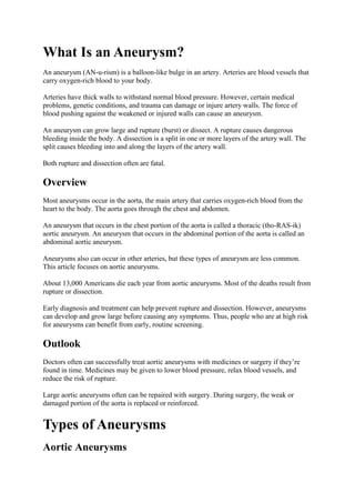
  • 2. The two types of aortic aneurysm are abdominal aortic aneurysm and thoracic aortic aneurysm. Some people have both types. Aortic Aneurysms Figure A shows a normal aorta. Figure B shows a thoracic aortic aneurysm, which is located behind the heart. Figure C shows an abdominal aortic aneurysm, which is located below the arteries that supply blood to the kidneys. Abdominal Aortic Aneurysms An aneurysm that occurs in the abdominal portion of the aorta is called an abdominal aortic aneurysm (AAA). Most aortic aneurysms are AAAs. These aneurysms are found more often now than in the past because of computed tomography (to-MOG-rah-fee) scans, or CT scans , done for other medical problems. Small AAAs rarely rupture. However, AAAs can grow very large without causing symptoms. Routine checkups and treatment for an AAA can help prevent growth and rupture.
  • 3. Thoracic Aortic Aneurysms An aneurysm that occurs in the chest portion of the aorta (above the diaphragm, a muscle that helps you breathe) is called a thoracic aortic aneurysm (TAA). TAAs don't always cause symptoms, even when they're large. Only half of all people who have TAAs notice any symptoms. TAAs are found more often now than in the past because of chest CT scans done for other medical problems. With a common type of TAA, the walls of the aorta weaken and a section close to the heart enlarges. As a result, the valve between the heart and the aorta can't close properly. This allows blood to leak back into the heart. A less common type of TAA can develop in the upper back, away from the heart. A TAA in this location may result from an injury to the chest, such as from a car crash. Other Types of Aneurysms Brain Aneurysms Aneurysms in the arteries of the brain are called cerebral (seh-RE-bral) aneurysms or brain aneurysms. Brain aneurysms also are called berry aneurysms because they're often the size of a small berry. Brain Aneurysm The illustration shows a typical location of a brain aneurysm in the arteries that supply blood to the brain. The inset image shows a closeup view of the sac-like aneurysm.
  • 4. Most brain aneurysms cause no symptoms until they become large, begin to leak blood, or rupture (burst). A ruptured brain aneurysm can cause a stroke. Peripheral Aneurysms Aneurysms that occur in arteries other than the aorta and the brain arteries are called peripheral (peh-RIF-eh-ral) aneurysms. Common locations for peripheral aneurysms include the popliteal (pop-li-TE-al), femoral (FEM-o-ral), and carotid (ka-ROT-id) arteries. The popliteal arteries run down the back of the thighs, behind the knees. The femoral arteries are the main arteries in the groin. The carotid arteries are the two main arteries on each side of your neck. Peripheral aneurysms aren’t as likely to rupture or dissect as aortic aneurysms. However, blood clots can form in peripheral aneurysms. If a blood clot breaks away from the aneurysm, it can block blood flow through the artery. If a peripheral aneurysm is large, it can press on a nearby nerve or vein and cause Other Names for Aneurysm Abdominal aortic aneurysm Aortic aneurysm Berry aneurysm Brain aneurysm Cerebral aneurysm Peripheral aneurysm Thoracic aortic aneurysm What Causes an Aneurysm? The force of blood pushing against the walls of an artery combined with damage or injury to the artery’s walls can cause an aneurysm. Many conditions and factors can damage and weaken the walls of the aorta and cause aortic aneurysms. Examples include aging, smoking, high blood pressure, and atherosclerosis (ath- er-o-skler-O-sis). Atherosclerosis is the hardening and narrowing of the arteries due to the buildup of a waxy substance called plaque (plak). Rarely, infections—such as untreated syphilis (a sexually transmitted infection)—can cause aortic aneurysms. Aortic aneurysms also can occur as a result of diseases that inflame the blood vessels, such as vasculitis (vas-kyu-LI-tis). A family history of aneurysms also may play a role in causing aortic aneurysms. In addition to the factors above, certain genetic conditions may cause thoracic aortic aneurysms (TAAs). Examples of these conditions include Marfan syndrome, Loeys-Dietz syndrome, Ehlers-Danlos syndrome (the vascular type), and Turner syndrome.
  • 5. These genetic conditions can weaken the body’s connective tissues and damage the aorta. People who have these conditions tend to develop aneurysms at a younger age than other people. They’re also at higher risk for rupture and dissection. Trauma, such as a car accident, also can damage the walls of the aorta and lead to TAAs. Researchers continue to look for other causes of aortic aneurysms. For example, they’re looking for genetic mutations (changes in the genes) that may contribute to or cause aneurysms. Who Is at Risk for an Aneurysm? Certain factors put you at higher risk for an aortic aneurysm. These factors include: Male gender. Men are more likely than women to have aortic aneurysms. Age. The risk for abdominal aortic aneurysms increases as you get older. These aneurysms are more likely to occur in people who are aged 65 or older. Smoking. Smoking can damage and weaken the walls of the aorta. A family history of aortic aneurysms. People who have family histories of aortic aneurysms are at higher risk for the condition, and they may have aneurysms before the age of 65. A history of aneurysms in the arteries of the legs. Certain diseases and conditions that weaken the walls of the aorta. Examples include high blood pressure and atherosclerosis. Having a bicuspid aortic valve can raise the risk of having a thoracic aortic aneurysm. A bicuspid aortic valve has two leaflets instead of the typical three. Car accidents or trauma also can injure the arteries and increase the risk for aneurysms. If you have any of these risk factors, talk with your doctor about whether you need screening for aneurysms. What Are the Signs and Symptoms of an Aneurysm? The signs and symptoms of an aortic aneurysm depend on the type and location of the aneurysm. Signs and symptoms also depend on whether the aneurysm has ruptured (burst) or is affecting other parts of the body. Aneurysms can develop and grow for years without causing any signs or symptoms. They often don't cause signs or symptoms until they rupture, grow large enough to press on nearby body parts, or block blood flow. Abdominal Aortic Aneurysms
  • 6. Most abdominal aortic aneurysms (AAAs) develop slowly over years. They often don't cause signs or symptoms unless they rupture. If you have an AAA, your doctor may feel a throbbing mass while checking your abdomen. When symptoms are present, they can include: A throbbing feeling in the abdomen Deep pain in your back or the side of your abdomen Steady, gnawing pain in your abdomen that lasts for hours or days If an AAA ruptures, symptoms may include sudden, severe pain in your lower abdomen and back; nausea (feeling sick to your stomach) and vomiting; constipation and problems with urination; clammy, sweaty skin; light-headedness; and a rapid heart rate when standing up. Internal bleeding from a ruptured AAA can send you into shock . Shock is a life- threatening condition in which blood pressure drops so low that the brain, kidneys, and other vital organs can't get enough blood to work well. Shock can be fatal if it’s not treated right away. Thoracic Aortic Aneurysms A thoracic aortic aneurysm (TAA) may not cause symptoms until it dissects or grows large. If you have symptoms, they may include: Pain in your jaw, neck, back, or chest Coughing and/or hoarseness Shortness of breath and/or trouble breathing or swallowing A dissection is a split in one or more layers of the artery wall. The split causes bleeding into and along the layers of the artery wall. If a TAA ruptures or dissects, you may feel sudden, severe, sharp or stabbing pain starting in your upper back and moving down into your abdomen. You may have pain in your chest and arms, and you can quickly go into shock. If you have any symptoms of TAA or aortic dissection, call 9–1–1. If left untreated, these conditions may lead to organ damage or death. How Is an Aneurysm Diagnosed? If you have an aortic aneurysm but no symptoms, your doctor may find it by chance during a routine physical exam. More often, doctors find aneurysms during tests done for other reasons, such as chest or abdominal pain. If you have an abdominal aortic aneurysm (AAA), your doctor may feel a throbbing mass in your abdomen. A rapidly growing aneurysm about to rupture (burst) can be tender and very painful when pressed. If you're overweight or obese, it may be hard for your doctor to feel even a large AAA.
  • 7. If you have an AAA, your doctor may hear rushing blood flow instead of the normal whooshing sound when listening to your abdomen with a stethoscope. Specialists Involved Your primary care doctor may refer you to a cardiothoracic or vascular surgeon for diagnosis and treatment of an aortic aneurysm. A cardiothoracic surgeon does surgery on the heart, lungs, and other organs and structures in the chest, including the aorta. A vascular surgeon does surgery on the aorta and other blood vessels, except those of the heart and brain. Diagnostic Tests and Procedures To diagnose and study an aneurysm, your doctor may recommend one or more of the following tests. Ultrasound and Echocardiography Ultrasound and echocardiography (echo) are simple, painless tests that use sound waves to create pictures of the structures inside your body. These tests can show the size of an aortic aneurysm, if one is found. Computed Tomography Scan A computed tomography scan, or CT scan , is a painless test that uses x rays to take clear, detailed pictures of your organs. During the test, your doctor will inject dye into a vein in your arm. The dye makes your arteries, including your aorta, visible on the CT scan pictures. Your doctor may recommend this test if he or she thinks you have an AAA or a thoracic aortic aneurysm (TAA). A CT scan can show the size and shape of an aneurysm. This test provides more detailed pictures than an ultrasound or echo. Magnetic Resonance Imaging Magnetic resonance imaging (MRI) uses magnets and radio waves to create pictures of the organs and structures in your body. This test works well for detecting aneurysms and pinpointing their size and exact location. Angiography Angiography (an-jee-OG-ra-fee) is a test that uses dye and special x rays to show the insides of your arteries. This test shows the amount of damage and blockage in blood vessels. Aortic angiography shows the inside of your aorta. The test may show the location and size of an aortic aneurysm.
  • 8. How Is an Aneurysm Treated? Aortic aneurysms are treated with medicines and surgery. Small aneurysms that are found early and aren’t causing symptoms may not need treatment. Other aneurysms need to be treated. The goals of treatment may include: Preventing the aneurysm from growing Preventing or reversing damage to other body structures Preventing or treating a rupture or dissection Allowing you to continue doing your normal daily activities Treatment for an aortic aneurysm is based on its size. Your doctor may recommend routine testing to make sure an aneurysm isn't getting bigger. This method usually is used for aneurysms that are smaller than 5 centimeters (about 2 inches) across. How often you need testing (for example, every few months or every year) is based on the size of the aneurysm and how fast it's growing. The larger it is and the faster it's growing, the more often you may need to be checked. Medicines If you have an aortic aneurysm, your doctor may prescribe medicines before surgery or instead of surgery. Medicines are used to lower blood pressure, relax blood vessels, and lower the risk that the aneurysm will rupture (burst). Beta blockers and calcium channel blockers are the medicines most commonly used. Surgery Your doctor may recommend surgery if your aneurysm is growing quickly or is at risk of rupture or dissection. The two main types of surgery to repair aortic aneurysms are open abdominal or open chest repair and endovascular repair. Open Abdominal or Open Chest Repair The standard and most common type of surgery for aortic aneurysms is open abdominal or open chest repair. This surgery involves a major incision (cut) in the abdomen or chest. General anesthesia (AN-es-THE-ze-ah) is used during this procedure. The term ―anesthesia‖ refers to a loss of feeling and awareness. General anesthesia temporarily puts you to sleep. During the surgery, the aneurysm is removed. Then, the section of aorta is replaced with a graft made of material such as Dacron® or Teflon.® The surgery takes 3 to 6 hours; you’ll remain in the hospital for 5 to 8 days.
  • 9. If needed, repair of the aortic heart valve also may be done during open abdominal or open chest surgery. It often takes a month to recover from open abdominal or open chest surgery and return to full activity. Most patients make a full recovery. Endovascular Repair In endovascular repair, the aneurysm isn't removed. Instead, a graft is inserted into the aorta to strengthen it. Surgeons do this type of surgery using catheters (tubes) inserted into the arteries; it doesn't require surgically opening the chest or abdomen. General anesthesia is used during this procedure. The surgeon first inserts a catheter into an artery in the groin (upper thigh) and threads it to the aneurysm. Then, using an x ray to see the artery, the surgeon threads the graft (also called a stent graft) into the aorta to the aneurysm. The graft is then expanded inside the aorta and fastened in place to form a stable channel for blood flow. The graft reinforces the weakened section of the aorta. This helps prevent the aneurysm from rupturing. Endovascular Repair The illustration shows the placement of a stent graft in an aortic aneurysm. In figure A, a catheter is inserted into an artery in the groin (upper thigh). The catheter is threaded to the abdominal aorta, and the stent graft is released from the catheter. In figure B, the stent graft allows blood to flow through the aneurysm.
  • 10. The recovery time for endovascular repair is less than the recovery time for open abdominal or open chest repair. However, doctors can’t repair all aortic aneurysms with endovascular repair. The location or size of an aneurysm may prevent the use of a stent graft. How Can an Aneurysm Be Prevented? The best way to prevent an aortic aneurysm is to avoid the factors that put you at higher risk for one. You can’t control all aortic aneurysm risk factors, but lifestyle changes can help you lower some risks. For example, if you smoke, try to quit. Talk with your doctor about programs and products that can help you quit smoking. Also, try to avoid secondhand smoke. For more information about how to quit smoking, go to the Health Topics Smoking and Your Heart article. Another important lifestyle change is following a healthy diet. A healthy diet includes a variety of fruits, vegetables, and whole grains. It also includes lean meats, poultry, fish, beans, and fat-free or low-fat milk or milk products. A healthy diet is low in saturated fat, trans fat, cholesterol, sodium (salt), and added sugar. For more information about following a healthy diet, go to the National Heart, Lung, and Blood Institute’s (NHLBI’s) Aim for a Healthy Weight Web site, "Your Guide to a Healthy Heart," and "Your Guide to Lowering Your Blood Pressure With DASH." All of these resources include general information about healthy eating. Be as physically active as you can. Talk with your doctor about the amounts and types of physical activity that are safe for you. For more information about physical activity, go to the Health Topics Physical Activity and Your Heart article and the NHLBI’s "Your Guide to Physical Activity and Your Heart." Work with your doctor to control medical conditions such as high blood pressure and high blood cholesterol. Follow your treatment plans and take all of your medicines as your doctor prescribes. Screening for Aneurysms Although you may not be able to prevent an aneurysm, early diagnosis and treatment can help prevent rupture and dissection. Aneurysms can develop and grow large before causing any signs or symptoms. Thus, people who are at high risk for aneurysms may benefit from early, routine screening. Your doctor may recommend routine screening if you’re: A man between the ages of 65 and 75 who has ever smoked A man or woman between the ages of 65 and 75 who has a family history of aneurysms
  • 11. If you’re at risk, but not in one of these high-risk groups, ask your doctor whether screening will benefit you. What Is Atherosclerosis? Atherosclerosis (ath-er-o-skler-O-sis) is a disease in which plaque (plak) builds up inside your arteries. Arteries are blood vessels that carry oxygen-rich blood to your heart and other parts of your body. Plaque is made up of fat, cholesterol, calcium, and other substances found in the blood. Over time, plaque hardens and narrows your arteries. This limits the flow of oxygen-rich blood to your organs and other parts of your body. Atherosclerosis can lead to serious problems, including heart attack, stroke, or even death. Atherosclerosis Figure A shows a normal artery with normal blood flow. Figure B shows an artery with plaque buildup. Atherosclerosis-Related Diseases
  • 12. Atherosclerosis can affect any artery in the body, including arteries in the heart, brain, arms, legs, pelvis, and kidneys. As a result, different diseases may develop based on which arteries are affected. Coronary Heart Disease Coronary heart disease (CHD), also called coronary artery disease, is the #1 killer of both men and women in the United States. CHD occurs if plaque builds up in the coronary arteries. These arteries supply oxygen-rich blood to your heart. Plaque narrows the coronary arteries and reduces blood flow to your heart muscle. Plaque buildup also makes it more likely that blood clots will form in your arteries. Blood clots can partially or completely block blood flow. If blood flow to your heart muscle is reduced or blocked, you may have angina (chest pain or discomfort) or a heart attack. Plaque also can form in the heart's smallest arteries. This disease is called coronary microvascular disease (MVD). In coronary MVD, plaque doesn't cause blockages in the arteries as it does in CHD. Carotid Artery Disease Carotid (ka-ROT-id) artery disease occurs if plaque builds up in the arteries on each side of your neck (the carotid arteries). These arteries supply oxygen-rich blood to your brain. If blood flow to your brain is reduced or blocked, you may have a stroke. Peripheral Arterial Disease Peripheral arterial disease (P.A.D.) occurs if plaque builds up in the major arteries that supply oxygen-rich blood to your legs, arms, and pelvis. If blood flow to these parts of your body is reduced or blocked, you may have numbness, pain, and, sometimes, dangerous infections. Chronic Kidney Disease Chronic kidney disease can occur if plaque builds up in the renal arteries. These arteries supply oxygen-rich blood to your kidneys. Over time, chronic kidney disease causes a slow loss of kidney function. The main function of the kidneys is to remove waste and extra water from the body. Overview The cause of atherosclerosis isn't known. However, certain traits, conditions, or habits may raise your risk for the disease. These conditions are known as risk factors. You can control some risk factors, such as lack of physical activity, smoking, and an unhealthy diet. Others you can't control, such as age and a family history of heart disease.
  • 13. Some people who have atherosclerosis have no signs or symptoms. They may not be diagnosed until after a heart attack or stroke. The main treatment for atherosclerosis is lifestyle changes. You also may need medicines and medical procedures. These treatments, along with ongoing medical care, can help you live a healthier life. Outlook Improved treatments have reduced the number of deaths from atherosclerosis-related diseases. These treatments also have improved the quality of life for people who have these diseases. However, atherosclerosis remains a common health problem. You may be able to prevent or delay atherosclerosis and the diseases it can cause. Making lifestyle changes and getting ongoing care can help you avoid the problems of atherosclerosis and live a long, healthy life. What Causes Atherosclerosis? The exact cause of atherosclerosis isn't known. However, studies show that atherosclerosis is a slow, complex disease that may start in childhood. It develops faster as you age. Atherosclerosis may start when certain factors damage the inner layers of the arteries. These factors include: Smoking High amounts of certain fats and cholesterol in the blood High blood pressure High amounts of sugar in the blood due to insulin resistance or diabetes Plaque may begin to build up where the arteries are damaged. Over time, plaque hardens and narrows the arteries. Eventually, an area of plaque can rupture (break open). When this happens, blood cell fragments called platelets (PLATE-lets) stick to the site of the injury. They may clump together to form blood clots. Clots narrow the arteries even more, limiting the flow of oxygen-rich blood to your body. Depending on which arteries are affected, blood clots can worsen angina (chest pain) or cause a heart attack or stroke. Researchers continue to look for the causes of atherosclerosis. They hope to find answers to questions such as: Why and how do the arteries become damaged? How does plaque develop and change over time? Why does plaque rupture and lead to blood clots?
  • 14. What Are the Signs and Symptoms of Atherosclerosis? Atherosclerosis usually doesn't cause signs and symptoms until it severely narrows or totally blocks an artery. Many people don't know they have the disease until they have a medical emergency, such as a heart attack or stroke. Some people may have signs and symptoms of the disease. Signs and symptoms will depend on which arteries are affected. Coronary Arteries The coronary arteries supply oxygen-rich blood to your heart. If plaque narrows or blocks these arteries (a disease called coronary heart disease, or CHD), a common symptom is angina. Angina is chest pain or discomfort that occurs when your heart muscle doesn't get enough oxygen-rich blood. Angina may feel like pressure or squeezing in your chest. You also may feel it in your shoulders, arms, neck, jaw, or back. Angina pain may even feel like indigestion. The pain tends to get worse with activity and go away with rest. Emotional stress also can trigger the pain. Other symptoms of CHD are shortness of breath and arrhythmias (ah-RITH-me-ahs). Arrhythmias are problems with the rate or rhythm of the heartbeat. Plaque also can form in the heart's smallest arteries. This disease is called coronary microvascular disease (MVD). Symptoms of coronary MVD include angina, shortness of breath, sleep problems, fatigue (tiredness), and lack of energy. Carotid Arteries The carotid arteries supply oxygen-rich blood to your brain. If plaque narrows or blocks these arteries (a disease called carotid artery disease), you may have symptoms of a stroke. These symptoms may include: Sudden weakness Paralysis (an inability to move) or numbness of the face, arms, or legs, especially on one side of the body Confusion Trouble speaking or understanding speech Trouble seeing in one or both eyes Problems breathing Dizziness, trouble walking, loss of balance or coordination, and unexplained falls Loss of consciousness Sudden and severe headache
  • 15. Peripheral Arteries Plaque also can build up in the major arteries that supply oxygen-rich blood to the legs, arms, and pelvis (a disease called peripheral arterial disease). If these major arteries are narrowed or blocked, you may have numbness, pain, and, sometimes, dangerous infections. Renal Arteries The renal arteries supply oxygen-rich blood to your kidneys. If plaque builds up in these arteries, you may develop chronic kidney disease . Over time, chronic kidney disease causes a slow loss of kidney function. Early kidney disease often has no signs or symptoms. As the disease gets worse it can cause tiredness, changes in how you urinate (more often or less often), loss of appetite, nausea (feeling sick to the stomach), swelling in the hands or feet, itchiness or numbness, and trouble concentrating. How Is Atherosclerosis Diagnosed? Your doctor will diagnose atherosclerosis based on your medical and family histories, a physical exam, and test results. Specialists Involved If you have atherosclerosis, a primary care doctor, such as an internist or family practitioner, may handle your care. Your doctor may recommend other health care specialists if you need expert care, such as: A cardiologist. This is a doctor who specializes in diagnosing and treating heart diseases and conditions. You may go to a cardiologist if you have coronary heart disease (CHD) or coronary microvascular disease (MVD). A vascular specialist. This is a doctor who specializes in diagnosing and treating blood vessel problems. You may go to a vascular specialist if you have peripheral arterial disease (P.A.D.). A neurologist. This is a doctor who specializes in diagnosing and treating nervous system disorders. You may see a neurologist if you've had a stroke due to carotid artery disease. A nephrologist. This is a doctor who specializes in diagnosing and treating kidney diseases and conditions. You may go to a nephrologist if you have chronic kidney disease . Physical Exam During the physical exam, your doctor may listen to your arteries for an abnormal whooshing sound called a bruit (broo-E). Your doctor can hear a bruit when placing a stethoscope over an affected artery. A bruit may indicate poor blood flow due to plaque buildup.
  • 16. Your doctor also may check to see whether any of your pulses (for example, in the leg or foot) are weak or absent. A weak or absent pulse can be a sign of a blocked artery. Diagnostic Tests Your doctor may recommend one or more tests to diagnose atherosclerosis. These tests also can help your doctor learn the extent of your disease and plan the best treatment. Blood Tests Blood tests check the levels of certain fats, cholesterol, sugar, and proteins in your blood. Abnormal levels may be a sign that you're at risk for atherosclerosis. EKG (Electrocardiogram) An EKG is a simple, painless test that detects and records the heart's electrical activity. The test shows how fast the heart is beating and its rhythm (steady or irregular). An EKG also records the strength and timing of electrical signals as they pass through the heart. An EKG can show signs of heart damage caused by CHD. The test also can show signs of a previous or current heart attack. Chest X Ray A chest x ray takes pictures of the organs and structures inside your chest, such as your heart, lungs, and blood vessels. A chest x ray can reveal signs of heart failure. Ankle/Brachial Index This test compares the blood pressure in your ankle with the blood pressure in your arm to see how well your blood is flowing. This test can help diagnose P.A.D. Echocardiography Echocardiography (echo) uses sound waves to create a moving picture of your heart. The test provides information about the size and shape of your heart and how well your heart chambers and valves are working. Echo also can identify areas of poor blood flow to the heart, areas of heart muscle that aren't contracting normally, and previous injury to the heart muscle caused by poor blood flow. Computed Tomography Scan A computed tomography (CT) scan creates computer-generated pictures of the heart, brain, or other areas of the body. The test can show hardening and narrowing of large arteries. A cardiac CT scan also can show whether calcium has built up in the walls of the coronary (heart) arteries. This may be an early sign of CHD.
  • 17. Stress Testing During stress testing, you exercise to make your heart work hard and beat fast while heart tests are done. If you can't exercise, you may be given medicine to make your heart work hard and beat fast. When your heart is working hard, it needs more blood and oxygen. Plaque-narrowed arteries can't supply enough oxygen-rich blood to meet your heart's needs. A stress test can show possible signs and symptoms of CHD, such as: Abnormal changes in your heart rate or blood pressure Shortness of breath or chest pain Abnormal changes in your heart rhythm or your heart's electrical activity As part of some stress tests, pictures are taken of your heart while you exercise and while you rest. These imaging stress tests can show how well blood is flowing in various parts of your heart. They also can show how well your heart pumps blood when it beats. Angiography Angiography (an-jee-OG-ra-fee) is a test that uses dye and special x rays to show the inside of your arteries. This test can show whether plaque is blocking your arteries and how severe the blockage is. A thin, flexible tube called a catheter is put into a blood vessel in your arm, groin (upper thigh), or neck. Dye that can be seen on an x-ray picture is injected through the catheter into the arteries. By looking at the x-ray picture, your doctor can see the flow of blood through your arteries. Other Tests Other tests are being studied to see whether they can give a better view of plaque buildup in the arteries. Examples of these tests include magnetic resonance imaging (MRI) and positron emission tomography (PET). How Is Atherosclerosis Treated? Treatments for atherosclerosis may include lifestyle changes, medicines, and medical procedures or surgery. The goals of treatment include: Relieving symptoms Reducing risk factors in an effort to slow or stop the buildup of plaque Lowering the risk of blood clots forming Widening or bypassing plaque-clogged arteries Preventing atherosclerosis-related diseases
  • 18. Lifestyle Changes Making lifestyle changes often can help prevent or treat atherosclerosis. For some people, these changes may be the only treatment needed. Follow a Healthy Diet A healthy diet is an important part of a healthy lifestyle. Following a healthy diet can prevent or reduce high blood pressure and high blood cholesterol and help you maintain a healthy weight. For information about healthy eating, go to the National Heart, Lung, and Blood Institute's (NHLBI's) Aim for a Healthy Weight Web site. This site provides practical tips on healthy eating, physical activity, and weight control. Therapeutic Lifestyle Changes (TLC). Your doctor may recommend TLC if you have high blood cholesterol. TLC is a three-part program that includes a healthy diet, physical activity, and weight management. With the TLC diet, less than 7 percent of your daily calories should come from saturated fat. This kind of fat is found in some meats, dairy products, chocolate, baked goods, and deep- fried and processed foods. No more than 25 to 35 percent of your daily calories should come from all fats, including saturated, trans, monounsaturated, and polyunsaturated fats. You also should have less than 200 mg a day of cholesterol. The amounts of cholesterol and the types of fat in prepared foods can be found on the foods' Nutrition Facts labels. Foods high in soluble fiber also are part of a healthy diet. They help prevent the digestive tract from absorbing cholesterol. These foods include: Whole-grain cereals such as oatmeal and oat bran Fruits such as apples, bananas, oranges, pears, and prunes Legumes such as kidney beans, lentils, chick peas, black-eyed peas, and lima beans A diet rich in fruits and vegetables can increase important cholesterol-lowering compounds in your diet. These compounds, called plant stanols or sterols, work like soluble fiber. A healthy diet also includes some types of fish, such as salmon, tuna (canned or fresh), and mackerel. These fish are a good source of omega-3 fatty acids. These acids may help protect the heart from blood clots and inflammation and reduce the risk for heart attack. Try to have about two fish meals every week. You should try to limit the amount of sodium (salt) that you eat. This means choosing low- salt and "no added salt" foods and seasonings at the table or while cooking. The Nutrition Facts label on food packaging shows the amount of sodium in the item.
  • 19. Try to limit drinks with alcohol. Too much alcohol will raise your blood pressure and triglyceride level. (Triglycerides are a type of fat found in the blood.) Alcohol also adds extra calories, which will cause weight gain. Men should have no more than two drinks containing alcohol a day. Women should have no more than one drink containing alcohol a day. One drink is a glass of wine, beer, or a small amount of hard liquor. For more information about TLC, go to the NHLBI's "Your Guide to Lowering Your Cholesterol With TLC." Dietary Approaches to Stop Hypertension (DASH). Your doctor may recommend the DASH eating plan if you have high blood pressure. The DASH eating plan focuses on fruits, vegetables, whole grains, and other foods that are heart healthy and low in fat, cholesterol, and salt. DASH also focuses on fat-free or low-fat milk and dairy products, fish, poultry, and nuts. The DASH eating plan is reduced in red meats (including lean red meats), sweets, added sugars, and sugar-containing beverages. The plan is rich in nutrients, protein, and fiber. The DASH eating plan is a good heart healthy eating plan, even for those who don't have high blood pressure. For more information, go to the NHLBI's "Your Guide to Lowering Your Blood Pressure With DASH." Be Physically Active Regular physical activity can lower many atherosclerosis risk factors, including LDL ("bad") cholesterol, high blood pressure, and excess weight. Physical activity also can lower your risk for diabetes and raise your HDL cholesterol level. HDL is the "good" cholesterol that helps prevent atherosclerosis. Talk with your doctor before you start a new exercise plan. Ask him or her how much and what kinds of physical activity are safe for you. People gain health benefits from as little as 60 minutes of moderate-intensity aerobic activity per week. For major health benefits, do at least 150 minutes (2 hours and 30 minutes) of moderate-intensity aerobic activity or 75 minutes (1 hour and 15 minutes) of vigorous- intensity aerobic activity each week. The more active you are, the more you will benefit. For more information about physical activity, go to the U.S. Department of Health and Human Services' "2008 Physical Activity Guidelines for Americans," the Health Topics Physical Activity and Your Heart article, and the NHLBI's "Your Guide to Physical Activity and Your Heart." Maintain a Healthy Weight Maintaining a healthy weight can lower your risk for atherosclerosis. A general goal to aim for is a body mass index (BMI) of less than 25.
  • 20. BMI measures your weight in relation to your height and gives an estimate of your total body fat. You can use the NHLBI's online BMI calculator to figure out your BMI, or your doctor can help you. A BMI between 25 and 29.9 is considered overweight. A BMI of 30 or more is considered obese. A BMI of less than 25 is the goal for preventing and treating atherosclerosis. Your doctor or health care provider can help you set an appropriate BMI goal. For more information about losing weight or maintaining your weight, go to the Health Topics Overweight and Obesity article. Quit Smoking If you smoke or use tobacco, quit. Smoking can damage and tighten blood vessels and raise your risk for atherosclerosis. Talk with your doctor about programs and products that can help you quit. Also, try to avoid secondhand smoke. If you have trouble quitting smoking on your own, consider joining a support group. Many hospitals, workplaces, and community groups offer classes to help people quit smoking. For more information about how to quit smoking, go to the Health Topics Smoking and Your Heart article and the NHLBI's "Your Guide to a Healthy Heart." Manage Stress Research shows that the most commonly reported "trigger" for a heart attack is an emotionally upsetting event—particularly one involving anger. Also, some of the ways people cope with stress—such as drinking, smoking, or overeating—aren't healthy. Learning how to manage stress , relax, and cope with problems can improve your emotional and physical health. Having supportive people in your life with whom you can share your feelings or concerns can help relieve stress. Physical activity, medicine, and relaxation therapy also can help relieve stress. You may want to consider taking part in a stress management program. Medicines To slow the progress of plaque buildup, your doctor may prescribe medicines to help lower your cholesterol level or blood pressure. He or she also may prescribe medicines to prevent blood clots from forming. For successful treatment, take all medicines as your doctor prescribes. Medical Procedures and Surgery If you have severe atherosclerosis, your doctor may recommend a medical procedure or surgery.
  • 21. Angioplasty (AN-jee-oh-plas-tee) is a procedure that's used to open blocked or narrowed coronary (heart) arteries. Angioplasty can improve blood flow to the heart and relieve chest pain. Sometimes a small mesh tube called a stent is placed in the artery to keep it open after the procedure. Coronary artery bypass grafting (CABG) is a type of surgery. In CABG, arteries or veins from other areas in your body are used to bypass (that is, go around) your narrowed coronary arteries. CABG can improve blood flow to your heart, relieve chest pain, and possibly prevent a heart attack. Bypass grafting also can be used for leg arteries. For this surgery, a healthy blood vessel is used to bypass a narrowed or blocked artery in one of the legs. The healthy blood vessel redirects blood around the blocked artery, improving blood flow to the leg. Carotid endarterectomy (END-ar-ter-EK-to-me) is surgery to remove plaque buildup from the carotid arteries in the neck. This procedure restores blood flow to the brain, which can help prevent a stroke.