SlideShare ist ein Scribd-Unternehmen logo
1 von 49
Macroscopic slides:
1. Arteriolosclerotic nephrosclerosis
2. Hypertensive heart
3. Atherosclerotic aorta
4. Recent myocardial infarction
5. Mitral valve – rheumatic fibrous endocarditis
6. Ulcero-polypotic endocarditis
7. Fibrinous pericarditis
8. Emphysematous bullae in lungs
9. Bronchopneumonia abscedens
10. Silicosis
11. Lung cancer
12. Diverticulitis of Esophagus
13. Acute stomach ulcer
14. Chronic gastric ulcer with perforation
15. Gastric cancer
16. Micronodular liver cirrhosis
17. Cholelithiasis
18. NHL
19. Porphyry spleen – HL
20. Sclerosing GN/ glomerulonephritic nephrosclerosis
21. Calculosal chronic pyelonephritis
22. Polycystic kidney disease
23. Carcinoma of kidney
Macro continued…
24. Hypertrophy of prostate
25. Bladder cancer
26. Seminoma of testis
27. Tubal abortion
28. Molar pregnancy
29. Carcinoma cervix
30. Carcinoma endometrium
31. Krukenberg metastasis in ovaries
32. Breast cancer
33. Macroglossia – acromegaly
34. Nodosal goitre
35. Diabetic glomerulosclerosis
36. Glioma
37. Meningioma
38. Neurinoma
39. Fibrocavernous TB lungs
40. Renal tuberculosis
41. Luetic aneurysm Aorta
1. Benign Nephrosclerosis
Arteriolosclerotica (Macroscopic):
 Patchy ischemic atrophy with focal
loss of parenchyma
 Granular appearance
 Diffuse Pin-point haemorrhages on
cortical surface.
 Kidneys are slightly smaller than
normal
 Essential hypertension/ diabetes
mellitus, benign because there is
little altered function of the
kidneys but increased risk for renal
failure.
Accelerated (malignant) stage of
essential HT
Left: Malignant hypertension
(300/150mmhg) – a
complication/sequelae of benign form,
leads to focal haemorrhaging which
obscures the corticomedullary junction
2. Hypertensive heart – macro:
• Increased peripheral vascular resistance
and cardiac workload induces
remodelling of the myocardium. This is
seen as concentric left ventricular
hypertrophy a.k.a “Cor hypertonicum”.
• Normal thickness = 5-9mm, borderline =
13/14mm, hypertrophic = > 15mm
• The rest of the heart is relatively
unchanged. Secondary right ventricular
hypertrophy may occur after
hypertension induced left sided heart
failure and pulmonary hypertension.
• Complications of hypertensive heart
disease: increased risk of myocardial
infarction (larger mass of muscle to be
supplied with O2), arrhythmias, heart
failure, aortic dissection, aneurysms,
accelerated atherosclerosis due to
endothelial injury, nephrosclerosis,
hypertensive retinopathy, cerebral
vascular accident (stroke).
3. Atherosclerosis Aortae (Macro):
FIRST STAGE - Fatty streak (white arrows)
 Thin, flat, yellow
 Non-protruding linear streaks
 Present from very young age, even
birth
 Increase in number until young
adulthood then stabilise or may
regress
 Reversible with lifestyle modification
 occur at areas of turbulent flow such
as around the orifices of branching
vessels of the aorta.
From top to bottom: Severe. Moderate and mild
atherosclerosis
THIRD STAGE – Atheromatous
plaque
 Reddish brown mural
thrombi
 Ulceration of intimal surface
 Irregular shaped white-
yellow atheromatous
plaques
 Calcifications
 Plaques become more
confluent/ join up the
further the disease
progresses.
FOURTH STAGE – complicated plaque (unstable)
Fates:
1. Plaque may grow around the branch points
of vasa vasorum (own vessels) of the aorta
leading to atrophy of tunica media. This
weakens the wall and can lead to
atherosclerotic dissection or aneurysm.
2. Hemorrhage of weak neovascularisation in
the plaque may lead to a hematoma which
expands the plaque to occlude the lumen,
subsequent infarction.
3. Ulceration and rupture of the plaque can
release cholesterol (fat) emboli leading to
stroke, MI or PE.
4. Dystrophic calcification which hardens the
artery leading to changes in pulse, blood
pressure and further endothelial damage.
Calcific fragments can break off into
circulation (calcific emboli) and cause
infarction.
5. Re-canalisation: rare but the plaque can be
invades by endothelial cells which re-open a
larger lumen. A compensatory response.
Common location of atherosclerosis:
1. Coronary As => IHD and MI
2. Carotids => vascular dementia, berry aneurysms, cerebral stroke
3. Renal Artery => Stenosis and Secondary hypertension, renal infarction.
4. Iliac As/ Femoral => dry gangrene of feet, intermittent claudication.
5. Aorta => Mesenteric ischemia, dissection, aneurysm
6. Retinal arteries => amaurosis fugax/ acute temporary vision loss
Retinal
embolus,
gangrene,
renal stenosis,
berry
aneurysm.
4. Myocardial Infarction (Macro):
Collagen deposition healed MI
Discrete pale
infarct with
hyperaemic border
Large transmural MI
 Often multi-focal lesions
 Initially; Red-purple blotchy
areas (hemorrhage and
reperfusion injury)
 Progression: Yellow border
which extends inwards
(collagen/ granulation tissue)
and hyperaemic border.
 Finally; Bright pale yellow/
white/ grey scar
 Can lead to HF, arrhythmia.
Aneurysm, cardiac tamponade
5. Fibrous endocarditis (Macroscopic):
Rheumatic (post-infectious) type endocarditis. The other two types
are 1). Infective (ulcero-polypotic) and 2). Non infective which is
further subdivided into a). NBTE (marantic) and b). Libman-sacks
(verrucous - SLE, anti-phospholipid syndrome)
Chronic valvulitis of mitral valve
In rheumatic
valvopathy we see
fusion of
commissures, sterile
fibrin vegetations and
shortened thickened,
fused chordae.
6. Ulcero-polypotic Endocarditis (infectious) – macroscopic:
 Bacterial/ septic endocarditis characterised by the
formation of pedunculated masses/ vegetations of fibrin
or thrombin mixed with bacterial colonies and
calcifications adhered to an ulcerative heart valve.
 Acute forms = S. aureus or GABHS infection
 Subacute forms = Strep. Viridans, Strep pyogenes and
Staph. Epidermidis
 2 predisposing factors are required:
1). BACTEREMIA (anything from poor oral hygiene/ teeth
brushing micro-traumas – full blown sepsis)
2). Damage to or structural abnormality of valve endocardium
– allows for adherence of passing bacterial colonies.
 Vegetations are multiple, large and friable
 Tendency to break off as septic- thrombo emboli
 More common on left side of heart – mitral and aortic
valves.
 Ulceration of valve cusps, rupture of chordae tendineae.
Local destruction may be rapid and severe
 Complications include: heart murmur, arrhythmia, AV
block, pericardial effusion, cardiogenic shock, acute heart
failure, stroke or pulmonary thromboembolism, brain,
heart or lung abscesses, meningitis, glomerulonephritis,
sepsis.
7. Fibrinous pericarditis (gross appearance);
 Dry course granular pericardial surface
 Fibrinous exudate with stringy adhesions between
external epicardial surface and the inner surface of the
pericardium.
 Fibrin has pale yellowish colour
 Shaggy rough “Bread and Butter” appearance.
 Clinically; pericardial friction rub can be heard.
Haemorrhagic
pericarditis
8. Emphysema bullosa (macro):
 5 classifications – 1. centri-acinar/ centrilobular, 2. pan-acinar/ panlobular, 3.
paraseptal (distal lobular), 4. irregular (paracictricial) and 5. mixed.
 Multiple, large dilations most commonly under the pleura
 Lungs are large, hyperinflated, pale and with little blood
 Evidence of chronic smoking often evident; tar deposits
 Complications: Spontaneous pneumothorax if they
burst, collapse of terminal bronchioles – pneumonia,
abscesses, pulmonary fibrosis, atelectasis, respiratory
failure, cor pulmonale, chronic bronchitis (often
concurrent), increased risk of lung cancer.
9. Bronchopneumonia (lobular pneumonia)
abscedens – macro:
• Patchy areas of consolidation
• Usually bilateral.
• Hemorrhage and brighter areas of alveolar
purulent exudate.
• Consolidation matches the distribution/ shape of
the pulmonary lobules hence the name.
• This type of pneumonia is most often acquired in
hospital settings.
• Staph Aureus, Klebsiella, E-coli and Pseudomonas
are common culprits.
• Poorly defined areas are usually grey-red to tan
yellow in colour.
• Cavitary lesions/ abscesses may develop amongst
the consolidation.
• Areas of consolidation are firmer than healthy
areas. They also appear raised on cut section.
10. Silicosis – Gross:
 Upper lobe is usually affected earlier in the disease
 Early lesions = multiple small pale to black ( if coal dust also present)
nodules progressing to hard collagenous scars.
 Fibrosis tends to occur more at the hilar lymphnodes and pleura.
 Nodules may have undulating/ irregular or stellate shaped edges due to
traction from the thick fibrosis. They may cavitate centrally due to
tuberculosis or ischemia.
 Nodules range from 1-5mm.
 Well circumscribed
 Pleura is thickened, may
have adhesions to chest wall
 If severe, or in concurrence
with COPD, may lead to cor
pulmonale.
 Silicosis does not seem to
increase risk of bronchogenic
carcinomas, unlike
asbestosis.
11. Lung Carcinoma - Gross:
 Most common and fatal cancer
 90% of lung cancers are associated with smoking
 Cancers most commonly arise in the hilum
(centrally) and less often in the periphery
Classification of lung cancers:
1). Bronchogenic (most common type)
TWO subtypes of bronchogenic Ca:
a). NSCLC (non small cell lung ca -85%)
b). SCLC (small cell lung cancer – 15%)
Of NSCLC, adenocarcinoma is most common = 40%
Followed by Squamous = 30% and Large cell = 15%
1. Adenocarcinoma:
 Strong assoc. with scarred areas and chronic
damage
 Lobulated or spiculated shaped mass
 May have central area of fibrosis or necrosis
 More common in upper lobes, prefers periphery.
 Pale, solid, whitish mass
 Well circumscribed but not encapsulated
 Anthracotic pigments (from smoking)
 Has four subtypes
Bronchogenic Lung Cancers (hilar/ centrally located – most
likely squamous cell carcinomas)
Peripherally located carcinomas – later identified to
be adenocarcinomas.
2. Squamous cell ca:
 Arises centrally from main
bronchi
 Massive necrosis
 Cavitation
 Hemorrhages
 Strong assoc. with smoking
 Grey-white fleshy tumour
 May cause bronchiectasis,
atelectasis, recurrent
pneumonia.
 Tendency to invade hilum and
surrounding parenchyma
12. Esophageal diverticulum – macroscopic:
 Visible outpouching of wall of Esophagus
 Contains ALL layers of wall (true diverticulum)
 Rare = pseudo diverticulum – only mucosa or
submucosa
 2 types by etiology: A). Congenital (more
common in upper Esophagus or at level of
carina) B). Acquired: 1. Pulsion (from
increased intraluminal pressure) or 2.
Traction (pulled by the formation of
adhesions, fibrosis)
 Three types by location: 1. proximal
(zenkers), 2. mid-esophageal (usually traction
type) and 3. epiphrenic (distal)
 Zenkers diverticulum is a pulsion type
acquired esophageal diverticulum in
hypopharyngeal location – due to esophageal
obstructions.
 Complications: diverticulitis (hyperaemia/
wall thickening, leucocyte infiltration, peri-
diverticular edema, muco-purulent exudates,
hemorrhage, perforation and subsequent
pneumomediastinum or subcutaneous
emphysema in neck, esophageal cancer.
Opened post-mortem specimen of the oesophagus. An oesophageal
diverticulum with creamy exudates and a hard foreign body (bone)
adherent to the oesophageal wall (arrow).
13. Acute stomach ulcer – gross :
 Less than 1cm
 Round shape
 Well defined margins
 Sometimes confluent
 Dark brown – black colour due to formation of hematin.
 Fibrinoid necrosis centrally in pit of ulcer
 Surrounding tissue is hyperaemic
 Most common location: lesser curvature of stomach, if
lesion occurs on greater curvature suspect ulcerating gastric
carcinoma.
 Subtypes of ulcer = A). Curling's ulcer (severe burns) and B).
Cushing's ulcer – due to hemodynamic changed due to brain
injury/ elevated ICP.
14. Chronic gastric ulcer with
perforation - gross :
 Round/ oval shape
 4-6 cm in diameter (larger than
acute ulcers)
 Depth variable – may extend
through all layers; submucosa,
muscle layer and adventitia
 Sides are hyperaemic, bulging
and overhanging
 Base is firm due to fibrosis –
callus of the ulcer.
 Floor may be relatively clean,
active hemorrhage or fully
perforating the gastric wall.
 Mucosal folds radiate outward
from ulcer margins.
15. Gastric carcinoma:
 Variable presentation; as large solitary mass,
non-healing ulcer or diffuse infiltrative
burrowing process (linitis plastica).
 Four types: 1). Polypoid/ papillary exophytic
growth 2). Fungating (raised margins with
central ulceration 3) ulcerative with infiltrating
margins 4). Linitis plastica (wall becomes thick
and rigid)
 Wall thickening, rugal fold flattening
 Hemorrhage and necrosis (signs of aggression)
16. Micronodular liver cirrhosis – gross:
 Nodules are less than 3mm (macro = 3mm
– 3cm).
 Diffuse involvement of all hepatic lobules.
 Fat deposition may be seen – although less
than in fatty liver disease, this gives the
organ a tawny brown-yellow appearance.
Normal liver is rich maroon red/brown.
 Surface is granular and studded with
nodules.
 Thick fibrose septae may be seen on cut-
section.
 In severe disease the organ shrinks.
17: Cholelithiasis - gross:
 Gallbladder wall may be hypertrophic (thick,
contracted, calcified and firm organ) or
atrophic (large, flaccid, distended, pale and
grey organ).
 Diffuse mural hyperaemia – mucosa is bright
red, mucinous or muco-purulent exudate
(empyema)
 Abscesses may form in the wall.
 Stones can be of FOUR kinds:
1). Cholesterol stones – oral or polygonal with
flattened facets, smooth, yellow-white.
2). Pigment stones – due to increased
unconjugated bilirubin often as a result of
haemolysis. These stones are small, numerous,
mulberry shape and jet black.
3). Calcium stones – grey/white, hard and small.
4). Mixed stones – most common type, a
laminated stone comprised of alternating deposits
of the contents of the other stones
18. Malignant Non H Lymphoma - (macro):
 Solid tumours of the lymphnodes – usually
cervical. One third of time they affect extranodal
sites – the tonsils, GIT and spleen.
 Grossly the appearance of HL and NHL are very
similar., the differences are in behaviour, labs and
microscopic examination.
 Normal lymphnode capsule invaded and destroyed
 Surrounding fat tissue is invaded forming thick
tracts investing multiple lymphnodes – matted,
firm texture upon palpation.
NHL in lymphnodes near pancreas
19. Hodgkin's lymphoma spleen (macro):
! DO NOT CONFUSE “Porphyry spleen” with “Sago” or “lardaceous”
spleen.
Porphyry spleen is caused by Hodgkin's lymphoma and is
characterised by multiple nodular leucocyte tumour infiltrates.
BOTH Sago spleen and lardaceous spleen are caused by amyloidosis
of the organ – characterised by diffuse granular amyloid deposits.
This can be due to hereditary amyloidosis (abnormal protein
synthesis) or secondary to chronic inflammatory conditions e.g.
rheumatoid arthritis, Inflammatory bowel disease etc, due to
plasma cell disorders that produce too much immunoglobulins or
senile amyloidosis– due to old age and other idiopathic causes.
These are shown on the next slide for comparison
Porphyry spleen:
 Scattered grey-
white nodules
 Can be singular
and large or
multiple and
smaller.
 Nodules coalesce
over course of
disease
 # of nodules =
prognostic (>5 =
bad).
:
SAGO
LARD
Lardaceous spleen
Sago spleen
20. Nephrosclerosis
(glomerulonephritic type - macro):
 A.k.a diffuse sclerosing GN.
 Symmetrically and severely
atrophic kidneys - shrunken and
only weigh 1/3rd of a healthy
kidney.
 Surface is finely granular and
cortex is thinned
 Pelvis and calices are relatively
spared, medulla unremarkable.
 Capsule may be adherent to
cortex.
21. Chronic pyelonephritis with
calculus – gross:
• Coarsely granular and nodular
surface of kidney.
• Kidney is asymmetrical and
irregular in shape.
• Kidney is heavily scarred and
shrunken, capsular scarring and
retractions correspond to
involvement and dilation/ blunting
of the underlying calyx.
• Blunting of papillae, papillary
necrosis is common.
• Abscesses and cysts may be seen
• Calculi are common in the renal
pelvis and proximal ureter, as a
result ureter may be dilated.
22. Polycystic kidney gross:
• A congenital/ genetic disease that
runs in families causing progressive
cystic change in the kidneys with age,
pain, high blood pressure and renal
failure.
• Kidneys are massively enlarged and
multi-cystic with replacement of the
entire cortex.
23. Renal Cell Carcinoma – gross:
• Large, round, well-defined
masses that usually occur and
one of the poles of the kidney.
• Pale beige-yellow appearance
against the healthy red kidney.
• Centred in the cortex, may have
pseudocapsule exteriorly and
disrupt the smooth contour of the
kidney while invading the
medulla or renal sinus interiorly.
• May be cystic in appearance
(bottom right image) or have
multiple hemorrhages, focal
necrosis or calcifications within
the mass – a sign of its aggressive
behaviour.
• Often invades the renal vein and
vena cava.
Kidney abscess – Macro:
• Round to ovoid pale yellow
lesions.
• Associated with purulent
infections of the kidney – either
due to hematogenous spread or
an ascending UTI, can also be
caused by renal tuberculosis
(bottom right image).
• Well circumscribed
• Frequently multitudinous –
especially in acute pyelonephritis
where many small abscesses are
dotted around the capsular
surface and cortex of enlarged
firm red kidneys (top left image).
24. Hypertrophy of prostate - gross:
 Benign prostatic hyperplasia mostly
affects the peri-urethral and transitional
area of the gland.
 Gland enlarges to over 5/6cm and can
weigh 100g+. Normal size = 3-4cm.
 Yellow – white nodules of variable size.
 Enlarged gland can be seen pushing
prominently out into bladder lumen.
 May compress urethra “slit-like
appearance” on cut section.
25. Carcinoma of urothelium of bladder
– Macro:
 Two classifications: 1). Epithelial
origin – more common 2). Non-
epithelial origin.
 Urothelial (transitional) cell
carcinoma constitutes 90% of
epithelial tumours. Others include
squamous cc, adenocarcinoma, small
cell and mixed.
 More common in males, 5th decade
of life.
 Non-papillary type usually bulky,
large and ulcerative.
Papillary type urothelial ca:
 fused and branched fern-like pattern.
 Exophytic, free-floating.
 Lateral wall most commonly
affected, then posterior wall and
trigone.
 Usually small – less than 2cm
 May be single or multiple
 May infiltrate lamina propria and
deep in detrusor muscle
26. Seminoma testis – macro:
• Most common malignant
tumour of testis (45%). It is of
the GERM CELL class of
tumours.
• Uniform solid tan to pale
cream, grey- white mass.
• Necrosis and hemorrhage are
uncommon and minimal.
• If spontaneous regression
occurs, scarring may be the
only indicator of its occurrence.
• Homogenous and lobulated,
well circumscribed.
• Testis is usually enlarged,
seminoma may replace entire
testicular tissue.
• Usually confined to just the
testis in 90% of cases.
27. Tubal abortion - macroscopic:
 Less than 1% of all pregnancies.
 Increased risk in patients with history of
pelvic inflammatory disease.
 Embryo, placenta and amniotic sac are
formed as normal.
 This is followed by rupture of the tube
around the 2nd – 6th week of gestation.
 May cause massive bleeding
intraperitoneally.
 Foetus either dies immediately or soon
after rupture, it may be absorbed,
calcified (lithopaedion), extruded into
peritoneum or the products of
conception may become septic.
 Grossly the affected ovarian tube is
usually dilated/ distended, hyperaemic.
 It may contain what appears to be a
hematoma.
 Products of conception are often visible
– embryo, chorionic villi
 Tube may be intact or ruptured.
28. Molar pregnancy – macro:
 There are four main categories of
trophoblastic gestational disease:
1) Partial mole
2) Complete mole
3) Invasive mole
4) Choriocarcinoma
Partial mole:
 Placental tissue exists but is immature and
abnormal – admixed with hydropic villi that
tend to be smaller and more numerous
than in complete mole.
 Fetal parts and gestational sac may be seen
Complete mole:
 Hydropic villi which appear as fluid-filled
semi-transparent vesicles or variable size
 “Bunch of grapes” appearance.
 Normal fetal parts are absent
 Normal placental structures are absent
Invasive mole: Fluid filled vesicles extend into
myometrium. Uterus may be perforated.
Appears as erosive haemorrhagic lesion.
Partial molar pregnancies
Complete molar
pregnancy
Partial mole
29. Carcinoma of cervix – macro:
Classification of cervical disease:
1). Inflammatory cervical lesions.
2). Non-neoplastic cervical proliferations (eg. squamous
metaplasia, polyps)
3). SIL (squamous intraepithelial lesion or CIN (cervical
intraepithelial lesion).
- CIN 1 = (Less than 1/3rd of epithelium affected or
Mild dysplasia) = low grade SIL
- CIN 2 (1-2/3rds involved or moderate dysplasia)
- CIN 3 (full thickness/ severe dysplasia or in situ
carcinoma – doesn’t invade basement membrane!)
[CIN 2+ 3 = High grade SIL]
Grossly: squamous cell carcinoma is most common
(75% of cases) followed by adenocarcinoma (20%).
Squamous Cell Carcinoma:
 Red, friable indurated lesion.
 Can be ulcerated or elevated granular lesion
 Variable morphology: Exophytic, papillary, polypoid,
nodular and ulcerative forms have been observed.
 Can be invasive and infiltrate surrounding
structures. May be necrotic and haemorrhagic.
 Associated with HPV 16 + 18 infection.
30. Carcinoma of endometrium – macro:
 Most common in post-menopausal women (55-65).
Previous pregnancies have a protective effect.
 Most present grossly as polypoid/ fungating masses.
Uterus is often asymmetrically enlarged
 Invasion into the muscular layer is usually present from
an early stage.
 Earliest symptom = abnormal uterine bleeding (AUB).
 Soft and homogenous growth. May sometimes be
heterogenous – with foci of necrosis and hemorrhage
on cut surface.
 Some cases present as simple endometrial thickening –
the lining in this case is red, abundant and friable to
touch.
 Usually has a favourable prognosis.
 Types include: 1), endometrioid 2). Serous 3). Clear cell
adenocarcinoma and 4). Mixed type
 Has five stages of progression:
STAGE 1 - body of uterus only
STAGE 2 – body and cervix
STAGE 3 – extra-uterine but remains in true pelvis
STAGE4 – Distant metastasis
31. Krukenberg metastasis in Ovaries - gross:
 Metastatic type tumour of the ovaries (5-10% of all
ovarian tumours are metastasis), krukenberg is almost
always bilateral (80%).
 It is a mucin rich signet- ring adenocarcinoma with
gastric origin.
 Other origins include breast and colon. Rarely the
primary tumour may be located in the
gallbladder, biliary tract, urinary bladder or
cervix.
 Prognosis of this tumour is extremely poor.
 Small, multinodular surface with extensive
extra-ovarian spread.
 Metastatic tumours are most difficult to
distinguish from mucinous type primary
tumours of the ovary.
 Scattered growth with intervening normal
ovarian parenchyma
 Multiple vascular emboli nearby
 Similar characteristics to primary tumour
– whichever that may be.
 May have multiple cysts.
 Lymph and blood vessel invasion
32. Breast Cancer - gross:
1. Invasive ductal carcinomas usually
form a solid mass with a variable
consistency.
2. Tumours with abundant
desmoplastic stroma are firm to
hard and grey-white in
appearance.
3. The presence of microcalcifications
may impart a gritty consistency.
4. There may be yellow-white streaks
of necrosis and foci of
hemorrhage.
5. Extensively necrotic tumours may
develop cystic change.
6. The tumours that are composed
largely of neoplastic and
inflammatory cells without much
stroma tend to be soft and
fleshy and bulge when they are
sectioned.
A B
A: Clearly visible nipple retraction in advanced stage breast cancer
B: Peau d’orange - skin appears
studded with dimples like an orange
peel.
2. Invasive
lobular
carcinoma -
gross:
33. Macroglossia (acromegaly) - gross:
 A form of “true” macroglossia (the tongue is indeed larger)
whereas “pseudo-macroglossia” is a relative macroglossia –
where the tongue appears larger due to the oral cavity or
mandible (micrognathia) being smaller the normal.
 Caused by muscular hypertrophy of tongue tissue in response to
excessive growth hormone. The oral cavity is often enlarged also.
 Most cases of acromegaly are caused by a benign
functional (secretory) adenoma of the pituitary gland.
 Acromegaly is usually undiagnosed until the age of
30 to 50. If it occurs before the fusing of the
epiphyseal growth plates at the end of puberty, it is
known as gigantism – due to prolonged bone growth.
 Differential diagnosis of macroglossia:
Congenital true macroglossia:
- Mucopolysaccharidosis
- Idiopathic tongue muscular hypertrophy
- Haemangioma
- Lymphangioma
Acquired true macroglossia:
- Amyloidosis (most common cause in adults)
- Hypothyroidism (myxoedema)
- Sarcoidosis, lymphoma, trauma, TB, syphilis, actinomycosis
34. Nodular goiter –
macroscopic:
 Unlike simple goiters,
multinodular goiters are
asymmetrical and very large,
weighing on average between
200-700g but can grow to be
as large as 2kg!
 Nodularity and poor
encapsulation
 Haemorrhages are common
 Scarring is abundant as is
cystic degeneration and focal
calcifications.
 Individual nodules are variable
in size with dominant ones
often larger than 10cm.
 Surface is bumpy and irregular
 Brown gelatinous colloid
collections may be seen on
cut surface.
35. Diabetic glomerulosclerosis - macroscopic:
 Renal lesions appear around 10-20 years
after onset of diabetes.
 Major symptoms include; proteinuria,
glucosuria, progressive decrease in renal
function, increased recurrence of
pyelonephritis, edema and hypertension
may manifest as late-stage symptoms.
 Gross features include:
- Slight to moderate symmetrical enlargement
of the size of the kidneys.
- Subcapsular cortical surface is finely granular
and dark red in colour.
- Cortex is thinned
- Atherosclerotic changes to renal vessels,
renal stenosis and subsequent renal atrophy.
- Pyelonephritic features may be observed
(due to the predisposition of glycosuria to
precipitate infections); deep irregular surface
scars and retractions, dilated and deformed
calyces, renal calculi, cysts and if active
infection was occurring; multiple micro-
abcesses.
36. Glioma – gross:
 Gliomas are tumours of the glial cells; astrocytoma,
glioblastoma, oligodendrogliomas, ependymomas and
choroid plexus papilloma. They are the most common
brain tumour group and comprise 40%.
 Astrocytoma is the most common glioma:
- Found in mid- late life (40-60yo).
- Most often located in cerebral hemispheres, sometimes
in spinal cord but can occur anywhere (optic nerves,
brainstem, cerebellum etc).
- Have a tendency to progress from low grade to high
grade and transform into glioblastoma multiforme.
- They are grey-white, cystic or solid masses, gradually
grow and therefore clinical symptoms are insidious.
- Signs of transformation to aggressive glioblastoma
includes acceleration in symptom progression, yellow,
hemorrhages, focal necrosis, poor margins and
heterogenous
soft consistency.
Astrocytoma
Glioblastoma
Glioblastoma
Glioblastoma
37. Meningioma – gross:
 Most common location is along falx cerebri
and parasagittal (25%), calvarium convexity
(20%), sphenoid wing (20%), suprasellar (10%),
posterior fossa (10%).
 It may occur in the orbit – congenital cause of
exophthalmos. May occasionally occur at
cerebellopontine angle and mimic acoustic
neuromas.
 Tan, quite spherical or hemi-spherical, well-
defined margins, may be lobulated, firm,
fibrose. Usually meningiomas are solitary.
 May contain focal calcific precipitates.
 1-10cm in size, overlying skull may have
reactive thickening (hyperostosis) and
sclerosis.
 Attached to dura (Note: it does not originate
from the dura, it arises from the arachnoid
layer!)
 Broad base, may be solid, cystic or gelatinous
in texture. When removed they leave a pit/
depression in the underlying cerebral
hemisphere. Clinical symptoms depend on
location; paresis, seizures, headaches, vision
changes, language impairment, memory loss.
38. Neurinoma – macroscopic:
 Does not occur in CNS only peripheral
nerves; spinal nerve roots, cranial nerves.
 Pontocerebellar angle near internal
acoustic meatus is a very common site –
affecting CN VIII (vestibulocochlear nerve).
 Arise from Schwann cells that make
myelin sheath on sensory, motor and
autonomic nerves.
 Encapsulated, solid, sometimes cystic,
eccentric enlargement.
 When they are rich in collagenous tissue
they are subclassified as neurofibromas
(neurofibromatosis).
 “Pepples in
shoe” pain
sensation is a
distinctive
symptom of
lower limb
neuroma.
39. Fibrocavernous TB of lungs –
gross:
 Apices of lungs are most heavily
affected. Usually bilateral
involvement. Unilateral hilar
lymphnodes involvement (kids).
 Thick fibrose non-compliant
upper lobes, pleura may be
thickened, bronchial stricture
and bronchiectasis due to
deformity, displacement and
traction.
 Multiple granulomas with
central caseous (cheese-like)
necrosis. There may be focal
hemorrhages and cavitation.
 Miliary type TB features
multiple diffuse micro-
abscesses throughout the lungs.
It indicates hematogenous
spread and may be associated
with TB meningitis, renal TB,
bone TB and has a poor
prognosis.
40. Renal Tuberculosis – gross:
 Young to middle aged adults.
 Renal involvement may be
indolent, insidious taking
anywhere from 10-30 years to
manifest after primary TB
infection.
 Onset of symptoms as unilateral
renal impairment and less often,
renal failure.
 Multiple large cavities filled with
thick, yellowish caseous necrosis.
 Liquefaction and cavity formation.
 Secondary or concomitant
infection - bacterial abscesses.
 Contraction and fibrosis may lead
to irregularity and deformity of the
cortical surface.
 Correlating clinical symptoms
include; flank pain, increased
frequency, blood and pus in urine,
fever, dysuria as well as signs of
renal insufficiency.
41. Luetic aneurysm – gross:
 Aortic intima has fine or
course, irregular foldings.
 Scattered foci of fibrosis
(pearly-white nodules) along
the intima creating
characteristic “tree-bark”
appearance.
 Typical feature of advanced or
tertiary stage of syphilis
infection.
 Ascending aorta and aortic
arch are most frequently
impacted.
 Superimposing
atherosclerotic changes are
frequent. The cerebral vessels
may also be affected by
syphilitic vasculitis
 Aorta lumen is dilated, walls
are hardened, fibrotic and
thickened, there is
replacement of the atrophied
tunica media muscle.

Weitere ähnliche Inhalte

Ähnlich wie PMU third/fourth year Clinical pathoanatomy Part 3

Pathology Slides For Exam (Kiev Medical University)
Pathology Slides For Exam (Kiev Medical University)Pathology Slides For Exam (Kiev Medical University)
Pathology Slides For Exam (Kiev Medical University)VIKRAM SINGH PANIHARIYA
 
EMBOLISM....Dr San Kauser (Pathology).pdf
EMBOLISM....Dr San Kauser (Pathology).pdfEMBOLISM....Dr San Kauser (Pathology).pdf
EMBOLISM....Dr San Kauser (Pathology).pdfansariabdullah8
 
2- ATHEROSCLEROSIS and its pathophysiology
2- ATHEROSCLEROSIS and its pathophysiology2- ATHEROSCLEROSIS and its pathophysiology
2- ATHEROSCLEROSIS and its pathophysiologywajidullah9551
 
Anatomy of mediastinum and its disorders
Anatomy of mediastinum and its disordersAnatomy of mediastinum and its disorders
Anatomy of mediastinum and its disordersGIREESH G
 
416456384-6-Thrombosis-ppt.ppt
416456384-6-Thrombosis-ppt.ppt416456384-6-Thrombosis-ppt.ppt
416456384-6-Thrombosis-ppt.pptAxmedAbdiHasen
 
Embolism (Dr. MURALEEDHARA)
Embolism  (Dr. MURALEEDHARA)Embolism  (Dr. MURALEEDHARA)
Embolism (Dr. MURALEEDHARA)MINDS MAHE
 
Thrombosis, embolism and infarction
Thrombosis, embolism and infarctionThrombosis, embolism and infarction
Thrombosis, embolism and infarctionDr. Varughese George
 
pathology of Atherosclerosis, dissection and aneurysm copy
pathology of Atherosclerosis, dissection and aneurysm   copypathology of Atherosclerosis, dissection and aneurysm   copy
pathology of Atherosclerosis, dissection and aneurysm copyranaelsaeedAboelfeto
 
CVS Pathology 5 Thromboembolism 2019, sufia husain
CVS Pathology 5 Thromboembolism 2019, sufia husainCVS Pathology 5 Thromboembolism 2019, sufia husain
CVS Pathology 5 Thromboembolism 2019, sufia husainSufia Husain
 
Rheumatic heart disease
Rheumatic heart diseaseRheumatic heart disease
Rheumatic heart diseaseadolescent4u
 
Mitral stenosis (propeduetics)
Mitral stenosis (propeduetics)Mitral stenosis (propeduetics)
Mitral stenosis (propeduetics)ajith joseph
 
15-HEMODYNAMIC DISORDERS.ppt
15-HEMODYNAMIC DISORDERS.ppt15-HEMODYNAMIC DISORDERS.ppt
15-HEMODYNAMIC DISORDERS.pptLawrenceshamboko
 
15-pathology of hemodynamicsHEMODYNAMIC DISORDERS.ppt
15-pathology of hemodynamicsHEMODYNAMIC DISORDERS.ppt15-pathology of hemodynamicsHEMODYNAMIC DISORDERS.ppt
15-pathology of hemodynamicsHEMODYNAMIC DISORDERS.pptLearta Asani
 
Pathology of hemodynamic disorders part 2 nov 2017 Dr. Sufia Husain
Pathology of hemodynamic disorders part 2 nov 2017 Dr. Sufia HusainPathology of hemodynamic disorders part 2 nov 2017 Dr. Sufia Husain
Pathology of hemodynamic disorders part 2 nov 2017 Dr. Sufia HusainSufia Husain
 
infarct-190709060623.pdf
infarct-190709060623.pdfinfarct-190709060623.pdf
infarct-190709060623.pdfssuser32a71d
 
ANEURYSMS.pptx
ANEURYSMS.pptxANEURYSMS.pptx
ANEURYSMS.pptxSAMOEINESH
 

Ähnlich wie PMU third/fourth year Clinical pathoanatomy Part 3 (20)

Pathology Slides For Exam (Kiev Medical University)
Pathology Slides For Exam (Kiev Medical University)Pathology Slides For Exam (Kiev Medical University)
Pathology Slides For Exam (Kiev Medical University)
 
EMBOLISM....Dr San Kauser (Pathology).pdf
EMBOLISM....Dr San Kauser (Pathology).pdfEMBOLISM....Dr San Kauser (Pathology).pdf
EMBOLISM....Dr San Kauser (Pathology).pdf
 
Infarction
InfarctionInfarction
Infarction
 
2- ATHEROSCLEROSIS and its pathophysiology
2- ATHEROSCLEROSIS and its pathophysiology2- ATHEROSCLEROSIS and its pathophysiology
2- ATHEROSCLEROSIS and its pathophysiology
 
Anatomy of mediastinum and its disorders
Anatomy of mediastinum and its disordersAnatomy of mediastinum and its disorders
Anatomy of mediastinum and its disorders
 
416456384-6-Thrombosis-ppt.ppt
416456384-6-Thrombosis-ppt.ppt416456384-6-Thrombosis-ppt.ppt
416456384-6-Thrombosis-ppt.ppt
 
Embolism (Dr. MURALEEDHARA)
Embolism  (Dr. MURALEEDHARA)Embolism  (Dr. MURALEEDHARA)
Embolism (Dr. MURALEEDHARA)
 
Thrombosis, embolism and infarction
Thrombosis, embolism and infarctionThrombosis, embolism and infarction
Thrombosis, embolism and infarction
 
pathology of Atherosclerosis, dissection and aneurysm copy
pathology of Atherosclerosis, dissection and aneurysm   copypathology of Atherosclerosis, dissection and aneurysm   copy
pathology of Atherosclerosis, dissection and aneurysm copy
 
CVS Pathology 5 Thromboembolism 2019, sufia husain
CVS Pathology 5 Thromboembolism 2019, sufia husainCVS Pathology 5 Thromboembolism 2019, sufia husain
CVS Pathology 5 Thromboembolism 2019, sufia husain
 
Cardiovascular system path docx
Cardiovascular system path docxCardiovascular system path docx
Cardiovascular system path docx
 
Ihd
IhdIhd
Ihd
 
Rheumatic heart disease
Rheumatic heart diseaseRheumatic heart disease
Rheumatic heart disease
 
Mitral stenosis (propeduetics)
Mitral stenosis (propeduetics)Mitral stenosis (propeduetics)
Mitral stenosis (propeduetics)
 
15-HEMODYNAMIC DISORDERS.ppt
15-HEMODYNAMIC DISORDERS.ppt15-HEMODYNAMIC DISORDERS.ppt
15-HEMODYNAMIC DISORDERS.ppt
 
15-pathology of hemodynamicsHEMODYNAMIC DISORDERS.ppt
15-pathology of hemodynamicsHEMODYNAMIC DISORDERS.ppt15-pathology of hemodynamicsHEMODYNAMIC DISORDERS.ppt
15-pathology of hemodynamicsHEMODYNAMIC DISORDERS.ppt
 
Pathology of hemodynamic disorders part 2 nov 2017 Dr. Sufia Husain
Pathology of hemodynamic disorders part 2 nov 2017 Dr. Sufia HusainPathology of hemodynamic disorders part 2 nov 2017 Dr. Sufia Husain
Pathology of hemodynamic disorders part 2 nov 2017 Dr. Sufia Husain
 
infarct-190709060623.pdf
infarct-190709060623.pdfinfarct-190709060623.pdf
infarct-190709060623.pdf
 
Infarct
InfarctInfarct
Infarct
 
ANEURYSMS.pptx
ANEURYSMS.pptxANEURYSMS.pptx
ANEURYSMS.pptx
 

Kürzlich hochgeladen

Call Girls Kanakapura Road Just Call 7001305949 Top Class Call Girl Service A...
Call Girls Kanakapura Road Just Call 7001305949 Top Class Call Girl Service A...Call Girls Kanakapura Road Just Call 7001305949 Top Class Call Girl Service A...
Call Girls Kanakapura Road Just Call 7001305949 Top Class Call Girl Service A...narwatsonia7
 
Russian Call Girl Brookfield - 7001305949 Escorts Service 50% Off with Cash O...
Russian Call Girl Brookfield - 7001305949 Escorts Service 50% Off with Cash O...Russian Call Girl Brookfield - 7001305949 Escorts Service 50% Off with Cash O...
Russian Call Girl Brookfield - 7001305949 Escorts Service 50% Off with Cash O...narwatsonia7
 
97111 47426 Call Girls In Delhi MUNIRKAA
97111 47426 Call Girls In Delhi MUNIRKAA97111 47426 Call Girls In Delhi MUNIRKAA
97111 47426 Call Girls In Delhi MUNIRKAAjennyeacort
 
Russian Call Girls Chickpet - 7001305949 Booking and charges genuine rate for...
Russian Call Girls Chickpet - 7001305949 Booking and charges genuine rate for...Russian Call Girls Chickpet - 7001305949 Booking and charges genuine rate for...
Russian Call Girls Chickpet - 7001305949 Booking and charges genuine rate for...narwatsonia7
 
Call Girls ITPL Just Call 7001305949 Top Class Call Girl Service Available
Call Girls ITPL Just Call 7001305949 Top Class Call Girl Service AvailableCall Girls ITPL Just Call 7001305949 Top Class Call Girl Service Available
Call Girls ITPL Just Call 7001305949 Top Class Call Girl Service Availablenarwatsonia7
 
Call Girl Surat Madhuri 7001305949 Independent Escort Service Surat
Call Girl Surat Madhuri 7001305949 Independent Escort Service SuratCall Girl Surat Madhuri 7001305949 Independent Escort Service Surat
Call Girl Surat Madhuri 7001305949 Independent Escort Service Suratnarwatsonia7
 
call girls in munirka DELHI 🔝 >༒9540349809 🔝 genuine Escort Service 🔝✔️✔️
call girls in munirka  DELHI 🔝 >༒9540349809 🔝 genuine Escort Service 🔝✔️✔️call girls in munirka  DELHI 🔝 >༒9540349809 🔝 genuine Escort Service 🔝✔️✔️
call girls in munirka DELHI 🔝 >༒9540349809 🔝 genuine Escort Service 🔝✔️✔️saminamagar
 
Housewife Call Girls Hsr Layout - Call 7001305949 Rs-3500 with A/C Room Cash ...
Housewife Call Girls Hsr Layout - Call 7001305949 Rs-3500 with A/C Room Cash ...Housewife Call Girls Hsr Layout - Call 7001305949 Rs-3500 with A/C Room Cash ...
Housewife Call Girls Hsr Layout - Call 7001305949 Rs-3500 with A/C Room Cash ...narwatsonia7
 
Call Girls Thane Just Call 9910780858 Get High Class Call Girls Service
Call Girls Thane Just Call 9910780858 Get High Class Call Girls ServiceCall Girls Thane Just Call 9910780858 Get High Class Call Girls Service
Call Girls Thane Just Call 9910780858 Get High Class Call Girls Servicesonalikaur4
 
Call Girls Jp Nagar Just Call 7001305949 Top Class Call Girl Service Available
Call Girls Jp Nagar Just Call 7001305949 Top Class Call Girl Service AvailableCall Girls Jp Nagar Just Call 7001305949 Top Class Call Girl Service Available
Call Girls Jp Nagar Just Call 7001305949 Top Class Call Girl Service Availablenarwatsonia7
 
VIP Call Girls Lucknow Nandini 7001305949 Independent Escort Service Lucknow
VIP Call Girls Lucknow Nandini 7001305949 Independent Escort Service LucknowVIP Call Girls Lucknow Nandini 7001305949 Independent Escort Service Lucknow
VIP Call Girls Lucknow Nandini 7001305949 Independent Escort Service Lucknownarwatsonia7
 
Noida Sector 135 Call Girls ( 9873940964 ) Book Hot And Sexy Girls In A Few C...
Noida Sector 135 Call Girls ( 9873940964 ) Book Hot And Sexy Girls In A Few C...Noida Sector 135 Call Girls ( 9873940964 ) Book Hot And Sexy Girls In A Few C...
Noida Sector 135 Call Girls ( 9873940964 ) Book Hot And Sexy Girls In A Few C...rajnisinghkjn
 
High Profile Call Girls Jaipur Vani 8445551418 Independent Escort Service Jaipur
High Profile Call Girls Jaipur Vani 8445551418 Independent Escort Service JaipurHigh Profile Call Girls Jaipur Vani 8445551418 Independent Escort Service Jaipur
High Profile Call Girls Jaipur Vani 8445551418 Independent Escort Service Jaipurparulsinha
 
Glomerular Filtration and determinants of glomerular filtration .pptx
Glomerular Filtration and  determinants of glomerular filtration .pptxGlomerular Filtration and  determinants of glomerular filtration .pptx
Glomerular Filtration and determinants of glomerular filtration .pptxDr.Nusrat Tariq
 
Call Girl Lucknow Mallika 7001305949 Independent Escort Service Lucknow
Call Girl Lucknow Mallika 7001305949 Independent Escort Service LucknowCall Girl Lucknow Mallika 7001305949 Independent Escort Service Lucknow
Call Girl Lucknow Mallika 7001305949 Independent Escort Service Lucknownarwatsonia7
 
Call Girl Bangalore Nandini 7001305949 Independent Escort Service Bangalore
Call Girl Bangalore Nandini 7001305949 Independent Escort Service BangaloreCall Girl Bangalore Nandini 7001305949 Independent Escort Service Bangalore
Call Girl Bangalore Nandini 7001305949 Independent Escort Service Bangalorenarwatsonia7
 
Call Girls Service Chennai Jiya 7001305949 Independent Escort Service Chennai
Call Girls Service Chennai Jiya 7001305949 Independent Escort Service ChennaiCall Girls Service Chennai Jiya 7001305949 Independent Escort Service Chennai
Call Girls Service Chennai Jiya 7001305949 Independent Escort Service ChennaiNehru place Escorts
 
Call Girls Frazer Town Just Call 7001305949 Top Class Call Girl Service Avail...
Call Girls Frazer Town Just Call 7001305949 Top Class Call Girl Service Avail...Call Girls Frazer Town Just Call 7001305949 Top Class Call Girl Service Avail...
Call Girls Frazer Town Just Call 7001305949 Top Class Call Girl Service Avail...narwatsonia7
 
Book Call Girls in Kasavanahalli - 7001305949 with real photos and phone numbers
Book Call Girls in Kasavanahalli - 7001305949 with real photos and phone numbersBook Call Girls in Kasavanahalli - 7001305949 with real photos and phone numbers
Book Call Girls in Kasavanahalli - 7001305949 with real photos and phone numbersnarwatsonia7
 
Book Call Girls in Yelahanka - For 7001305949 Cheap & Best with original Photos
Book Call Girls in Yelahanka - For 7001305949 Cheap & Best with original PhotosBook Call Girls in Yelahanka - For 7001305949 Cheap & Best with original Photos
Book Call Girls in Yelahanka - For 7001305949 Cheap & Best with original Photosnarwatsonia7
 

Kürzlich hochgeladen (20)

Call Girls Kanakapura Road Just Call 7001305949 Top Class Call Girl Service A...
Call Girls Kanakapura Road Just Call 7001305949 Top Class Call Girl Service A...Call Girls Kanakapura Road Just Call 7001305949 Top Class Call Girl Service A...
Call Girls Kanakapura Road Just Call 7001305949 Top Class Call Girl Service A...
 
Russian Call Girl Brookfield - 7001305949 Escorts Service 50% Off with Cash O...
Russian Call Girl Brookfield - 7001305949 Escorts Service 50% Off with Cash O...Russian Call Girl Brookfield - 7001305949 Escorts Service 50% Off with Cash O...
Russian Call Girl Brookfield - 7001305949 Escorts Service 50% Off with Cash O...
 
97111 47426 Call Girls In Delhi MUNIRKAA
97111 47426 Call Girls In Delhi MUNIRKAA97111 47426 Call Girls In Delhi MUNIRKAA
97111 47426 Call Girls In Delhi MUNIRKAA
 
Russian Call Girls Chickpet - 7001305949 Booking and charges genuine rate for...
Russian Call Girls Chickpet - 7001305949 Booking and charges genuine rate for...Russian Call Girls Chickpet - 7001305949 Booking and charges genuine rate for...
Russian Call Girls Chickpet - 7001305949 Booking and charges genuine rate for...
 
Call Girls ITPL Just Call 7001305949 Top Class Call Girl Service Available
Call Girls ITPL Just Call 7001305949 Top Class Call Girl Service AvailableCall Girls ITPL Just Call 7001305949 Top Class Call Girl Service Available
Call Girls ITPL Just Call 7001305949 Top Class Call Girl Service Available
 
Call Girl Surat Madhuri 7001305949 Independent Escort Service Surat
Call Girl Surat Madhuri 7001305949 Independent Escort Service SuratCall Girl Surat Madhuri 7001305949 Independent Escort Service Surat
Call Girl Surat Madhuri 7001305949 Independent Escort Service Surat
 
call girls in munirka DELHI 🔝 >༒9540349809 🔝 genuine Escort Service 🔝✔️✔️
call girls in munirka  DELHI 🔝 >༒9540349809 🔝 genuine Escort Service 🔝✔️✔️call girls in munirka  DELHI 🔝 >༒9540349809 🔝 genuine Escort Service 🔝✔️✔️
call girls in munirka DELHI 🔝 >༒9540349809 🔝 genuine Escort Service 🔝✔️✔️
 
Housewife Call Girls Hsr Layout - Call 7001305949 Rs-3500 with A/C Room Cash ...
Housewife Call Girls Hsr Layout - Call 7001305949 Rs-3500 with A/C Room Cash ...Housewife Call Girls Hsr Layout - Call 7001305949 Rs-3500 with A/C Room Cash ...
Housewife Call Girls Hsr Layout - Call 7001305949 Rs-3500 with A/C Room Cash ...
 
Call Girls Thane Just Call 9910780858 Get High Class Call Girls Service
Call Girls Thane Just Call 9910780858 Get High Class Call Girls ServiceCall Girls Thane Just Call 9910780858 Get High Class Call Girls Service
Call Girls Thane Just Call 9910780858 Get High Class Call Girls Service
 
Call Girls Jp Nagar Just Call 7001305949 Top Class Call Girl Service Available
Call Girls Jp Nagar Just Call 7001305949 Top Class Call Girl Service AvailableCall Girls Jp Nagar Just Call 7001305949 Top Class Call Girl Service Available
Call Girls Jp Nagar Just Call 7001305949 Top Class Call Girl Service Available
 
VIP Call Girls Lucknow Nandini 7001305949 Independent Escort Service Lucknow
VIP Call Girls Lucknow Nandini 7001305949 Independent Escort Service LucknowVIP Call Girls Lucknow Nandini 7001305949 Independent Escort Service Lucknow
VIP Call Girls Lucknow Nandini 7001305949 Independent Escort Service Lucknow
 
Noida Sector 135 Call Girls ( 9873940964 ) Book Hot And Sexy Girls In A Few C...
Noida Sector 135 Call Girls ( 9873940964 ) Book Hot And Sexy Girls In A Few C...Noida Sector 135 Call Girls ( 9873940964 ) Book Hot And Sexy Girls In A Few C...
Noida Sector 135 Call Girls ( 9873940964 ) Book Hot And Sexy Girls In A Few C...
 
High Profile Call Girls Jaipur Vani 8445551418 Independent Escort Service Jaipur
High Profile Call Girls Jaipur Vani 8445551418 Independent Escort Service JaipurHigh Profile Call Girls Jaipur Vani 8445551418 Independent Escort Service Jaipur
High Profile Call Girls Jaipur Vani 8445551418 Independent Escort Service Jaipur
 
Glomerular Filtration and determinants of glomerular filtration .pptx
Glomerular Filtration and  determinants of glomerular filtration .pptxGlomerular Filtration and  determinants of glomerular filtration .pptx
Glomerular Filtration and determinants of glomerular filtration .pptx
 
Call Girl Lucknow Mallika 7001305949 Independent Escort Service Lucknow
Call Girl Lucknow Mallika 7001305949 Independent Escort Service LucknowCall Girl Lucknow Mallika 7001305949 Independent Escort Service Lucknow
Call Girl Lucknow Mallika 7001305949 Independent Escort Service Lucknow
 
Call Girl Bangalore Nandini 7001305949 Independent Escort Service Bangalore
Call Girl Bangalore Nandini 7001305949 Independent Escort Service BangaloreCall Girl Bangalore Nandini 7001305949 Independent Escort Service Bangalore
Call Girl Bangalore Nandini 7001305949 Independent Escort Service Bangalore
 
Call Girls Service Chennai Jiya 7001305949 Independent Escort Service Chennai
Call Girls Service Chennai Jiya 7001305949 Independent Escort Service ChennaiCall Girls Service Chennai Jiya 7001305949 Independent Escort Service Chennai
Call Girls Service Chennai Jiya 7001305949 Independent Escort Service Chennai
 
Call Girls Frazer Town Just Call 7001305949 Top Class Call Girl Service Avail...
Call Girls Frazer Town Just Call 7001305949 Top Class Call Girl Service Avail...Call Girls Frazer Town Just Call 7001305949 Top Class Call Girl Service Avail...
Call Girls Frazer Town Just Call 7001305949 Top Class Call Girl Service Avail...
 
Book Call Girls in Kasavanahalli - 7001305949 with real photos and phone numbers
Book Call Girls in Kasavanahalli - 7001305949 with real photos and phone numbersBook Call Girls in Kasavanahalli - 7001305949 with real photos and phone numbers
Book Call Girls in Kasavanahalli - 7001305949 with real photos and phone numbers
 
Book Call Girls in Yelahanka - For 7001305949 Cheap & Best with original Photos
Book Call Girls in Yelahanka - For 7001305949 Cheap & Best with original PhotosBook Call Girls in Yelahanka - For 7001305949 Cheap & Best with original Photos
Book Call Girls in Yelahanka - For 7001305949 Cheap & Best with original Photos
 

PMU third/fourth year Clinical pathoanatomy Part 3

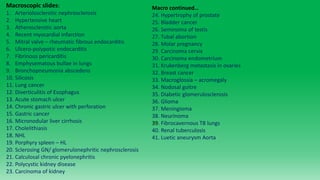
  • 1. Macroscopic slides: 1. Arteriolosclerotic nephrosclerosis 2. Hypertensive heart 3. Atherosclerotic aorta 4. Recent myocardial infarction 5. Mitral valve – rheumatic fibrous endocarditis 6. Ulcero-polypotic endocarditis 7. Fibrinous pericarditis 8. Emphysematous bullae in lungs 9. Bronchopneumonia abscedens 10. Silicosis 11. Lung cancer 12. Diverticulitis of Esophagus 13. Acute stomach ulcer 14. Chronic gastric ulcer with perforation 15. Gastric cancer 16. Micronodular liver cirrhosis 17. Cholelithiasis 18. NHL 19. Porphyry spleen – HL 20. Sclerosing GN/ glomerulonephritic nephrosclerosis 21. Calculosal chronic pyelonephritis 22. Polycystic kidney disease 23. Carcinoma of kidney Macro continued… 24. Hypertrophy of prostate 25. Bladder cancer 26. Seminoma of testis 27. Tubal abortion 28. Molar pregnancy 29. Carcinoma cervix 30. Carcinoma endometrium 31. Krukenberg metastasis in ovaries 32. Breast cancer 33. Macroglossia – acromegaly 34. Nodosal goitre 35. Diabetic glomerulosclerosis 36. Glioma 37. Meningioma 38. Neurinoma 39. Fibrocavernous TB lungs 40. Renal tuberculosis 41. Luetic aneurysm Aorta
  • 2. 1. Benign Nephrosclerosis Arteriolosclerotica (Macroscopic):  Patchy ischemic atrophy with focal loss of parenchyma  Granular appearance  Diffuse Pin-point haemorrhages on cortical surface.  Kidneys are slightly smaller than normal  Essential hypertension/ diabetes mellitus, benign because there is little altered function of the kidneys but increased risk for renal failure. Accelerated (malignant) stage of essential HT Left: Malignant hypertension (300/150mmhg) – a complication/sequelae of benign form, leads to focal haemorrhaging which obscures the corticomedullary junction
  • 3. 2. Hypertensive heart – macro: • Increased peripheral vascular resistance and cardiac workload induces remodelling of the myocardium. This is seen as concentric left ventricular hypertrophy a.k.a “Cor hypertonicum”. • Normal thickness = 5-9mm, borderline = 13/14mm, hypertrophic = > 15mm • The rest of the heart is relatively unchanged. Secondary right ventricular hypertrophy may occur after hypertension induced left sided heart failure and pulmonary hypertension. • Complications of hypertensive heart disease: increased risk of myocardial infarction (larger mass of muscle to be supplied with O2), arrhythmias, heart failure, aortic dissection, aneurysms, accelerated atherosclerosis due to endothelial injury, nephrosclerosis, hypertensive retinopathy, cerebral vascular accident (stroke).
  • 4. 3. Atherosclerosis Aortae (Macro): FIRST STAGE - Fatty streak (white arrows)  Thin, flat, yellow  Non-protruding linear streaks  Present from very young age, even birth  Increase in number until young adulthood then stabilise or may regress  Reversible with lifestyle modification  occur at areas of turbulent flow such as around the orifices of branching vessels of the aorta. From top to bottom: Severe. Moderate and mild atherosclerosis THIRD STAGE – Atheromatous plaque  Reddish brown mural thrombi  Ulceration of intimal surface  Irregular shaped white- yellow atheromatous plaques  Calcifications  Plaques become more confluent/ join up the further the disease progresses.
  • 5. FOURTH STAGE – complicated plaque (unstable) Fates: 1. Plaque may grow around the branch points of vasa vasorum (own vessels) of the aorta leading to atrophy of tunica media. This weakens the wall and can lead to atherosclerotic dissection or aneurysm. 2. Hemorrhage of weak neovascularisation in the plaque may lead to a hematoma which expands the plaque to occlude the lumen, subsequent infarction. 3. Ulceration and rupture of the plaque can release cholesterol (fat) emboli leading to stroke, MI or PE. 4. Dystrophic calcification which hardens the artery leading to changes in pulse, blood pressure and further endothelial damage. Calcific fragments can break off into circulation (calcific emboli) and cause infarction. 5. Re-canalisation: rare but the plaque can be invades by endothelial cells which re-open a larger lumen. A compensatory response. Common location of atherosclerosis: 1. Coronary As => IHD and MI 2. Carotids => vascular dementia, berry aneurysms, cerebral stroke 3. Renal Artery => Stenosis and Secondary hypertension, renal infarction. 4. Iliac As/ Femoral => dry gangrene of feet, intermittent claudication. 5. Aorta => Mesenteric ischemia, dissection, aneurysm 6. Retinal arteries => amaurosis fugax/ acute temporary vision loss Retinal embolus, gangrene, renal stenosis, berry aneurysm.
  • 6. 4. Myocardial Infarction (Macro): Collagen deposition healed MI Discrete pale infarct with hyperaemic border Large transmural MI  Often multi-focal lesions  Initially; Red-purple blotchy areas (hemorrhage and reperfusion injury)  Progression: Yellow border which extends inwards (collagen/ granulation tissue) and hyperaemic border.  Finally; Bright pale yellow/ white/ grey scar  Can lead to HF, arrhythmia. Aneurysm, cardiac tamponade
  • 7. 5. Fibrous endocarditis (Macroscopic): Rheumatic (post-infectious) type endocarditis. The other two types are 1). Infective (ulcero-polypotic) and 2). Non infective which is further subdivided into a). NBTE (marantic) and b). Libman-sacks (verrucous - SLE, anti-phospholipid syndrome) Chronic valvulitis of mitral valve In rheumatic valvopathy we see fusion of commissures, sterile fibrin vegetations and shortened thickened, fused chordae.
  • 8. 6. Ulcero-polypotic Endocarditis (infectious) – macroscopic:  Bacterial/ septic endocarditis characterised by the formation of pedunculated masses/ vegetations of fibrin or thrombin mixed with bacterial colonies and calcifications adhered to an ulcerative heart valve.  Acute forms = S. aureus or GABHS infection  Subacute forms = Strep. Viridans, Strep pyogenes and Staph. Epidermidis  2 predisposing factors are required: 1). BACTEREMIA (anything from poor oral hygiene/ teeth brushing micro-traumas – full blown sepsis) 2). Damage to or structural abnormality of valve endocardium – allows for adherence of passing bacterial colonies.  Vegetations are multiple, large and friable  Tendency to break off as septic- thrombo emboli  More common on left side of heart – mitral and aortic valves.  Ulceration of valve cusps, rupture of chordae tendineae. Local destruction may be rapid and severe  Complications include: heart murmur, arrhythmia, AV block, pericardial effusion, cardiogenic shock, acute heart failure, stroke or pulmonary thromboembolism, brain, heart or lung abscesses, meningitis, glomerulonephritis, sepsis.
  • 9. 7. Fibrinous pericarditis (gross appearance);  Dry course granular pericardial surface  Fibrinous exudate with stringy adhesions between external epicardial surface and the inner surface of the pericardium.  Fibrin has pale yellowish colour  Shaggy rough “Bread and Butter” appearance.  Clinically; pericardial friction rub can be heard. Haemorrhagic pericarditis
  • 10. 8. Emphysema bullosa (macro):  5 classifications – 1. centri-acinar/ centrilobular, 2. pan-acinar/ panlobular, 3. paraseptal (distal lobular), 4. irregular (paracictricial) and 5. mixed.  Multiple, large dilations most commonly under the pleura  Lungs are large, hyperinflated, pale and with little blood  Evidence of chronic smoking often evident; tar deposits  Complications: Spontaneous pneumothorax if they burst, collapse of terminal bronchioles – pneumonia, abscesses, pulmonary fibrosis, atelectasis, respiratory failure, cor pulmonale, chronic bronchitis (often concurrent), increased risk of lung cancer.
  • 11. 9. Bronchopneumonia (lobular pneumonia) abscedens – macro: • Patchy areas of consolidation • Usually bilateral. • Hemorrhage and brighter areas of alveolar purulent exudate. • Consolidation matches the distribution/ shape of the pulmonary lobules hence the name. • This type of pneumonia is most often acquired in hospital settings. • Staph Aureus, Klebsiella, E-coli and Pseudomonas are common culprits. • Poorly defined areas are usually grey-red to tan yellow in colour. • Cavitary lesions/ abscesses may develop amongst the consolidation. • Areas of consolidation are firmer than healthy areas. They also appear raised on cut section.
  • 12. 10. Silicosis – Gross:  Upper lobe is usually affected earlier in the disease  Early lesions = multiple small pale to black ( if coal dust also present) nodules progressing to hard collagenous scars.  Fibrosis tends to occur more at the hilar lymphnodes and pleura.  Nodules may have undulating/ irregular or stellate shaped edges due to traction from the thick fibrosis. They may cavitate centrally due to tuberculosis or ischemia.  Nodules range from 1-5mm.  Well circumscribed  Pleura is thickened, may have adhesions to chest wall  If severe, or in concurrence with COPD, may lead to cor pulmonale.  Silicosis does not seem to increase risk of bronchogenic carcinomas, unlike asbestosis.
  • 13. 11. Lung Carcinoma - Gross:  Most common and fatal cancer  90% of lung cancers are associated with smoking  Cancers most commonly arise in the hilum (centrally) and less often in the periphery Classification of lung cancers: 1). Bronchogenic (most common type) TWO subtypes of bronchogenic Ca: a). NSCLC (non small cell lung ca -85%) b). SCLC (small cell lung cancer – 15%) Of NSCLC, adenocarcinoma is most common = 40% Followed by Squamous = 30% and Large cell = 15% 1. Adenocarcinoma:  Strong assoc. with scarred areas and chronic damage  Lobulated or spiculated shaped mass  May have central area of fibrosis or necrosis  More common in upper lobes, prefers periphery.  Pale, solid, whitish mass  Well circumscribed but not encapsulated  Anthracotic pigments (from smoking)  Has four subtypes Bronchogenic Lung Cancers (hilar/ centrally located – most likely squamous cell carcinomas)
  • 14. Peripherally located carcinomas – later identified to be adenocarcinomas. 2. Squamous cell ca:  Arises centrally from main bronchi  Massive necrosis  Cavitation  Hemorrhages  Strong assoc. with smoking  Grey-white fleshy tumour  May cause bronchiectasis, atelectasis, recurrent pneumonia.  Tendency to invade hilum and surrounding parenchyma
  • 15. 12. Esophageal diverticulum – macroscopic:  Visible outpouching of wall of Esophagus  Contains ALL layers of wall (true diverticulum)  Rare = pseudo diverticulum – only mucosa or submucosa  2 types by etiology: A). Congenital (more common in upper Esophagus or at level of carina) B). Acquired: 1. Pulsion (from increased intraluminal pressure) or 2. Traction (pulled by the formation of adhesions, fibrosis)  Three types by location: 1. proximal (zenkers), 2. mid-esophageal (usually traction type) and 3. epiphrenic (distal)  Zenkers diverticulum is a pulsion type acquired esophageal diverticulum in hypopharyngeal location – due to esophageal obstructions.  Complications: diverticulitis (hyperaemia/ wall thickening, leucocyte infiltration, peri- diverticular edema, muco-purulent exudates, hemorrhage, perforation and subsequent pneumomediastinum or subcutaneous emphysema in neck, esophageal cancer. Opened post-mortem specimen of the oesophagus. An oesophageal diverticulum with creamy exudates and a hard foreign body (bone) adherent to the oesophageal wall (arrow).
  • 16. 13. Acute stomach ulcer – gross :  Less than 1cm  Round shape  Well defined margins  Sometimes confluent  Dark brown – black colour due to formation of hematin.  Fibrinoid necrosis centrally in pit of ulcer  Surrounding tissue is hyperaemic  Most common location: lesser curvature of stomach, if lesion occurs on greater curvature suspect ulcerating gastric carcinoma.  Subtypes of ulcer = A). Curling's ulcer (severe burns) and B). Cushing's ulcer – due to hemodynamic changed due to brain injury/ elevated ICP.
  • 17. 14. Chronic gastric ulcer with perforation - gross :  Round/ oval shape  4-6 cm in diameter (larger than acute ulcers)  Depth variable – may extend through all layers; submucosa, muscle layer and adventitia  Sides are hyperaemic, bulging and overhanging  Base is firm due to fibrosis – callus of the ulcer.  Floor may be relatively clean, active hemorrhage or fully perforating the gastric wall.  Mucosal folds radiate outward from ulcer margins.
  • 18. 15. Gastric carcinoma:  Variable presentation; as large solitary mass, non-healing ulcer or diffuse infiltrative burrowing process (linitis plastica).  Four types: 1). Polypoid/ papillary exophytic growth 2). Fungating (raised margins with central ulceration 3) ulcerative with infiltrating margins 4). Linitis plastica (wall becomes thick and rigid)  Wall thickening, rugal fold flattening  Hemorrhage and necrosis (signs of aggression)
  • 19. 16. Micronodular liver cirrhosis – gross:  Nodules are less than 3mm (macro = 3mm – 3cm).  Diffuse involvement of all hepatic lobules.  Fat deposition may be seen – although less than in fatty liver disease, this gives the organ a tawny brown-yellow appearance. Normal liver is rich maroon red/brown.  Surface is granular and studded with nodules.  Thick fibrose septae may be seen on cut- section.  In severe disease the organ shrinks.
  • 20. 17: Cholelithiasis - gross:  Gallbladder wall may be hypertrophic (thick, contracted, calcified and firm organ) or atrophic (large, flaccid, distended, pale and grey organ).  Diffuse mural hyperaemia – mucosa is bright red, mucinous or muco-purulent exudate (empyema)  Abscesses may form in the wall.  Stones can be of FOUR kinds: 1). Cholesterol stones – oral or polygonal with flattened facets, smooth, yellow-white. 2). Pigment stones – due to increased unconjugated bilirubin often as a result of haemolysis. These stones are small, numerous, mulberry shape and jet black. 3). Calcium stones – grey/white, hard and small. 4). Mixed stones – most common type, a laminated stone comprised of alternating deposits of the contents of the other stones
  • 21. 18. Malignant Non H Lymphoma - (macro):  Solid tumours of the lymphnodes – usually cervical. One third of time they affect extranodal sites – the tonsils, GIT and spleen.  Grossly the appearance of HL and NHL are very similar., the differences are in behaviour, labs and microscopic examination.  Normal lymphnode capsule invaded and destroyed  Surrounding fat tissue is invaded forming thick tracts investing multiple lymphnodes – matted, firm texture upon palpation. NHL in lymphnodes near pancreas
  • 22. 19. Hodgkin's lymphoma spleen (macro): ! DO NOT CONFUSE “Porphyry spleen” with “Sago” or “lardaceous” spleen. Porphyry spleen is caused by Hodgkin's lymphoma and is characterised by multiple nodular leucocyte tumour infiltrates. BOTH Sago spleen and lardaceous spleen are caused by amyloidosis of the organ – characterised by diffuse granular amyloid deposits. This can be due to hereditary amyloidosis (abnormal protein synthesis) or secondary to chronic inflammatory conditions e.g. rheumatoid arthritis, Inflammatory bowel disease etc, due to plasma cell disorders that produce too much immunoglobulins or senile amyloidosis– due to old age and other idiopathic causes. These are shown on the next slide for comparison Porphyry spleen:  Scattered grey- white nodules  Can be singular and large or multiple and smaller.  Nodules coalesce over course of disease  # of nodules = prognostic (>5 = bad).
  • 24. 20. Nephrosclerosis (glomerulonephritic type - macro):  A.k.a diffuse sclerosing GN.  Symmetrically and severely atrophic kidneys - shrunken and only weigh 1/3rd of a healthy kidney.  Surface is finely granular and cortex is thinned  Pelvis and calices are relatively spared, medulla unremarkable.  Capsule may be adherent to cortex.
  • 25. 21. Chronic pyelonephritis with calculus – gross: • Coarsely granular and nodular surface of kidney. • Kidney is asymmetrical and irregular in shape. • Kidney is heavily scarred and shrunken, capsular scarring and retractions correspond to involvement and dilation/ blunting of the underlying calyx. • Blunting of papillae, papillary necrosis is common. • Abscesses and cysts may be seen • Calculi are common in the renal pelvis and proximal ureter, as a result ureter may be dilated.
  • 26. 22. Polycystic kidney gross: • A congenital/ genetic disease that runs in families causing progressive cystic change in the kidneys with age, pain, high blood pressure and renal failure. • Kidneys are massively enlarged and multi-cystic with replacement of the entire cortex.
  • 27. 23. Renal Cell Carcinoma – gross: • Large, round, well-defined masses that usually occur and one of the poles of the kidney. • Pale beige-yellow appearance against the healthy red kidney. • Centred in the cortex, may have pseudocapsule exteriorly and disrupt the smooth contour of the kidney while invading the medulla or renal sinus interiorly. • May be cystic in appearance (bottom right image) or have multiple hemorrhages, focal necrosis or calcifications within the mass – a sign of its aggressive behaviour. • Often invades the renal vein and vena cava.
  • 28. Kidney abscess – Macro: • Round to ovoid pale yellow lesions. • Associated with purulent infections of the kidney – either due to hematogenous spread or an ascending UTI, can also be caused by renal tuberculosis (bottom right image). • Well circumscribed • Frequently multitudinous – especially in acute pyelonephritis where many small abscesses are dotted around the capsular surface and cortex of enlarged firm red kidneys (top left image).
  • 29. 24. Hypertrophy of prostate - gross:  Benign prostatic hyperplasia mostly affects the peri-urethral and transitional area of the gland.  Gland enlarges to over 5/6cm and can weigh 100g+. Normal size = 3-4cm.  Yellow – white nodules of variable size.  Enlarged gland can be seen pushing prominently out into bladder lumen.  May compress urethra “slit-like appearance” on cut section.
  • 30. 25. Carcinoma of urothelium of bladder – Macro:  Two classifications: 1). Epithelial origin – more common 2). Non- epithelial origin.  Urothelial (transitional) cell carcinoma constitutes 90% of epithelial tumours. Others include squamous cc, adenocarcinoma, small cell and mixed.  More common in males, 5th decade of life.  Non-papillary type usually bulky, large and ulcerative. Papillary type urothelial ca:  fused and branched fern-like pattern.  Exophytic, free-floating.  Lateral wall most commonly affected, then posterior wall and trigone.  Usually small – less than 2cm  May be single or multiple  May infiltrate lamina propria and deep in detrusor muscle
  • 31. 26. Seminoma testis – macro: • Most common malignant tumour of testis (45%). It is of the GERM CELL class of tumours. • Uniform solid tan to pale cream, grey- white mass. • Necrosis and hemorrhage are uncommon and minimal. • If spontaneous regression occurs, scarring may be the only indicator of its occurrence. • Homogenous and lobulated, well circumscribed. • Testis is usually enlarged, seminoma may replace entire testicular tissue. • Usually confined to just the testis in 90% of cases.
  • 32. 27. Tubal abortion - macroscopic:  Less than 1% of all pregnancies.  Increased risk in patients with history of pelvic inflammatory disease.  Embryo, placenta and amniotic sac are formed as normal.  This is followed by rupture of the tube around the 2nd – 6th week of gestation.  May cause massive bleeding intraperitoneally.  Foetus either dies immediately or soon after rupture, it may be absorbed, calcified (lithopaedion), extruded into peritoneum or the products of conception may become septic.  Grossly the affected ovarian tube is usually dilated/ distended, hyperaemic.  It may contain what appears to be a hematoma.  Products of conception are often visible – embryo, chorionic villi  Tube may be intact or ruptured.
  • 33. 28. Molar pregnancy – macro:  There are four main categories of trophoblastic gestational disease: 1) Partial mole 2) Complete mole 3) Invasive mole 4) Choriocarcinoma Partial mole:  Placental tissue exists but is immature and abnormal – admixed with hydropic villi that tend to be smaller and more numerous than in complete mole.  Fetal parts and gestational sac may be seen Complete mole:  Hydropic villi which appear as fluid-filled semi-transparent vesicles or variable size  “Bunch of grapes” appearance.  Normal fetal parts are absent  Normal placental structures are absent Invasive mole: Fluid filled vesicles extend into myometrium. Uterus may be perforated. Appears as erosive haemorrhagic lesion. Partial molar pregnancies Complete molar pregnancy Partial mole
  • 34. 29. Carcinoma of cervix – macro: Classification of cervical disease: 1). Inflammatory cervical lesions. 2). Non-neoplastic cervical proliferations (eg. squamous metaplasia, polyps) 3). SIL (squamous intraepithelial lesion or CIN (cervical intraepithelial lesion). - CIN 1 = (Less than 1/3rd of epithelium affected or Mild dysplasia) = low grade SIL - CIN 2 (1-2/3rds involved or moderate dysplasia) - CIN 3 (full thickness/ severe dysplasia or in situ carcinoma – doesn’t invade basement membrane!) [CIN 2+ 3 = High grade SIL] Grossly: squamous cell carcinoma is most common (75% of cases) followed by adenocarcinoma (20%). Squamous Cell Carcinoma:  Red, friable indurated lesion.  Can be ulcerated or elevated granular lesion  Variable morphology: Exophytic, papillary, polypoid, nodular and ulcerative forms have been observed.  Can be invasive and infiltrate surrounding structures. May be necrotic and haemorrhagic.  Associated with HPV 16 + 18 infection.
  • 35. 30. Carcinoma of endometrium – macro:  Most common in post-menopausal women (55-65). Previous pregnancies have a protective effect.  Most present grossly as polypoid/ fungating masses. Uterus is often asymmetrically enlarged  Invasion into the muscular layer is usually present from an early stage.  Earliest symptom = abnormal uterine bleeding (AUB).  Soft and homogenous growth. May sometimes be heterogenous – with foci of necrosis and hemorrhage on cut surface.  Some cases present as simple endometrial thickening – the lining in this case is red, abundant and friable to touch.  Usually has a favourable prognosis.  Types include: 1), endometrioid 2). Serous 3). Clear cell adenocarcinoma and 4). Mixed type  Has five stages of progression: STAGE 1 - body of uterus only STAGE 2 – body and cervix STAGE 3 – extra-uterine but remains in true pelvis STAGE4 – Distant metastasis
  • 36. 31. Krukenberg metastasis in Ovaries - gross:  Metastatic type tumour of the ovaries (5-10% of all ovarian tumours are metastasis), krukenberg is almost always bilateral (80%).  It is a mucin rich signet- ring adenocarcinoma with gastric origin.  Other origins include breast and colon. Rarely the primary tumour may be located in the gallbladder, biliary tract, urinary bladder or cervix.  Prognosis of this tumour is extremely poor.  Small, multinodular surface with extensive extra-ovarian spread.  Metastatic tumours are most difficult to distinguish from mucinous type primary tumours of the ovary.  Scattered growth with intervening normal ovarian parenchyma  Multiple vascular emboli nearby  Similar characteristics to primary tumour – whichever that may be.  May have multiple cysts.  Lymph and blood vessel invasion
  • 37. 32. Breast Cancer - gross: 1. Invasive ductal carcinomas usually form a solid mass with a variable consistency. 2. Tumours with abundant desmoplastic stroma are firm to hard and grey-white in appearance. 3. The presence of microcalcifications may impart a gritty consistency. 4. There may be yellow-white streaks of necrosis and foci of hemorrhage. 5. Extensively necrotic tumours may develop cystic change. 6. The tumours that are composed largely of neoplastic and inflammatory cells without much stroma tend to be soft and fleshy and bulge when they are sectioned.
  • 38. A B A: Clearly visible nipple retraction in advanced stage breast cancer B: Peau d’orange - skin appears studded with dimples like an orange peel.
  • 40. 33. Macroglossia (acromegaly) - gross:  A form of “true” macroglossia (the tongue is indeed larger) whereas “pseudo-macroglossia” is a relative macroglossia – where the tongue appears larger due to the oral cavity or mandible (micrognathia) being smaller the normal.  Caused by muscular hypertrophy of tongue tissue in response to excessive growth hormone. The oral cavity is often enlarged also.  Most cases of acromegaly are caused by a benign functional (secretory) adenoma of the pituitary gland.  Acromegaly is usually undiagnosed until the age of 30 to 50. If it occurs before the fusing of the epiphyseal growth plates at the end of puberty, it is known as gigantism – due to prolonged bone growth.  Differential diagnosis of macroglossia: Congenital true macroglossia: - Mucopolysaccharidosis - Idiopathic tongue muscular hypertrophy - Haemangioma - Lymphangioma Acquired true macroglossia: - Amyloidosis (most common cause in adults) - Hypothyroidism (myxoedema) - Sarcoidosis, lymphoma, trauma, TB, syphilis, actinomycosis
  • 41. 34. Nodular goiter – macroscopic:  Unlike simple goiters, multinodular goiters are asymmetrical and very large, weighing on average between 200-700g but can grow to be as large as 2kg!  Nodularity and poor encapsulation  Haemorrhages are common  Scarring is abundant as is cystic degeneration and focal calcifications.  Individual nodules are variable in size with dominant ones often larger than 10cm.  Surface is bumpy and irregular  Brown gelatinous colloid collections may be seen on cut surface.
  • 42. 35. Diabetic glomerulosclerosis - macroscopic:  Renal lesions appear around 10-20 years after onset of diabetes.  Major symptoms include; proteinuria, glucosuria, progressive decrease in renal function, increased recurrence of pyelonephritis, edema and hypertension may manifest as late-stage symptoms.  Gross features include: - Slight to moderate symmetrical enlargement of the size of the kidneys. - Subcapsular cortical surface is finely granular and dark red in colour. - Cortex is thinned - Atherosclerotic changes to renal vessels, renal stenosis and subsequent renal atrophy. - Pyelonephritic features may be observed (due to the predisposition of glycosuria to precipitate infections); deep irregular surface scars and retractions, dilated and deformed calyces, renal calculi, cysts and if active infection was occurring; multiple micro- abcesses.
  • 43.
  • 44. 36. Glioma – gross:  Gliomas are tumours of the glial cells; astrocytoma, glioblastoma, oligodendrogliomas, ependymomas and choroid plexus papilloma. They are the most common brain tumour group and comprise 40%.  Astrocytoma is the most common glioma: - Found in mid- late life (40-60yo). - Most often located in cerebral hemispheres, sometimes in spinal cord but can occur anywhere (optic nerves, brainstem, cerebellum etc). - Have a tendency to progress from low grade to high grade and transform into glioblastoma multiforme. - They are grey-white, cystic or solid masses, gradually grow and therefore clinical symptoms are insidious. - Signs of transformation to aggressive glioblastoma includes acceleration in symptom progression, yellow, hemorrhages, focal necrosis, poor margins and heterogenous soft consistency. Astrocytoma Glioblastoma Glioblastoma Glioblastoma
  • 45. 37. Meningioma – gross:  Most common location is along falx cerebri and parasagittal (25%), calvarium convexity (20%), sphenoid wing (20%), suprasellar (10%), posterior fossa (10%).  It may occur in the orbit – congenital cause of exophthalmos. May occasionally occur at cerebellopontine angle and mimic acoustic neuromas.  Tan, quite spherical or hemi-spherical, well- defined margins, may be lobulated, firm, fibrose. Usually meningiomas are solitary.  May contain focal calcific precipitates.  1-10cm in size, overlying skull may have reactive thickening (hyperostosis) and sclerosis.  Attached to dura (Note: it does not originate from the dura, it arises from the arachnoid layer!)  Broad base, may be solid, cystic or gelatinous in texture. When removed they leave a pit/ depression in the underlying cerebral hemisphere. Clinical symptoms depend on location; paresis, seizures, headaches, vision changes, language impairment, memory loss.
  • 46. 38. Neurinoma – macroscopic:  Does not occur in CNS only peripheral nerves; spinal nerve roots, cranial nerves.  Pontocerebellar angle near internal acoustic meatus is a very common site – affecting CN VIII (vestibulocochlear nerve).  Arise from Schwann cells that make myelin sheath on sensory, motor and autonomic nerves.  Encapsulated, solid, sometimes cystic, eccentric enlargement.  When they are rich in collagenous tissue they are subclassified as neurofibromas (neurofibromatosis).  “Pepples in shoe” pain sensation is a distinctive symptom of lower limb neuroma.
  • 47. 39. Fibrocavernous TB of lungs – gross:  Apices of lungs are most heavily affected. Usually bilateral involvement. Unilateral hilar lymphnodes involvement (kids).  Thick fibrose non-compliant upper lobes, pleura may be thickened, bronchial stricture and bronchiectasis due to deformity, displacement and traction.  Multiple granulomas with central caseous (cheese-like) necrosis. There may be focal hemorrhages and cavitation.  Miliary type TB features multiple diffuse micro- abscesses throughout the lungs. It indicates hematogenous spread and may be associated with TB meningitis, renal TB, bone TB and has a poor prognosis.
  • 48. 40. Renal Tuberculosis – gross:  Young to middle aged adults.  Renal involvement may be indolent, insidious taking anywhere from 10-30 years to manifest after primary TB infection.  Onset of symptoms as unilateral renal impairment and less often, renal failure.  Multiple large cavities filled with thick, yellowish caseous necrosis.  Liquefaction and cavity formation.  Secondary or concomitant infection - bacterial abscesses.  Contraction and fibrosis may lead to irregularity and deformity of the cortical surface.  Correlating clinical symptoms include; flank pain, increased frequency, blood and pus in urine, fever, dysuria as well as signs of renal insufficiency.
  • 49. 41. Luetic aneurysm – gross:  Aortic intima has fine or course, irregular foldings.  Scattered foci of fibrosis (pearly-white nodules) along the intima creating characteristic “tree-bark” appearance.  Typical feature of advanced or tertiary stage of syphilis infection.  Ascending aorta and aortic arch are most frequently impacted.  Superimposing atherosclerotic changes are frequent. The cerebral vessels may also be affected by syphilitic vasculitis  Aorta lumen is dilated, walls are hardened, fibrotic and thickened, there is replacement of the atrophied tunica media muscle.