
Recommended
More Related Content
Similar to ANA 211 Imaging.pdf
Similar to ANA 211 Imaging.pdf (20)
Recently uploaded
Recently uploaded (20)
ANA 211 Imaging.pdf
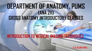
- 1. DEPARTMENT OF ANATOMY, PUMS (ANA 211) GROSS ANATOMY INTRODUCTORY CLASSES TOPIC: INTRODUCTION TO MEDICAL IMAGING TECHNIQUES S. O. ELIJAH (PhD)
- 2. INTRODUCTION • Medical imaging is the technique of producing visual representations of areas inside the human body to diagnose medical problems and monitor treatment. Types of Medical Imaging • The major types of imaging used in modern medicine include: • Radiography • Computer Tomography (CT) • Magnetic resonance imaging (MRI) • Nuclear medicine • Ultrasound.
- 3. RADIOGRAPHY • Radiography uses electromagnetic radiation to take images of the body. • Most known and common form of radiography is x-ray. Procedure: - • Machine beams high-energy waves onto the body. • The soft tissues, such as skin and organs, do not absorb waves, appear black on film. • Hard tissue like bones absorb waves, appear white on film.
- 4. USES OF X-RAY •Mostly used to check for bone fractures. •Chest x-ray can spot pneumonia •Mammograms - breast cancer
- 5. COMPUTED TOMOGRAPHY (CT) • Scans show radiographic images of the body that resemble transverse anatomical sections. Technique – • A beam of X-rays passes through the body as the X-ray tube and detector rotate around the axis of the body. • The computer maps the voxels into a planar image (slice) that is displayed on a monitor or printout. • Areas of great absorption (e.g., bone) are relatively transparent (white) and those with little absorption are black.
- 6. • Scans are always displayed as if the viewer were standing at a supine patient’s feet—i.e., from an inferior view. USES OF COMPUTED TOMOGRAPHY • Diagnostic examinations of diseases and injuries • Plan medical, surgical or radiation treatment. • Dictate internal bleeding
- 7. MAGNETIC RESONANCE IMAGING (MRI) • Magnetic resonance imaging involves radio waves and magnetic fields to look at the organs and other structures in the body. Procedure requires an MRI scanner, (a large tube that contains a massive circular magnet). • Magnet creates a powerful magnetic field that aligns the protons of hydrogen atoms in the body. • Protons are then exposed to radio waves, causing the protons to rotate. • When the radio waves are turned off, the protons relax and realign themselves, emitting radio waves in the recovery process that can be detected by the machine to create an image.
- 8. USES OF MAGNETIC RESONANCE IMAGING (MRI) •Mainly for diagnostic examinations of the brain and spinal cord. •Can be used to diagnose torn ligaments or even tumors
- 9. NUCLEAR MEDICAL IMAGING • Nuclear medical imaging refers to the use of radioactive tracers (radioactive materials). • Materials are injected or swallowed so they can travel through the digestive or circulatory system. • The radiation produced by the material can then be detected to create an image.
- 10. USES OF NUCLEAR MEDICAL IMAGING • Used mainly in the diagnoses and treatment of abnormalities very early in the progression of a disease,such as thyroid cancer. • NMI provides unique information that is often unattainable using other imaging procedures.
- 11. ULTRASOUND • Ultrasound utilizes high-frequency sound waves, which • Sounds are reflected off tissue to create images of organs, muscles, joints, and other soft tissues. • It's more like shining a light on the inside of the body, except that this light travels through the skin layers and can only be viewed using electronic sensors.
- 12. USES OF ULTRASOUND • Viewing uterus and overies during pregnancy and monitor the developing baby’s health. • Diagnose gallbladder disease • Evaluate blood flow • Guid a needle for biopsy or tumor ytreatment • Examine breast lump • Check thyroid gland • Detect genital and prostate problems.
- 13. THANK YOU