

Recommended
More Related Content
What's hot
What's hot (20)
Similar to Mcqs images cvs
Similar to Mcqs images cvs (20)
More from DOCTOR WHO
More from DOCTOR WHO (20)
Recently uploaded
Recently uploaded (20)
Mcqs images cvs
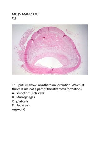
- 1. MCQS IMAGES CVS Q1 This picture shows an atheroma formation. Which of the cells are not a part of the atheroma formation? A Smooth muscle cells B Macrophages C glial cells D Foam cells Answer C
- 2. Q2 There is atheroma formation starting with a fatty streak and then plaque formation. An atheroma formation can lead to A Thrombosis B Infarction C Hemorrhage D All of the above Answer D
- 3. Q3 This area shows a slight pallor with neutrophils begin to show up. Which one is the right time for the neutrophils to show up in a Myocardial infection? A 1 to 12 hrs B 18 to 72 hrs C After 3 days D 4 to 7 days Answer B
- 4. Q4 The above picture is arterial wall from a 70 year old man suffering from essential hypertension shows A Hyaline arteriosclerosis B Hyperplastic arteriosclerosis C Vasculitis D Degenerative vasculopathy Answer A
- 5. Q5 Hyperplastic arteriosclerosis is commonly associated With A Malignant Hypertension B Nephrotic Syndrome C Nephritic Syndrome D Infective endocarditis Answer A
- 6. Q6 Multinucleated macrophages in rheumatic heart disease are known as A Aschoff Cells B Antischkow cells C MacCallum cells D Libman-sack cells
- 7. Q7 The above picture shows microscopic features of Hypersensitivity Myocarditis it reveals A Interstitial inflammatory infiltrate composed largely of eosinophils and mononuclear inflammatory cells. B Interstitial inflammatory infiltrate composed largely of neutrophils. C Interstitial inflammatory infiltrate composed largely of plasma cells. D Interstitial inflammatory infiltrate composed largely of plasma cells Answer A
- 8. Q8 Infective endocarditis showing irregular large friable valvular vegetation. These are caused mainly by A Streptococcus viridian B Streptococcus aureus C Pseudomonas D Viruses Answer A
- 9. Q9 Temporal (giant cell) arteritis microscopically shows A Giant cells at the degenerated external elastic membrane and intimal thickening. B Giant cells at the degenerated internal elastic membrane in active arteritis and intimal thickening. C Degenerated external elastic membrane in active arteritis and intimal thickening. D Degenerated internal elastic membrane and intimal thinning. Answer B
- 10. Q10 The above picture shows dense clumps of irregular, moderate anaplastic cells and distinct vascular lumens. which of the vascular tumor reveal these features? A Myxoma of Atrium B Kaposisarcoma C Angiosarcoma D Capillary Heamangioma Answer C