SlideShare ist ein Scribd-Unternehmen logo
1 von 26
General Features of Cerebellum :
Lateral aspect of brain
stem & cerebellum ,
showing cerebellar
peduncles.
It controls equilibrium, it influences
posture & muscle tone and coordinates
the movements
Its surface is high convoluted, forming
folds or folia, being oriented transversely
It lies behind Pons & M.O. , separated
from them by the cavity of 4th ventricle.
It is connected to brain stem (medulla,
pons& midbrain) by inferior, middle &
superior cerebellar pedunclesrespectively.
The cerebellum consists of a midline
vermis and 2-lateral hemispheres.
Anatomically , it is divided into anterior
, posterior & flocculo-nodular lobes.
External Features of Cerebellum :
It has anterior notch ,which is
wider and lodging the back of pons
& medulla. It is separated from
them by cavity of 4thventricle
 It has also posterior notch
occupied by falx cerebelli, which
separates the 2 cerebellar H.
Inferior surface : rounded on
each side and presents :
a deep groove (vallecula) between
the 2-cerebellar hemispheres,which
is occupied by the inferior vermis.
-Tonsil is a small part of cerebellar
hemisphere that lies lateral to
inferior vermis.
Superior surface
Inferior surface
External Features of Cerebellum :
Superior surface : lies beneath
tentorium cerebelli and has a raised superior
vermis + a large cerebellar hemisphere on
each side + primary & horizontal fissures.
1- Primary fissure V-shaped,well defined
fissure, lies on superior surface and
separates the small anterior lobe from the
larger middle lobe (or posterior lobe).
2- Horizontal fissure lies along the sides of
cerebellum, extending from anterior notch
to posterior notch, separates the superior
from the inferior surfaces.
3- Secondary (posterolateral) fissure
lies on inferior surface and separates
flocculo-nodular lobe from the ramainder
of cerebellum.
Schematic drawing of cerebellum
showing the relationships between the
anatomical & functional divisions of
cerebellum.
Green =archi-cerebellum,
blue=paleo-cerebellum.
Pink= neo-cerebellum
Functional subdivision of
cerebellum :
1- Archi-cerebellum =
posterior lobe
(Vestibular part) :
_ It is formed of the flocculo-
nodular lobe + associated fastigial
nuclei, lying on inf. Surface in front
of postero-lateral fissure.
_Embryologically, it is the oldest
part of cerebellum.
_It receives afferent Fs. From
vestibular apparatus of internal ear
Via vestibulo-cerebellar tracts.
_It is concerned with equlibrium.
Connections of
archicerebellum
I- Archicerebellum
It is concerned with equilibrium.
It represents flocculo-nodular lobe.
It has connections with vestibular &
reticular nuclei of brain stem
through the inferior cerebellar
peduncle.
Afferent vestibular Fs. Pass from
vestibular nuclei in pons & medulla to the
cortex of ipsilateral flocculo-nodular lobe.
Efferent cortical (purkinje cell) Fs.
Project to fastigial nucleus, which projects to
vestibular nuclei & reticular formation.
It affects the L.M.system bilaterally via
descending vestibulo-spinal & reticulo-spinal
Schematic drawing of cerebellum
showing the relationships between the
anatomical & functional divisions of
cerebellum.
Green =archi-cerebellum,
blue=paleo-cerebellum.
Pink= neo-cerebellum
2- Paleo-cerebellum=
(spinal part) :
-_it is formed of midline vermis
+ surrounding paravermis +
globose & emboliform nuclei.
_It receives afferent proprio-
ceptive impulses from Ms.&
tendons Via spino-cerebellar
tracts (dorsal & ventral) mainly.
-it sends efferents to red nucleus
of midbrain.
-it is concerned with muscle tone
Connections of Paleo-cerebellum.
2-Paleo-cerebellum
It is concerned with muscle tone
& posture.
Afferents spinal Fs. consist of
dorsal & ventral spino-cerebellar
tract from muscle, joint &
cutaneous receptors to enter the
cortex of ipsilateral vermis & para
vermis Via inferior & superior
cerebellar peduncles .
Efferents cortical fibres pass to
globose & emboliform nuclei, then
Via sup. C. peduncle to contra-
lateral red nucleus of midbrain to
give rise descending rubro-spinal
tract.
Schematic drawing of cerebellum showing
the relationships between the anatomical &
functional divisions of cerebellum.
Green =archi-cerebellum,
blue=paleo-cerebellum.
Pink= neo-cerebellum
3- Neo-cerebellum=
(cerebral part) :
_It is the remaining largest part
of cerebellum.
_It includes the most 2-cerebellar
hemispheres + dendate nuclei.
_It receives afferent impulses
from the cerebral cortex+pons
Via cerebro-ponto- cerebellar
pathway.
-it sends efferents to V.L.nucleus
of thalamus.
-it controls voluntary movements
(muscle coordination).
Connections of Neo-cerebellum.
3- Neo-cerebellum
It is concerned with muscular
coordination.
It receives afferents from cerebral
cortex involved in planning of
movement- to pontine nuclei ,cross to
opposite side Via middle Cerebellar
peduncle to end in lateral parts of
cerebellum (cerebro-ponto-cerebellar
tract).
Neo-cerebellar efferents project to
dendate nucleus,which in turn projects to
contra-lateral red nucleus & ventral
lateral nucleus of thalamus ,then to
motor cortex of frontal lobe, giving rise
descending cortico-spinal & cortico-
bulbar pathways.
Efferents of dentate nucleus form a
major part of superior C. peduncle.
Cerebellar Lesions
 Are usually vascular, may be traumatic or tumour.
 Manifestations of unilateral cerebellar lesions :
1-ipsilateral incoordination of (U.L) arm = intention tremors : it is a
terminal tremors at the end of movement as in touching nose or button
the shirt.
2-Or ipsilateral cerebellar ataxia affects (L.L.) leg, causing wide-based
unsteady gait.
 Manifestations of bilateral cerebellar lesions (caused by alcoholic
intoxication, hypothyrodism, cerebellar degeneration & multiple
sclerosis) :
1-dysarthria : slowness & slurring of speech.
2-Incoordination of both arms.= intention tremors.
3-Cerebellar ataxia : intermittent jerky movements or staggering ,
wide-based, unsteady gait.
4-Nystagmus : is a very common feature of multiple sclerosis. It is due
to impairment coordination of eye movements /so, incoordination of
eye movements occurs and eyes exhibit a to-and-fro motion.
 Combination of nystagmus+ dysarthria + intension tremors constitutes
Chacot’triad, which is highly diagnostic of the disease.
Internal Structure of cerebellum :
Sagittal section of cerebellum.
T.S.of cerebellum & brain at level
of 4th V. to show cerebellar nuclei.
It consists of an outer
layer of grey matter
(cerebellar cortex) , &
inner layer of white matter
containing 4-pairs of
cerebellar nuclei :
above roof of 4th V. from
medial to lateral :
1-Fastigial nucleus.
2-Globose nucleus.
3-Emboliform nucleus.
4-Dendate nucleus.
(the only one that can be
seen clearly with the naked
eye).
Cerebellar cortex
It is highly convoluted,
forming numerous transversely
oriented folia.
It contains nerve cells,
dendrites and synaptic
connections of cellular
neurones.
The cellular organization of
the cortex consists of 3-layers :
1-Outer molecular layer.
2-Intermediate, purkinje cell
layer.
3-Inner granular layer, which is
dominated by granule cell.
T.S of cerebellar folia showing
layers of cerebellar cortex.
Afferent & Efferent connecltions and
their relationships to principal cells of
cerebellar cortex.
Cerebellar cortex
Molecular layer : contains
1-Cells : molecular cells (stellate cells) &
basket cells.
2-Nerve Fibres :
a- dendrites of Purkinje cells
(arborisations).
B-axons of granule cells. ( bifurcate to
produce 2-parallel fibres , oriented
along long axis of folium).
C-ending of climbing fibers.
Purkinje cell layer : it is formed
of one layer (unicellular) of large flask-
shaped purkinje cells. Their arborisations
are at right angles to long axis to folium.
Granular layer : it is formed of
small granule cells & ending of mossy
fibres.
MThere are 3-types of
Nerve Fibres in white
Matter :
1-Axons of purkinje cells :
the only axons to leave cerebellar
cortex to end in deep cerebellar
nuclei specially dendate nucleus.
2-Mossy Fibres : end in the
granular layer.
3-Climbing Fibres : end in the
molecular layer.
Afferent Fibres to cerebellum :
Mostly end in cerebellar cortex,
excitatory to cortical neurones,
as mossy or climbing Fs. passing
through the cerebellar peduncles.
The following are Afferent fibres:
1-dorsal & ventral spino-cerebellar
tract. (passing via I.C.P & S.C.P)
2-vestibulo-cerebellar Fs. (via I.C.P)
3-olivo-cerebellar Fs. (via I.C.P)/
(extrapyramidal fibres), (end as
climbing or mossy fibres)
4-ponto-cerebellar Fs. (via M.C.P).
(In M.O)
M
Efferent Fibres of the
cerebellum :
It sends the following fibres :
1-Cerebello-vestibular Fs. to
vestibular nuclei of pons & M.O.
2-Cerebello-olivary Fs. To M.O.
3-Dendato-rubro-thalamic tract To
red nucleus of midbrain & ventro-
lateral nucleus of the thalamus and
finally to motor cortex of frontal
lobe to coordinate movement via
cortico-spinal & corticobulbar
tracts.
The Fourth Ventricle
It is a cavity of hindbrain.
Position : lies between pons & M.O.
anteriorly and the cerebellum posteriorly.
It is a diamond-shaped space which is
lined by ependyma.
Its superior angle is continuous with
cerebral aqueduct of midbrain.
inferior angle is continuous with
centeral canal of closed M.O.
Its lateral angles extend laterally to
form a lateral recess on each side to
open into subarachnoid space.
The Boundaries of 4th Ventricle
Superiolateral boundary :
-it is formed by superior cerebellar
peduncle on each side.
Inferiolateral boundary :
-it is formed by inferior cerebellar
peduncle + gracile & cuneate
tubercles on each side.
The Roof of 4th Ventricle
-it is a tent-shaped when seen
laterally, and diamond-shaped when
seen behind.
-it is formed of superior & inferior
medullary vela, which are thin sheets
of white matter /consists of :
ependyma covered by pia mater.
-Sup.medullary velum connects the 2
sup.cerebellar peduncles.
-Inf.medullary velum connects the 2
inf.cerebellar peduncles.
-Inferior vermis of cerebellum : lies
in the middle of roof of 4th ventricle.
The Roof of 4th Ventricle
The lower part of roof is
invaginated by tela choroidea of 4th
ventricle.
The tela choroidea is a double
layer of pia mater which encloses the
choroid plexus of 4th ventricle.
The choroid plexus is a vascular
capillary tuft covered by ependymal
cells and secretes C.S.F. into the
lumen of 4th ventricle.
The Openings of 4th Ventricle
The roof contains 3 aperatures
which transmit C.S.F. from ventricular
lumen to subarachnoid space.
Median aperature (foramen of
Magendie) : lies in the median plane at
lower end of inferior medullary velum,
and opens into subarachnoid space at
cistrna magna at cerebello-medullary
angle
2 lateral openings (foramina of
Luschka) : each one lies at the lateral end
of lateral recess to open into
subarachnoid space at cerebello-pontine
angle. choroid plexus partly protrudes out
through each lateral aperture.
The Floor of 4th Ventricle
A diagram to show the floor & lateral
boundaries of 4th ventricle.
It is called rhomboid fossa.
It is diamond-shaped and is
divided into right & left halves
by the median sulcus.
It is crossed in the middle by
transvere Fs. (ponto-cerebellar
Fs.)called medullary stria,
which divide floor of 4th
ventricle into upper (pontine)
& lower (medullary) part.
The Floor of 4th Ventricle
A diagram to show the floor & lateral
boundaries of 4th ventricle.
Upper pontine part : presents
on each side of median sulcus.
1-Medial eminence : a rounded
elevation produced by the abducent
nucleus.
2-Facial colliculus : an elevation
on the top of lower part of medial
eminence. It is produced by the
fibres of facial nerve which
surround abducent nucleus.
3-Superior fovea : a groove lateral
to facial colliculus.
4-Vestibular area : lateral to
superior fovea. It overlies superior,
medial & lateral vestibular nuclei.
The Floor of 4th Ventricle
A diagram to show the floor & lateral
boundaries of 4th ventricle.
Lower medullary part :
presents on each side of the
median sulcus. 1-Inferior fovea :
inverted V-shaped groove.
2-Hypoglossal area : medial to
inferior fovea. It overlies
hypo-glossal nucleus.
3-Vagal area (triangle) : between
limbs of inferior fovea.It overlies
dorsal nucleus of vagus.
4-Vestibular area : lateral to
inferior fovea. It overlies inferior
vestibular nucleus.
10-cerebellum.ppt

Weitere ähnliche Inhalte

Ähnlich wie 10-cerebellum.ppt

Neuroanatomy Cerebellum-5 (Muhadharaty).pdf
Neuroanatomy Cerebellum-5 (Muhadharaty).pdfNeuroanatomy Cerebellum-5 (Muhadharaty).pdf
Neuroanatomy Cerebellum-5 (Muhadharaty).pdfmaktabaalarbaeen
 
Cerebellum 2010
Cerebellum 2010Cerebellum 2010
Cerebellum 2010PS Deb
 
Cerebellum by dr.gourav thakre 20 03-2012
Cerebellum by dr.gourav thakre 20 03-2012Cerebellum by dr.gourav thakre 20 03-2012
Cerebellum by dr.gourav thakre 20 03-2012Gourav Thakre
 
Cerebellum parts and functions
Cerebellum parts and functionsCerebellum parts and functions
Cerebellum parts and functionsKokachi KP
 
Embryology development of central nervous system
Embryology   development of central nervous systemEmbryology   development of central nervous system
Embryology development of central nervous systemMBBS IMS MSU
 
Anatomy & functions of the Brainstem & Cerebellum
Anatomy & functions of the Brainstem & CerebellumAnatomy & functions of the Brainstem & Cerebellum
Anatomy & functions of the Brainstem & CerebellumRafid Rashid
 
Central nervous system
Central nervous system Central nervous system
Central nervous system Sunil Pahari
 
Nervous System- Comparative account of Brain.pdf
Nervous System- Comparative account of Brain.pdfNervous System- Comparative account of Brain.pdf
Nervous System- Comparative account of Brain.pdfPRIMEPRIME4
 
Cerebellum Physiology
Cerebellum  PhysiologyCerebellum  Physiology
Cerebellum PhysiologyRaghu Veer
 
Embryology developmentofcentralnervoussystem-100708132545-phpapp02
Embryology developmentofcentralnervoussystem-100708132545-phpapp02Embryology developmentofcentralnervoussystem-100708132545-phpapp02
Embryology developmentofcentralnervoussystem-100708132545-phpapp02PTCHANDAN221
 
embryology_developmentofcentralnervoussystem_100708132545_phpapp02.pdf
embryology_developmentofcentralnervoussystem_100708132545_phpapp02.pdfembryology_developmentofcentralnervoussystem_100708132545_phpapp02.pdf
embryology_developmentofcentralnervoussystem_100708132545_phpapp02.pdfJoharseid1
 

Ähnlich wie 10-cerebellum.ppt (20)

Neuroanatomy Cerebellum-5 (Muhadharaty).pdf
Neuroanatomy Cerebellum-5 (Muhadharaty).pdfNeuroanatomy Cerebellum-5 (Muhadharaty).pdf
Neuroanatomy Cerebellum-5 (Muhadharaty).pdf
 
Nervous systems in vertebrates: T.Y.B.Sc. Sem VI Notes
Nervous systems in vertebrates: T.Y.B.Sc. Sem VI NotesNervous systems in vertebrates: T.Y.B.Sc. Sem VI Notes
Nervous systems in vertebrates: T.Y.B.Sc. Sem VI Notes
 
Cerebellum 2010
Cerebellum 2010Cerebellum 2010
Cerebellum 2010
 
Cerebellum by dr.gourav thakre 20 03-2012
Cerebellum by dr.gourav thakre 20 03-2012Cerebellum by dr.gourav thakre 20 03-2012
Cerebellum by dr.gourav thakre 20 03-2012
 
Cerebellum parts and functions
Cerebellum parts and functionsCerebellum parts and functions
Cerebellum parts and functions
 
Embryology development of central nervous system
Embryology   development of central nervous systemEmbryology   development of central nervous system
Embryology development of central nervous system
 
Anatomy of Cerebellum(Part- 2)
Anatomy of Cerebellum(Part- 2)Anatomy of Cerebellum(Part- 2)
Anatomy of Cerebellum(Part- 2)
 
Cerebellum.ppt
Cerebellum.pptCerebellum.ppt
Cerebellum.ppt
 
Anatomy & functions of the Brainstem & Cerebellum
Anatomy & functions of the Brainstem & CerebellumAnatomy & functions of the Brainstem & Cerebellum
Anatomy & functions of the Brainstem & Cerebellum
 
Central nervous system
Central nervous system Central nervous system
Central nervous system
 
Anatomy of Cerebellum 1
Anatomy of Cerebellum  1Anatomy of Cerebellum  1
Anatomy of Cerebellum 1
 
Nervous System- Comparative account of Brain.pdf
Nervous System- Comparative account of Brain.pdfNervous System- Comparative account of Brain.pdf
Nervous System- Comparative account of Brain.pdf
 
Medulla oblongata
Medulla oblongata Medulla oblongata
Medulla oblongata
 
Cerebellum
CerebellumCerebellum
Cerebellum
 
Cerebellum Physiology
Cerebellum  PhysiologyCerebellum  Physiology
Cerebellum Physiology
 
Embryology developmentofcentralnervoussystem-100708132545-phpapp02
Embryology developmentofcentralnervoussystem-100708132545-phpapp02Embryology developmentofcentralnervoussystem-100708132545-phpapp02
Embryology developmentofcentralnervoussystem-100708132545-phpapp02
 
embryology_developmentofcentralnervoussystem_100708132545_phpapp02.pdf
embryology_developmentofcentralnervoussystem_100708132545_phpapp02.pdfembryology_developmentofcentralnervoussystem_100708132545_phpapp02.pdf
embryology_developmentofcentralnervoussystem_100708132545_phpapp02.pdf
 
Ch11 eoc
Ch11 eocCh11 eoc
Ch11 eoc
 
Cns bs2
Cns bs2Cns bs2
Cns bs2
 
Cns bss
Cns bssCns bss
Cns bss
 

Kürzlich hochgeladen

Music 9 - 4th quarter - Vocal Music of the Romantic Period.pptx
Music 9 - 4th quarter - Vocal Music of the Romantic Period.pptxMusic 9 - 4th quarter - Vocal Music of the Romantic Period.pptx
Music 9 - 4th quarter - Vocal Music of the Romantic Period.pptxleah joy valeriano
 
GRADE 4 - SUMMATIVE TEST QUARTER 4 ALL SUBJECTS
GRADE 4 - SUMMATIVE TEST QUARTER 4 ALL SUBJECTSGRADE 4 - SUMMATIVE TEST QUARTER 4 ALL SUBJECTS
GRADE 4 - SUMMATIVE TEST QUARTER 4 ALL SUBJECTSJoshuaGantuangco2
 
ICS2208 Lecture6 Notes for SL spaces.pdf
ICS2208 Lecture6 Notes for SL spaces.pdfICS2208 Lecture6 Notes for SL spaces.pdf
ICS2208 Lecture6 Notes for SL spaces.pdfVanessa Camilleri
 
MULTIDISCIPLINRY NATURE OF THE ENVIRONMENTAL STUDIES.pptx
MULTIDISCIPLINRY NATURE OF THE ENVIRONMENTAL STUDIES.pptxMULTIDISCIPLINRY NATURE OF THE ENVIRONMENTAL STUDIES.pptx
MULTIDISCIPLINRY NATURE OF THE ENVIRONMENTAL STUDIES.pptxAnupkumar Sharma
 
Full Stack Web Development Course for Beginners
Full Stack Web Development Course  for BeginnersFull Stack Web Development Course  for Beginners
Full Stack Web Development Course for BeginnersSabitha Banu
 
INTRODUCTION TO CATHOLIC CHRISTOLOGY.pptx
INTRODUCTION TO CATHOLIC CHRISTOLOGY.pptxINTRODUCTION TO CATHOLIC CHRISTOLOGY.pptx
INTRODUCTION TO CATHOLIC CHRISTOLOGY.pptxHumphrey A Beña
 
Virtual-Orientation-on-the-Administration-of-NATG12-NATG6-and-ELLNA.pdf
Virtual-Orientation-on-the-Administration-of-NATG12-NATG6-and-ELLNA.pdfVirtual-Orientation-on-the-Administration-of-NATG12-NATG6-and-ELLNA.pdf
Virtual-Orientation-on-the-Administration-of-NATG12-NATG6-and-ELLNA.pdfErwinPantujan2
 
How to Add Barcode on PDF Report in Odoo 17
How to Add Barcode on PDF Report in Odoo 17How to Add Barcode on PDF Report in Odoo 17
How to Add Barcode on PDF Report in Odoo 17Celine George
 
ENGLISH 7_Q4_LESSON 2_ Employing a Variety of Strategies for Effective Interp...
ENGLISH 7_Q4_LESSON 2_ Employing a Variety of Strategies for Effective Interp...ENGLISH 7_Q4_LESSON 2_ Employing a Variety of Strategies for Effective Interp...
ENGLISH 7_Q4_LESSON 2_ Employing a Variety of Strategies for Effective Interp...JhezDiaz1
 
4.16.24 21st Century Movements for Black Lives.pptx
4.16.24 21st Century Movements for Black Lives.pptx4.16.24 21st Century Movements for Black Lives.pptx
4.16.24 21st Century Movements for Black Lives.pptxmary850239
 
Student Profile Sample - We help schools to connect the data they have, with ...
Student Profile Sample - We help schools to connect the data they have, with ...Student Profile Sample - We help schools to connect the data they have, with ...
Student Profile Sample - We help schools to connect the data they have, with ...Seán Kennedy
 
4.18.24 Movement Legacies, Reflection, and Review.pptx
4.18.24 Movement Legacies, Reflection, and Review.pptx4.18.24 Movement Legacies, Reflection, and Review.pptx
4.18.24 Movement Legacies, Reflection, and Review.pptxmary850239
 
Influencing policy (training slides from Fast Track Impact)
Influencing policy (training slides from Fast Track Impact)Influencing policy (training slides from Fast Track Impact)
Influencing policy (training slides from Fast Track Impact)Mark Reed
 
USPS® Forced Meter Migration - How to Know if Your Postage Meter Will Soon be...
USPS® Forced Meter Migration - How to Know if Your Postage Meter Will Soon be...USPS® Forced Meter Migration - How to Know if Your Postage Meter Will Soon be...
USPS® Forced Meter Migration - How to Know if Your Postage Meter Will Soon be...Postal Advocate Inc.
 
How to do quick user assign in kanban in Odoo 17 ERP
How to do quick user assign in kanban in Odoo 17 ERPHow to do quick user assign in kanban in Odoo 17 ERP
How to do quick user assign in kanban in Odoo 17 ERPCeline George
 

Kürzlich hochgeladen (20)

Music 9 - 4th quarter - Vocal Music of the Romantic Period.pptx
Music 9 - 4th quarter - Vocal Music of the Romantic Period.pptxMusic 9 - 4th quarter - Vocal Music of the Romantic Period.pptx
Music 9 - 4th quarter - Vocal Music of the Romantic Period.pptx
 
GRADE 4 - SUMMATIVE TEST QUARTER 4 ALL SUBJECTS
GRADE 4 - SUMMATIVE TEST QUARTER 4 ALL SUBJECTSGRADE 4 - SUMMATIVE TEST QUARTER 4 ALL SUBJECTS
GRADE 4 - SUMMATIVE TEST QUARTER 4 ALL SUBJECTS
 
YOUVE GOT EMAIL_FINALS_EL_DORADO_2024.pptx
YOUVE GOT EMAIL_FINALS_EL_DORADO_2024.pptxYOUVE GOT EMAIL_FINALS_EL_DORADO_2024.pptx
YOUVE GOT EMAIL_FINALS_EL_DORADO_2024.pptx
 
ICS2208 Lecture6 Notes for SL spaces.pdf
ICS2208 Lecture6 Notes for SL spaces.pdfICS2208 Lecture6 Notes for SL spaces.pdf
ICS2208 Lecture6 Notes for SL spaces.pdf
 
MULTIDISCIPLINRY NATURE OF THE ENVIRONMENTAL STUDIES.pptx
MULTIDISCIPLINRY NATURE OF THE ENVIRONMENTAL STUDIES.pptxMULTIDISCIPLINRY NATURE OF THE ENVIRONMENTAL STUDIES.pptx
MULTIDISCIPLINRY NATURE OF THE ENVIRONMENTAL STUDIES.pptx
 
Full Stack Web Development Course for Beginners
Full Stack Web Development Course  for BeginnersFull Stack Web Development Course  for Beginners
Full Stack Web Development Course for Beginners
 
INTRODUCTION TO CATHOLIC CHRISTOLOGY.pptx
INTRODUCTION TO CATHOLIC CHRISTOLOGY.pptxINTRODUCTION TO CATHOLIC CHRISTOLOGY.pptx
INTRODUCTION TO CATHOLIC CHRISTOLOGY.pptx
 
Virtual-Orientation-on-the-Administration-of-NATG12-NATG6-and-ELLNA.pdf
Virtual-Orientation-on-the-Administration-of-NATG12-NATG6-and-ELLNA.pdfVirtual-Orientation-on-the-Administration-of-NATG12-NATG6-and-ELLNA.pdf
Virtual-Orientation-on-the-Administration-of-NATG12-NATG6-and-ELLNA.pdf
 
LEFT_ON_C'N_ PRELIMS_EL_DORADO_2024.pptx
LEFT_ON_C'N_ PRELIMS_EL_DORADO_2024.pptxLEFT_ON_C'N_ PRELIMS_EL_DORADO_2024.pptx
LEFT_ON_C'N_ PRELIMS_EL_DORADO_2024.pptx
 
How to Add Barcode on PDF Report in Odoo 17
How to Add Barcode on PDF Report in Odoo 17How to Add Barcode on PDF Report in Odoo 17
How to Add Barcode on PDF Report in Odoo 17
 
YOUVE_GOT_EMAIL_PRELIMS_EL_DORADO_2024.pptx
YOUVE_GOT_EMAIL_PRELIMS_EL_DORADO_2024.pptxYOUVE_GOT_EMAIL_PRELIMS_EL_DORADO_2024.pptx
YOUVE_GOT_EMAIL_PRELIMS_EL_DORADO_2024.pptx
 
ENGLISH 7_Q4_LESSON 2_ Employing a Variety of Strategies for Effective Interp...
ENGLISH 7_Q4_LESSON 2_ Employing a Variety of Strategies for Effective Interp...ENGLISH 7_Q4_LESSON 2_ Employing a Variety of Strategies for Effective Interp...
ENGLISH 7_Q4_LESSON 2_ Employing a Variety of Strategies for Effective Interp...
 
4.16.24 21st Century Movements for Black Lives.pptx
4.16.24 21st Century Movements for Black Lives.pptx4.16.24 21st Century Movements for Black Lives.pptx
4.16.24 21st Century Movements for Black Lives.pptx
 
Student Profile Sample - We help schools to connect the data they have, with ...
Student Profile Sample - We help schools to connect the data they have, with ...Student Profile Sample - We help schools to connect the data they have, with ...
Student Profile Sample - We help schools to connect the data they have, with ...
 
4.18.24 Movement Legacies, Reflection, and Review.pptx
4.18.24 Movement Legacies, Reflection, and Review.pptx4.18.24 Movement Legacies, Reflection, and Review.pptx
4.18.24 Movement Legacies, Reflection, and Review.pptx
 
Influencing policy (training slides from Fast Track Impact)
Influencing policy (training slides from Fast Track Impact)Influencing policy (training slides from Fast Track Impact)
Influencing policy (training slides from Fast Track Impact)
 
USPS® Forced Meter Migration - How to Know if Your Postage Meter Will Soon be...
USPS® Forced Meter Migration - How to Know if Your Postage Meter Will Soon be...USPS® Forced Meter Migration - How to Know if Your Postage Meter Will Soon be...
USPS® Forced Meter Migration - How to Know if Your Postage Meter Will Soon be...
 
How to do quick user assign in kanban in Odoo 17 ERP
How to do quick user assign in kanban in Odoo 17 ERPHow to do quick user assign in kanban in Odoo 17 ERP
How to do quick user assign in kanban in Odoo 17 ERP
 
Raw materials used in Herbal Cosmetics.pptx
Raw materials used in Herbal Cosmetics.pptxRaw materials used in Herbal Cosmetics.pptx
Raw materials used in Herbal Cosmetics.pptx
 
FINALS_OF_LEFT_ON_C'N_EL_DORADO_2024.pptx
FINALS_OF_LEFT_ON_C'N_EL_DORADO_2024.pptxFINALS_OF_LEFT_ON_C'N_EL_DORADO_2024.pptx
FINALS_OF_LEFT_ON_C'N_EL_DORADO_2024.pptx
 

10-cerebellum.ppt

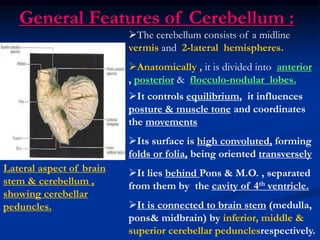
  • 1. General Features of Cerebellum : Lateral aspect of brain stem & cerebellum , showing cerebellar peduncles. It controls equilibrium, it influences posture & muscle tone and coordinates the movements Its surface is high convoluted, forming folds or folia, being oriented transversely It lies behind Pons & M.O. , separated from them by the cavity of 4th ventricle. It is connected to brain stem (medulla, pons& midbrain) by inferior, middle & superior cerebellar pedunclesrespectively. The cerebellum consists of a midline vermis and 2-lateral hemispheres. Anatomically , it is divided into anterior , posterior & flocculo-nodular lobes.
  • 2. External Features of Cerebellum : It has anterior notch ,which is wider and lodging the back of pons & medulla. It is separated from them by cavity of 4thventricle  It has also posterior notch occupied by falx cerebelli, which separates the 2 cerebellar H. Inferior surface : rounded on each side and presents : a deep groove (vallecula) between the 2-cerebellar hemispheres,which is occupied by the inferior vermis. -Tonsil is a small part of cerebellar hemisphere that lies lateral to inferior vermis. Superior surface Inferior surface
  • 3. External Features of Cerebellum : Superior surface : lies beneath tentorium cerebelli and has a raised superior vermis + a large cerebellar hemisphere on each side + primary & horizontal fissures. 1- Primary fissure V-shaped,well defined fissure, lies on superior surface and separates the small anterior lobe from the larger middle lobe (or posterior lobe). 2- Horizontal fissure lies along the sides of cerebellum, extending from anterior notch to posterior notch, separates the superior from the inferior surfaces. 3- Secondary (posterolateral) fissure lies on inferior surface and separates flocculo-nodular lobe from the ramainder of cerebellum.
  • 4. Schematic drawing of cerebellum showing the relationships between the anatomical & functional divisions of cerebellum. Green =archi-cerebellum, blue=paleo-cerebellum. Pink= neo-cerebellum Functional subdivision of cerebellum : 1- Archi-cerebellum = posterior lobe (Vestibular part) : _ It is formed of the flocculo- nodular lobe + associated fastigial nuclei, lying on inf. Surface in front of postero-lateral fissure. _Embryologically, it is the oldest part of cerebellum. _It receives afferent Fs. From vestibular apparatus of internal ear Via vestibulo-cerebellar tracts. _It is concerned with equlibrium.
  • 5. Connections of archicerebellum I- Archicerebellum It is concerned with equilibrium. It represents flocculo-nodular lobe. It has connections with vestibular & reticular nuclei of brain stem through the inferior cerebellar peduncle. Afferent vestibular Fs. Pass from vestibular nuclei in pons & medulla to the cortex of ipsilateral flocculo-nodular lobe. Efferent cortical (purkinje cell) Fs. Project to fastigial nucleus, which projects to vestibular nuclei & reticular formation. It affects the L.M.system bilaterally via descending vestibulo-spinal & reticulo-spinal
  • 6. Schematic drawing of cerebellum showing the relationships between the anatomical & functional divisions of cerebellum. Green =archi-cerebellum, blue=paleo-cerebellum. Pink= neo-cerebellum 2- Paleo-cerebellum= (spinal part) : -_it is formed of midline vermis + surrounding paravermis + globose & emboliform nuclei. _It receives afferent proprio- ceptive impulses from Ms.& tendons Via spino-cerebellar tracts (dorsal & ventral) mainly. -it sends efferents to red nucleus of midbrain. -it is concerned with muscle tone
  • 7. Connections of Paleo-cerebellum. 2-Paleo-cerebellum It is concerned with muscle tone & posture. Afferents spinal Fs. consist of dorsal & ventral spino-cerebellar tract from muscle, joint & cutaneous receptors to enter the cortex of ipsilateral vermis & para vermis Via inferior & superior cerebellar peduncles . Efferents cortical fibres pass to globose & emboliform nuclei, then Via sup. C. peduncle to contra- lateral red nucleus of midbrain to give rise descending rubro-spinal tract.
  • 8. Schematic drawing of cerebellum showing the relationships between the anatomical & functional divisions of cerebellum. Green =archi-cerebellum, blue=paleo-cerebellum. Pink= neo-cerebellum 3- Neo-cerebellum= (cerebral part) : _It is the remaining largest part of cerebellum. _It includes the most 2-cerebellar hemispheres + dendate nuclei. _It receives afferent impulses from the cerebral cortex+pons Via cerebro-ponto- cerebellar pathway. -it sends efferents to V.L.nucleus of thalamus. -it controls voluntary movements (muscle coordination).
  • 9. Connections of Neo-cerebellum. 3- Neo-cerebellum It is concerned with muscular coordination. It receives afferents from cerebral cortex involved in planning of movement- to pontine nuclei ,cross to opposite side Via middle Cerebellar peduncle to end in lateral parts of cerebellum (cerebro-ponto-cerebellar tract). Neo-cerebellar efferents project to dendate nucleus,which in turn projects to contra-lateral red nucleus & ventral lateral nucleus of thalamus ,then to motor cortex of frontal lobe, giving rise descending cortico-spinal & cortico- bulbar pathways. Efferents of dentate nucleus form a major part of superior C. peduncle.
  • 10. Cerebellar Lesions  Are usually vascular, may be traumatic or tumour.  Manifestations of unilateral cerebellar lesions : 1-ipsilateral incoordination of (U.L) arm = intention tremors : it is a terminal tremors at the end of movement as in touching nose or button the shirt. 2-Or ipsilateral cerebellar ataxia affects (L.L.) leg, causing wide-based unsteady gait.  Manifestations of bilateral cerebellar lesions (caused by alcoholic intoxication, hypothyrodism, cerebellar degeneration & multiple sclerosis) : 1-dysarthria : slowness & slurring of speech. 2-Incoordination of both arms.= intention tremors. 3-Cerebellar ataxia : intermittent jerky movements or staggering , wide-based, unsteady gait. 4-Nystagmus : is a very common feature of multiple sclerosis. It is due to impairment coordination of eye movements /so, incoordination of eye movements occurs and eyes exhibit a to-and-fro motion.  Combination of nystagmus+ dysarthria + intension tremors constitutes Chacot’triad, which is highly diagnostic of the disease.
  • 11.
  • 12. Internal Structure of cerebellum : Sagittal section of cerebellum. T.S.of cerebellum & brain at level of 4th V. to show cerebellar nuclei. It consists of an outer layer of grey matter (cerebellar cortex) , & inner layer of white matter containing 4-pairs of cerebellar nuclei : above roof of 4th V. from medial to lateral : 1-Fastigial nucleus. 2-Globose nucleus. 3-Emboliform nucleus. 4-Dendate nucleus. (the only one that can be seen clearly with the naked eye).
  • 13. Cerebellar cortex It is highly convoluted, forming numerous transversely oriented folia. It contains nerve cells, dendrites and synaptic connections of cellular neurones. The cellular organization of the cortex consists of 3-layers : 1-Outer molecular layer. 2-Intermediate, purkinje cell layer. 3-Inner granular layer, which is dominated by granule cell. T.S of cerebellar folia showing layers of cerebellar cortex. Afferent & Efferent connecltions and their relationships to principal cells of cerebellar cortex.
  • 14. Cerebellar cortex Molecular layer : contains 1-Cells : molecular cells (stellate cells) & basket cells. 2-Nerve Fibres : a- dendrites of Purkinje cells (arborisations). B-axons of granule cells. ( bifurcate to produce 2-parallel fibres , oriented along long axis of folium). C-ending of climbing fibers. Purkinje cell layer : it is formed of one layer (unicellular) of large flask- shaped purkinje cells. Their arborisations are at right angles to long axis to folium. Granular layer : it is formed of small granule cells & ending of mossy fibres.
  • 15. MThere are 3-types of Nerve Fibres in white Matter : 1-Axons of purkinje cells : the only axons to leave cerebellar cortex to end in deep cerebellar nuclei specially dendate nucleus. 2-Mossy Fibres : end in the granular layer. 3-Climbing Fibres : end in the molecular layer.
  • 16. Afferent Fibres to cerebellum : Mostly end in cerebellar cortex, excitatory to cortical neurones, as mossy or climbing Fs. passing through the cerebellar peduncles. The following are Afferent fibres: 1-dorsal & ventral spino-cerebellar tract. (passing via I.C.P & S.C.P) 2-vestibulo-cerebellar Fs. (via I.C.P) 3-olivo-cerebellar Fs. (via I.C.P)/ (extrapyramidal fibres), (end as climbing or mossy fibres) 4-ponto-cerebellar Fs. (via M.C.P). (In M.O)
  • 17. M Efferent Fibres of the cerebellum : It sends the following fibres : 1-Cerebello-vestibular Fs. to vestibular nuclei of pons & M.O. 2-Cerebello-olivary Fs. To M.O. 3-Dendato-rubro-thalamic tract To red nucleus of midbrain & ventro- lateral nucleus of the thalamus and finally to motor cortex of frontal lobe to coordinate movement via cortico-spinal & corticobulbar tracts.
  • 18. The Fourth Ventricle It is a cavity of hindbrain. Position : lies between pons & M.O. anteriorly and the cerebellum posteriorly. It is a diamond-shaped space which is lined by ependyma. Its superior angle is continuous with cerebral aqueduct of midbrain. inferior angle is continuous with centeral canal of closed M.O. Its lateral angles extend laterally to form a lateral recess on each side to open into subarachnoid space.
  • 19. The Boundaries of 4th Ventricle Superiolateral boundary : -it is formed by superior cerebellar peduncle on each side. Inferiolateral boundary : -it is formed by inferior cerebellar peduncle + gracile & cuneate tubercles on each side.
  • 20. The Roof of 4th Ventricle -it is a tent-shaped when seen laterally, and diamond-shaped when seen behind. -it is formed of superior & inferior medullary vela, which are thin sheets of white matter /consists of : ependyma covered by pia mater. -Sup.medullary velum connects the 2 sup.cerebellar peduncles. -Inf.medullary velum connects the 2 inf.cerebellar peduncles. -Inferior vermis of cerebellum : lies in the middle of roof of 4th ventricle.
  • 21. The Roof of 4th Ventricle The lower part of roof is invaginated by tela choroidea of 4th ventricle. The tela choroidea is a double layer of pia mater which encloses the choroid plexus of 4th ventricle. The choroid plexus is a vascular capillary tuft covered by ependymal cells and secretes C.S.F. into the lumen of 4th ventricle.
  • 22. The Openings of 4th Ventricle The roof contains 3 aperatures which transmit C.S.F. from ventricular lumen to subarachnoid space. Median aperature (foramen of Magendie) : lies in the median plane at lower end of inferior medullary velum, and opens into subarachnoid space at cistrna magna at cerebello-medullary angle 2 lateral openings (foramina of Luschka) : each one lies at the lateral end of lateral recess to open into subarachnoid space at cerebello-pontine angle. choroid plexus partly protrudes out through each lateral aperture.
  • 23. The Floor of 4th Ventricle A diagram to show the floor & lateral boundaries of 4th ventricle. It is called rhomboid fossa. It is diamond-shaped and is divided into right & left halves by the median sulcus. It is crossed in the middle by transvere Fs. (ponto-cerebellar Fs.)called medullary stria, which divide floor of 4th ventricle into upper (pontine) & lower (medullary) part.
  • 24. The Floor of 4th Ventricle A diagram to show the floor & lateral boundaries of 4th ventricle. Upper pontine part : presents on each side of median sulcus. 1-Medial eminence : a rounded elevation produced by the abducent nucleus. 2-Facial colliculus : an elevation on the top of lower part of medial eminence. It is produced by the fibres of facial nerve which surround abducent nucleus. 3-Superior fovea : a groove lateral to facial colliculus. 4-Vestibular area : lateral to superior fovea. It overlies superior, medial & lateral vestibular nuclei.
  • 25. The Floor of 4th Ventricle A diagram to show the floor & lateral boundaries of 4th ventricle. Lower medullary part : presents on each side of the median sulcus. 1-Inferior fovea : inverted V-shaped groove. 2-Hypoglossal area : medial to inferior fovea. It overlies hypo-glossal nucleus. 3-Vagal area (triangle) : between limbs of inferior fovea.It overlies dorsal nucleus of vagus. 4-Vestibular area : lateral to inferior fovea. It overlies inferior vestibular nucleus.