

Recommended
More Related Content
What's hot
What's hot (20)
Viewers also liked
Viewers also liked (18)
Similar to Cervical spine
Similar to Cervical spine (20)
More from AlAhly sporting club
Recently uploaded
Recently uploaded (20)
Cervical spine
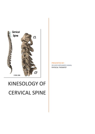
- 1. KINESOLOGY OF CERVICAL SPINE PRESENTED BY : DR.ASER MOHAMED KAMAL PHYSICAL THERAPIST
- 2. Biomechanics of Cervical Spine Made up of two anatomically and functionally distinct segments. 1. Superior segment/suboccipital segment:- A -consist of c1 /atlas and c2/axis B -connected to eachother and occiput with complex chain of joints. C -having 3 axes and 3 degrees of freedom. 2. Inferior segment- A -stretching from inferior surface of axis to the superior surface of T1. B -In total there are 7 cervical vertebras- c1-c2 c3-c6 c7 atypical typical transitional Structure of a atypical cervical vertebra 1- Atlas /C1-its ring shaped Transverse diameter greater than AP diameter Has two lateral faces oval in shape running obliquely anteriorly and medially Which bear biconcave superior articulate facet superiorly and medially meant to articulate with occipital condyles Inferior articular facet –facing inferiorly and medially Convex AP Corresponds to superior facet of axis Anterior arch consist of small cartilagenous oval shaped articular facets for the odontoid process of axis Posterior arch is initially flattened but becomes thicker posteriorly to form posterior tubercle on the midline. Transeverse process No spinous process No intervertebral disc
- 3. 2- The axis /C2-is atypicsl Superior surface of the body carries centrally the odomtoid process which acts as a pivot for atlantoodontoid joint . Laterally possess 2 articular facets facing superior and laterally Facets are convex AP and flat transversely Posterior arch consist of narrow laminae The cartilage lined inferior articular process corresponds to the superior articular process of c3 The cartilage lined inferior articular process corresponds to the superior articular process of c3 Transverse process The atlanto-axial joint complex it is a plane synovial joint comprises of 3 mechanically linked joints The central joint is the atlanto odontoid joint Two lateral joints-atlanto axial joint Atlanto-odointoid joint it is synovial trochoid /pivot joint Jointsurfaces-anterior articular facet of odontoid and posterior articular facet of the anterior arch of the atlas Movements at atlantoaxial and atlanto-odontoid joint Flexion-point of contact b/w two convex surface moves forward interspace of atlanto odontoid joint opens superiorly Extention- point of contact b/w two convex surface moves backward Interspace of atlanto odontoid jointopens inferiorly Radiological findingas does not shoe opening of interspaces
- 4. This is due to transverse ligament and keeps the anterior arch and odontoid process in close contact During flxn and extn tha inferior surface of atlas rols and sides over superior articular surface of axis Rotation Left to right rotation-The left lateral mass of the atlas moves forward Right lateral mass recedes in rotation from left to right and vice versa from right to left Movement of atlanto occipital joint Formed b/w superior articular facets of atlas and the occipital condyles. It is an enarthodrial kind of joint Gives 3 degrees of freedom Axial rotation-about vertical axis Flexion/extension-about transverse axis Lateral flexion-about AP axis. Flexion:The occipital condyles recede on the lateral masses of the atlas. The occipital bone moves away from the posterior archof the atlas Limited by tension developed in the articular capsules and the ligament Extension: Occipital condyles slides anteriorly on the lateral masses of the atlas. Occipital bone moves neatrer to the posterior arch of the atlas Posterior arch of the atlas and axis are approximated Limited by those 3 bony pieces Flexion/extension-15 degrees Lateral flexion Movement only occurs b/w c0-c1 and c2-c3 Left lateral flexion-slipping of occipital condyles on right of atlas Right lateral flexion-vice versa Ther is asmall range of motionTotal ROM-C0-C3=8 degrees C0-C1=3 degrees,C2-C3=5 degrees
- 5. Rotation When occiput rotates on atlas its rotation is secondary to rotation of atlas on axis Around vertical axis passing through the centre of odontoid Causes right anterior displacement of oright occipital condyle on right lateral mass of the atlas Lateral atlanto occipoital ligamenr is streched Thus rotation of occiput to left is associated with – Linear displacement of 2-3 mm to the left Lateral flexion to the right Structure of a typical cervical vertebra Vertebral body-superior plateau is raised on either sides by 2 buttresses. which is called as unciform process. It is concave transversely and convex anteroposteriorly- resembling a saddle . Unciform processes guoides the AP movements during flexion and extension ut limits lateral flexion Pedicals-connects the vertebral body to the transverse process. Project posterolaterally. Lamina-part of the posterior arch Meets in the midline to form the bifid spinous process Projects posteromedially and are thin and slightly curved. Spinous process-short slender and extend horizontally The tip is bifurcated Face superiorly and medially The length of spinous process decreases from c2-c3 C3-c5 remains constant And undergoes a significant increase at c7. Vertebral foramen –is large and triangular Transverse process They are peculiar in orientation They are hollowed in to a gutter AP and they point AL. The posteromedial end of the gutter lines the intervertebral foramen. The AL end is bifid giving attachment to scalene muscles. Possess foramen transversarium
- 6. Articular processes-they bear superior and inferior articular facets. Superior facets face superiorly and medially Inferior facets face anteriorly and laterally Movements at the lower cervical vertebral column Extension-ovrlying vertebral body tilts and slides posteriorly IV space is compressed posteriorly and opened wide anteriorly Nucleus palposus is driven slightly anteriorly Anterior fibers of annulus fibrosus is streched Superiorly articulating facet slides inferiorly posteriorly and tilts posteriorly Limited by anterior longitudinal ligament and by the impact of the posterior arches through ligaments Flexion-upper vertebral body tilts and slides anteriorly Intervertebral space is compressed anteriorly and opened wide posteriorly Nucleus pulposus is driven posteriorly Posterior fibres of annulus fiberosus is streched Limited by the tension developed in the posterior longitudinal ligament By the capsular ligament,ligamentum flavum,ligamentum nuchae, Combined lateral flexion and rotation- Does not occur as pure motions Governed by orientation of articular facets which are oblique inferiorly and posteriorly Rotation is always coupeled with lateral flexion Considering the whole cervical column from C2-T1 extension component is also added to these movements Any movement b/C7 and T1 amounts for mixed rotation and lateral flexion of C7 Where as any movement b/w C6-C7 also adds up extension component Thus three composite movement occurs in 3 planes- Lateral flexion –frontal plane Extension-sagittal plane Rotation-transverse plane
- 7. RANGE OF MOTION FROM- WHITE AND PUNJABI stability Cervical region bears less weoight and are more mobile Stability is provided by bony configuration, muscles , ligaments Ligaments Anterior atlnatoaxial ligament,posterior atlantoaxial ligament,tectorial membrane,ligamentum nuchae Transverse atlantal ligament-21.9 mm in length Also refered as atlantal cruciform ligament Holds dense in closed approximation against the anterior ring of the atlas Also serves as an articular surface for dense Prevents anterior displacement of C1 on C2 Alar ligaments-arise from axis on either side of dens Approx.1cm in legth Are taut in flexion Axial rotation of head and neck tightens both alar ligaments Prevents distraction of C1 on C2 Apical ligaments-of the dens connects the axis and occioital bone of the skull JOINT COMBINED FLEXION EXTENSION ONE SIDE LAT BENDING ONE SIDE AXIAL ROTATION C2-C3 10 10 3 C3-C4 15 11 7 C4-C5 20 11 7 C5-C6 20 8 7 C6-C7 17 7 6 C7-T1 9 4 2
- 8. Muscles of cervical vertebrae *Muscles-flexion of head and neck- Depends on anterior muscles of the neck They are: 1. rectus capitis major, rectus capitis minor 2. Longus cervicis which plays an important role in straightening the cervical column and holding it rigid 3. Scalene anterior posterior and medius 4. Suprahyoid and infrahyoid muscles helps in supporting the cervical column at rest 5. sternocliedomastoid Rectus Capitis Posterior Major Origin: Spinous process of axis (C2) Insertion: Lateral half of the inferior nuchal line of the occipital bone Action: Extension of the head Innervation: Posterior Primary Ramus of C1 Primary Actions of the Rectus Capitis Posterior Major: 1. Extension of the head on the neck when acting bilaterally 2. Ipsilateral rotation of the cervical spine when acting unilaterally Rectus Capitis Posterior Minor: Origin: Posterior tubercle of atlas (C1) Insertion: Lateral half of the inferior nuchal line of the occipital bone. Action: Extension of the head Innervation: Posterior Primary Ramus of C1 Primary Actions Rectus Capitis Posterior Minor: 1. Extension of the head on the neck when acting bilaterally 2. Ipsilateral rotation of the cervical spine when acting unilaterally
- 9. Splenius Cervicis Origin: Spinous processes and supraspinous ligaments of T3 to T6 Insertion: Posterior tubercles of transverse processes of C1 to C3 Action: Extention of the cervical spine,Lateral flexion of the cervical spine, Rotation of the cervical spine Innervation: Dorsal primary rami of C5 to C7 Primary Action of the splenius cervicis: 1. Extension of the cervical spine when acting bilaterally 2. Lateral Flexion of the cervical spine when acting unilaterally Secondary Action of the splenius cervicis: 1. Assists with ipsilateral rotation of the cervical spine when acting unilaterally Scalenes Scalene Muscle Group: Scalenus Anterior Origin: Anterior tubercles of the transverse processes of (C3-C6) Insertion: Scalene tubercle and cranial crest of first rib Action: Flexion of the cervical spine, Lateral flexion of the cervical spine, Innervation: Ventral rami of(C3-C8) Primary Actions of the Scalenus Anterior: 1. Flexion of the cervical spine when acting bilaterally 2. Lateral Flexion of the cervical spine when acting unilaterally Secondary Actions of the Scalenus Anterior: 1. Assists with contralateral rotation of the cervical spine when acting unilaterally 2. Assists with forced inspiration by elevating first rib
- 10. Scalenus Medius Origin: Posterior tubercles of the transverse processes of (C2-C7) Insertion: Cranial surface of the first rib Action: Flexion of the cervical spine, Lateral flexion of cervical spine Innervation: Ventral rami of (C3-C8) Primary Actions of the Scalenus Medius: 1. Flexion of the cervical spine when acting bilaterally 2. Lateral flexion of the cervical spine when acting unilaterally Secondary Actions of the Scalenus Medius: 1. Assists with contralateral rotation of the cervical spine when acting unilaterally 2. Assists with forced inspiration by elevating the first rib Scalenus Posterior Origin: By tendons from the posterior tubercles of the transverse processes of (C4–C6) Insertion: Outer surface of 2nd rib Action: Lateral flexion of the neck Innervation: Ventral rami of (C3-C8) Primary Actions of the Scalenus Posterior: 1. Lateral flexion of the cervical spine when acting unilaterally Secondary Actions of the Scalenus: 1. Assists with contralateral rotation of the cervical spine when acting unilaterally 2. Assists with forced inspiration by elevating the second rib Sternocleidomastoid Sternal Head: Origin: Upper part of the anterior surface of the manubrium Clavicular Head: Origin: Superior surface of the medial one third of clavicle Insertion: Lateral surface of the mastoid process of the occipital bone, from its apex to its superior border, and by a thin aponeurosis to the lateral half of the superior nuchal line
- 11. Action: Bilaterally: flexion of the head and neck, extension of the head and neck Unilaterally: rotation of head to opposite side, lateral flexion Innervation: Accessory nerve: cranial nerve XII and ventral rami of the (C2, C3) Primary Actions of the Sternocleidomastiod: 1. Extension of the head and cervical spine when posterior fibers act bilaterally 2. Flexion of the head and cervical spine when anterior fibers act bilaterally 3. Lateral flexion of the head and cervical spine when acting unilaterally 4. Contralateral rotation of the head and cervical spine when acting unilaterally *Extension of head and neck- Brought about by posterior neck muscles They are: 1- splenius cervicis, 2- semispinalis cervicis, 3- leavator scapulae, 4- transverso spinalis, 5- longismus capiis, 6- spenius capitis, 7- trapezius These muscles helps in maintaining the cervical lordosis When contract unilaterally they produce extension rotation and lateral flexion on the same side Both flexors and extensor group of muscles are responsible to maintain cervical column rigid in neutral position Essential in balancing the head and in supporting weights carried on head Splenius Cervicis Origin: Spinous processes and supraspinous ligaments of T3 to T6 Insertion: Posterior tubercles of transverse processes of C1 to C3 Action: Extention of the cervical spine,Lateral flexion of the cervical spine, Rotation of the cervical spine Innervation: Dorsal primary rami of C5 to C7 Primary Action of the splenius cervicis: 1. Extension of the cervical spine when acting bilaterally 2. Lateral Flexion of the cervical spine when acting unilaterally
- 12. Secondary Action of the splenius cervicis: 1. Assists with ipsilateral rotation of the cervical spine when acting unilaterally Semispinalis Cervicis : Origin: By fleshy and tendinous fibers to the transverse processes of (T1 – T6) Insertion: Spinous processes of C2 to C5, spanning four to six levels between attachment points Action: Extension of the cervical spine, lateral flexion of the cervical spine, rotation of the cervical spine Innervation: Dorsal primary rami of (C3-C5) Primary Actions of the Semispinalis Cervicis: 1. Extension of the cervical spine when acting bilaterally 2. Lateral flexion of the cervical spine when acting unilaterally 3. Contralateral rotation of the cervical spine when acting unilaterally 4. Extension of the thoracic spine when acting bilaterally 5. Contralateral rotation of the trunk when acting unilaterally Secondary Actions of the Semispinalis Cervicis: 1. Assists with lateral flexion of the thoracic spine when acting unilaterally Levator Scapulae Origin: Posterior tubercles of the transverse processes of C1-C4 Insertion: Superior part of the medial border of the scapula Action: Elevation of the scapula, downward rotation of the scapula Innervation: Anterior primary rami of (C3 and C4), dorsal scapular nerve (C5) Primary Actions of the Levator Scapulae: 1. Elevation of the scapula Secondary Actions of the Levator Scapulae: 1. Downward rotation of the scapula
- 13. Transverso spinalis ORIGIN Transverse processes INSERTION Spinous processes above and occipital bone (only semispinalis capitis part shown) NERVE Posterior primary rami ACTION: 1. One side - Lateral flexion of the cervical and thoracic spine 2. Both Sides - Extension and hyperextension of atlantooccipital joint and thoracic spine. Longissimus Capitis Origin: By tendons from the posterior surfaces of the transverse processes of T1 to T5 and the articular processes of C4 or C5-7 Insertion: Posterior margin of the mastoid process of the occipital bone Action: Extension of the head and cervical spine Innervation: Dorsal primary rami of C4 to T5 Primary Actions Longissimus Capitis: 1. Extension of the head and cervical spine when acting bilaterally Secondary Actions Longissimus Capitis: 1. Assists with ipsilateral rotation of the cervical spine and head when acting unilaterally SPLENIUS CAPITIS ORIGIN Lower ligament nuchae, spinous processes and supraspinous ligaments T1-3 INSERTION Lateral occiput between superior and inferior nuchal lines ACTION Extends and rotates cervical spine NERVE Posterior primary rami of C3, 4 Trapezius
- 14. Origin: Upper part: External occipital proturberance, medial third of the superior nuchal line, the ligamentum nuchae, and the spinous process of C7 Medial Part: Spinous processes of T1 to T5. Lower Part: Spinous processes of T6 to T12 Insertion: Upper Part: Lateral third of the clavicle and the medial aspect of the acromion process of the scapula Middle Part: Medial edge of the superior surface of the acromion process of the scapula and the superior edge of the scapular spine. Lower Part: Tubercles of the apex of the scapular spine Action: Upper Part: Upward rotation of the scapula, elevation of the scapula Middle Part: Retraction of the scapula Lower Part: Upper rotation of the scapula, depression of the scapula Innervation: Spinal Accessory Cranial XI , Ventral Rami C2-C4 Primary Actions of the Upper Trapezius 1. Upward rotation of the scapula 2. Elevation of scapula Secondary Actions: 1. Assists with elevation of the cervical spine (distal attachment fixed, acting bilaterally) 2. Assists with lateral flexion of the cervical spine (distal attachment fixed, acting unilaterally) 3. Assists with contralateral rotation of the head and cervical spine when acting unilaterally Primary Actions of the Middle Trapezius 1. Retraction of scapula Primary Actions of the Lower Trapezius 1. Upward rotation of the scapula 2. Depression of the scapula