SlideShare a Scribd company logo
1 of 7
Download to read offline
Ahmad Amro Baradee
General view on the skull:
The skull is divided intotwo parts: 1) Cranium 2) Facial skeleton
There are 22 bones forming the skull:
8 Cranial bones: Base: 1-sphenoid 2-ethmoid
Vertex: singular: 3-Frontal 4-Occipital
Pair: 5-6-parietal 7-8-temporal
14 Facial bones: mobile: 1-mandible
Immobile: singular: 2-vomer
Pair:3-4-inferior concha
5-6-maxilla 7-8-lacrimal
9-10-zygomatic 11-12-nasal
13-14-palatine
The Ophthalmic nerve:
Frontal Supraorbital(through supraorbital foramen)
Supratrochlear (through supratrochler notch)
Ophthalmic Lacrimal Infratochlear
Nasociliary anterior ethmoid
Posterior ethmoid
The ophthalmic nerve gives collateral branches (
before the three terminal branches ) these are: recurrent meningeal branch
, and anastomosis branches with the third and fourth cranial nerves and
with the carotid plexus.
Ahmad Amro Baradee
The external carotid artery:
Superior thyroid
Anteroir lingual
facial
External medial: ascending pharyngeal
Carotid
Artery posterior occipital
Posterior auricular
terminal Maxillary
Superficial temporal Transverse facial
Middle temporal
Zygomatico-orbital
Anterior auricular
2 terminal br.: frontal
parietal
The Maxillary Artery:
Classification "1":
We say the maxillary artery gives 14 collateral branches , and they are
classified among their directions into 4 categories:
A. Three ascending branches heading to the cranium:
1) Tympanic artery
2) Middle meningeal artery
3) Accessory meningeal artery
B. Three lateral branches heading to the muscles:
1) Masseteric artery
2) deep posterior temporal artery
3) deep anterior temporal artery
C. Four descending branches heading to masticating system:
1) inferior dental artery
2) pterygoid artery
3) orobuccal artery (oro=mouth )
4) alveolar artery
D. Four deep branches evolve in the pterygopalatine fossa:
1) Infraorbital artery
Ahmad Amro Baradee
2) Vidian artery ( the artery of pterygoid canal )
3) Descending palatine artery
4) Pterygopalatine artery
Classification "2": ( I, myself, prefer this way of classification)
We say the maxillary artery gives 16 branches , and they are classified
into 3 categories:
A. First category: 1) middle meningeal 2) accessory meningeal
3)inferior alveolar 4) deep auricular 5) anterior tympanic
B. Second category: 1) lateral pterygoid 2) medial pterygoid
3)masseter 4) temporal 5) buccinator
C. Third category: 1) posterior superior alveolar 2) Vidian (artery of
pterygoid canal) 3)greater palatine 4)lesser palatine
5)sphenopalatine 6)infraorbital
Temoral bone's fissures:
Squamotympanic f.
Tympanic Squamos
part part
Petrotympanic f. petrosal squamopetrosal f.
prominence
Only the tympanomastoid fissure is left to be pointed out
Ahmad Amro Baradee
The paranasal sinuses (air sinuses):
These sinuses:
1) Reduce the weight of the skull
2) Give the ability to make sounds
3) Give the shape of the face
Opens into:The paranasal sinus
Semilunar hiatus , middle meatusMaxillary
Semilunar hiatus , middle meatusFrontal
Semilunar hiatus , middle meatusAnterior and middle ethmoid
Superior meatusPosterior ethmoid
Sphenoethmoidal recess,above the superior meatusSphenoid
Don't open..Mastoid air cells
Remember: the nasolacrimal duct carries tears into the inferior meatus
Note: "meatus" refers to the space under the concha , means: the inferior
meatus is the space under the inferior concha
The venouses sinuses:
Singular:
 Superior sagittal
 Inferior sagittal
 Straight
 Occipital
 intercavernous
Pair:
 superior petrosal
 inferior petrosal
 transverse
 sigmoid
 cavernous
Ahmad Amro Baradee
EndsVenouses sinus
Right transverseSuperior sagittal
StraightInferior sagittal
Left transverseStraight
Continues as: the sigmoidTransverse (right or left)
Jugular bulbSigmoid
SigmoidSuperior petrosal
Jugular bulbInferior petrosal
The Sphenoid bone:
 The anterior aspect of the sphenoid bone we find the sphenoidal
crest , and on each side we see the apertute of the sphenoidal sinus,
and lateral to that aperture we see semi air cells which present the
place of articulation between the ethmoid labyrinth and the
sphenoid bone.
 The anterior part of the sphenoidal concha articulates with the
orbital process of the palatine bone.
 The posterior part of the orbital process of the palatine bone gives
an opening to connect with the sphenoidal sinus , whereas the
margins of this opening articulates with the sphenoidal concha.
 The inferior surface of the sphenoidal concha articulates with the
superior surface of the sphenoidal process of the palatine bone.
 We can recognise the following landmarks in an intercranial aspect
of the sphenoid bone: (in this exact order postero-anteriorly):
Dorsum sellae Pituitary gland's fossa Groove for
intercavernous sinus Tuberculum sellae (on each of its sides we find the
middle clinoid process ) Chiasmatic groove Sphenoidal limbus
Sphenoidal jugum ethmoidal process
Ahmad Amro Baradee
The pterygopalatine fossa:
It’s a pyramidal fossa
1) It's boundaries:
 Lateral: the internal surface of the ramus of mandible
 Anterior: maxillary tuber
 Medial: the perpendicular plate of palatine bone (at it's top
end we see the sphenopalatine foramen )
 Posterior: the anterior surface of the pterygoid process
 Superior: the inferior surface of the greater wing of the
sphenoid bone (the maxillary surface)
and the apex of this pyramidal fossa is at the inferior end (at the place of
meeting: the pterygoid process with the pyramidal process of palatine
bone together with the tuber of maxillary bone)
2) the foramina that open into the fossa:
 Foramen rotundom
 Foramen ovale
 The aperture of the pterygoid canal
 The aperture of the Palatovaginal canal
 the sphenopalatine foramen (connects the fossa with the
nasal cavity )
 the inferior orbital fissure (connects it with the orbit)
 the pterygomaxillary (pterygopalatine) fissure (connects it
with the infratemporal fossa)
 it connects with the oral cavity through the greater palatine
Canal (the pterygopalatine canal) which has the (Mercedes
sign shape:
the posterior part of the canal is formed by the pterygoid
process , the anterio-medial is formed by the perpendicular
plate of palatine bone (with making a groove on it) , the
anterio-lateral part is formed by the posterior part of the
nasal surface of the maxillary bone (also with making a
groove on it )
Ahmad Amro Baradee
Note: the term (pterygopalatine canal) can refer to one of two
canals:
1) the greater palatine canal
2) OR:the palatovaginal canal
3) It's contents:
 The two pterygoid muscles
 The maxillary artery and it's branches
 The maxillary nerve and it's branches
 The mandibular nerve and it's branches
 The pterygoid venouses plexus
 The pterygopalatine ganglion: lies at the bottom of the fossa
4) The branches of the pterygopalatine ganglion:
 Orbital branches(passes through the inferior orbital
fissure)
 nasopalatine nerve
 posterior superior nasal branches
 greater palatine nerve
 lesser palatine nerve
 Bock's nerve ( the pterygopalatine nerve) (passes through
the palatovaginal canal)
o The vidian nerve (the nerve of pterygoid canal) , and two sturdy
branches of the maxillary nerve: end up at the ganglion.
I hope this was helpful 
By: Ahmad Amro Baradee

More Related Content

What's hot

7938279 bone-of-the-cranial-cavity
7938279 bone-of-the-cranial-cavity7938279 bone-of-the-cranial-cavity
7938279 bone-of-the-cranial-cavity
Ngaire Taylor
 

What's hot (20)

Norma basalis (skull base).pps
Norma basalis (skull base).ppsNorma basalis (skull base).pps
Norma basalis (skull base).pps
 
Head and neck anatomy
Head and neck anatomyHead and neck anatomy
Head and neck anatomy
 
Norma basalis
Norma basalisNorma basalis
Norma basalis
 
Cranial fossa
Cranial fossaCranial fossa
Cranial fossa
 
Bones of Skull (Human Anatomy)
Bones of Skull (Human Anatomy)Bones of Skull (Human Anatomy)
Bones of Skull (Human Anatomy)
 
Cranial bones
Cranial bonesCranial bones
Cranial bones
 
7938279 bone-of-the-cranial-cavity
7938279 bone-of-the-cranial-cavity7938279 bone-of-the-cranial-cavity
7938279 bone-of-the-cranial-cavity
 
Topographic anatomy of the brain and brain part of the skull
Topographic anatomy of the brain and brain part of the skullTopographic anatomy of the brain and brain part of the skull
Topographic anatomy of the brain and brain part of the skull
 
Anatomy of the Temporal region & Temporomandibular joint
Anatomy of the Temporal region & Temporomandibular jointAnatomy of the Temporal region & Temporomandibular joint
Anatomy of the Temporal region & Temporomandibular joint
 
Cranial fossa
Cranial fossaCranial fossa
Cranial fossa
 
Normas of skull
Normas of skullNormas of skull
Normas of skull
 
Temporal & infratemporal region
Temporal & infratemporal regionTemporal & infratemporal region
Temporal & infratemporal region
 
Cranial cavity ( Department of Anatomy)
Cranial cavity ( Department of Anatomy)Cranial cavity ( Department of Anatomy)
Cranial cavity ( Department of Anatomy)
 
Anatomy of pterygopalatine fossa, infra temporal space
Anatomy of pterygopalatine fossa, infra temporal spaceAnatomy of pterygopalatine fossa, infra temporal space
Anatomy of pterygopalatine fossa, infra temporal space
 
Anterior cranial-fossa
Anterior cranial-fossaAnterior cranial-fossa
Anterior cranial-fossa
 
Anatomy of Skull
Anatomy of SkullAnatomy of Skull
Anatomy of Skull
 
Cranial nerves pathway update
Cranial  nerves  pathway updateCranial  nerves  pathway update
Cranial nerves pathway update
 
Head and Neck
Head and NeckHead and Neck
Head and Neck
 
Head and neck anatomy
Head and neck anatomyHead and neck anatomy
Head and neck anatomy
 
Topographic anatomy and operative surgery of head deepak pawar
Topographic anatomy and operative surgery of head   deepak pawarTopographic anatomy and operative surgery of head   deepak pawar
Topographic anatomy and operative surgery of head deepak pawar
 

Viewers also liked (7)

Head And Neck
Head And NeckHead And Neck
Head And Neck
 
Anatomy of head and neck
Anatomy of head and neckAnatomy of head and neck
Anatomy of head and neck
 
Head neck ppt
Head neck pptHead neck ppt
Head neck ppt
 
Head and neck rs
Head and neck  rsHead and neck  rs
Head and neck rs
 
Head and neck anatomy
Head and neck anatomyHead and neck anatomy
Head and neck anatomy
 
Complete Summary Of the Head and Neck Anatomy
Complete Summary Of the Head and Neck AnatomyComplete Summary Of the Head and Neck Anatomy
Complete Summary Of the Head and Neck Anatomy
 
Anatomy Lect 3 Head & Neck
Anatomy Lect 3 Head & NeckAnatomy Lect 3 Head & Neck
Anatomy Lect 3 Head & Neck
 

Similar to Review of Head and Neck Anatomy

Side kick git anatomy practical explained
Side kick git anatomy practical explainedSide kick git anatomy practical explained
Side kick git anatomy practical explained
sallamahmed1
 
ANATOMY OF NOSE AND PNS.pptx
ANATOMY OF NOSE AND PNS.pptxANATOMY OF NOSE AND PNS.pptx
ANATOMY OF NOSE AND PNS.pptx
Satishray9
 
Anatomy of nose and paranasal sinuses
Anatomy of nose and paranasal sinusesAnatomy of nose and paranasal sinuses
Anatomy of nose and paranasal sinuses
Vinay Bhat
 
Bone_Skull.pdf
Bone_Skull.pdfBone_Skull.pdf
Bone_Skull.pdf
JoehadaSalon
 
anatomyofnoseandparanasalsinuses-copy-230629065517-07a23e0b.pdf
anatomyofnoseandparanasalsinuses-copy-230629065517-07a23e0b.pdfanatomyofnoseandparanasalsinuses-copy-230629065517-07a23e0b.pdf
anatomyofnoseandparanasalsinuses-copy-230629065517-07a23e0b.pdf
AliyahJohanis1
 
Anatomy Of Nose And Paranasal Sinuses - Copy.pptx
Anatomy Of Nose And Paranasal Sinuses - Copy.pptxAnatomy Of Nose And Paranasal Sinuses - Copy.pptx
Anatomy Of Nose And Paranasal Sinuses - Copy.pptx
Htet Ko
 

Similar to Review of Head and Neck Anatomy (20)

Nose and PNS Anatomy.pptx
Nose and PNS Anatomy.pptxNose and PNS Anatomy.pptx
Nose and PNS Anatomy.pptx
 
Lec 5 skull
Lec 5 skullLec 5 skull
Lec 5 skull
 
skull_2.pdf
skull_2.pdfskull_2.pdf
skull_2.pdf
 
Side kick git anatomy practical explained
Side kick git anatomy practical explainedSide kick git anatomy practical explained
Side kick git anatomy practical explained
 
ANATOMY OF NOSE AND PNS.pptx
ANATOMY OF NOSE AND PNS.pptxANATOMY OF NOSE AND PNS.pptx
ANATOMY OF NOSE AND PNS.pptx
 
Anatomy of temporal bone final
Anatomy of temporal bone finalAnatomy of temporal bone final
Anatomy of temporal bone final
 
Osteology of head n neck ppt 1
Osteology of head n neck ppt 1Osteology of head n neck ppt 1
Osteology of head n neck ppt 1
 
Lecture 2 (The parotid gland and the extracranial part of the facial nerve)
Lecture 2 (The parotid gland and the extracranial part of the facial nerve)Lecture 2 (The parotid gland and the extracranial part of the facial nerve)
Lecture 2 (The parotid gland and the extracranial part of the facial nerve)
 
Nose and pns 25.04.16 dr.sithandandhakumar
Nose and pns 25.04.16 dr.sithandandhakumarNose and pns 25.04.16 dr.sithandandhakumar
Nose and pns 25.04.16 dr.sithandandhakumar
 
Anatomy of nose and para nasal sinuses . by DR. MD. KHURSHID PERVEJ. GMC PATIALA
Anatomy of nose and para nasal sinuses . by DR. MD. KHURSHID PERVEJ. GMC PATIALAAnatomy of nose and para nasal sinuses . by DR. MD. KHURSHID PERVEJ. GMC PATIALA
Anatomy of nose and para nasal sinuses . by DR. MD. KHURSHID PERVEJ. GMC PATIALA
 
norma b extrnaالثالثه تشريح راس.pptx
norma b extrnaالثالثه تشريح راس.pptxnorma b extrnaالثالثه تشريح راس.pptx
norma b extrnaالثالثه تشريح راس.pptx
 
Anatomy of nose and paranasal sinuses
Anatomy of nose and paranasal sinusesAnatomy of nose and paranasal sinuses
Anatomy of nose and paranasal sinuses
 
Bone_Skull.pdf
Bone_Skull.pdfBone_Skull.pdf
Bone_Skull.pdf
 
Lateral skull base anatomy and applied science by Dr, bomkar bam
Lateral skull base anatomy and applied science by Dr, bomkar bamLateral skull base anatomy and applied science by Dr, bomkar bam
Lateral skull base anatomy and applied science by Dr, bomkar bam
 
anatomyofnoseandparanasalsinuses-copy-230629065517-07a23e0b.pdf
anatomyofnoseandparanasalsinuses-copy-230629065517-07a23e0b.pdfanatomyofnoseandparanasalsinuses-copy-230629065517-07a23e0b.pdf
anatomyofnoseandparanasalsinuses-copy-230629065517-07a23e0b.pdf
 
Anatomy of Nose And Paranasal Sinuses - Copy.pptx
Anatomy of Nose And Paranasal Sinuses - Copy.pptxAnatomy of Nose And Paranasal Sinuses - Copy.pptx
Anatomy of Nose And Paranasal Sinuses - Copy.pptx
 
Temporal & Infratemporal Region -I
Temporal &  Infratemporal  Region -ITemporal &  Infratemporal  Region -I
Temporal & Infratemporal Region -I
 
Rhinoplasty (Nose Anatomy)
Rhinoplasty  (Nose Anatomy) Rhinoplasty  (Nose Anatomy)
Rhinoplasty (Nose Anatomy)
 
Anatomy of Internal Nose
Anatomy of Internal Nose  Anatomy of Internal Nose
Anatomy of Internal Nose
 
Anatomy Of Nose And Paranasal Sinuses - Copy.pptx
Anatomy Of Nose And Paranasal Sinuses - Copy.pptxAnatomy Of Nose And Paranasal Sinuses - Copy.pptx
Anatomy Of Nose And Paranasal Sinuses - Copy.pptx
 

More from Ahmad Amro Baradee

More from Ahmad Amro Baradee (9)

The Maxilla - Anatomy تشريح عظم الفك العلوي
The Maxilla - Anatomy تشريح عظم الفك العلويThe Maxilla - Anatomy تشريح عظم الفك العلوي
The Maxilla - Anatomy تشريح عظم الفك العلوي
 
Failure of Dental Implants أسباب فشل الغرسات السنية
Failure of Dental Implants أسباب فشل الغرسات السنيةFailure of Dental Implants أسباب فشل الغرسات السنية
Failure of Dental Implants أسباب فشل الغرسات السنية
 
Todd Snyder's Provisionalization Technique طريقة لصنع التعويض المؤقت
Todd Snyder's Provisionalization Technique طريقة لصنع التعويض المؤقت Todd Snyder's Provisionalization Technique طريقة لصنع التعويض المؤقت
Todd Snyder's Provisionalization Technique طريقة لصنع التعويض المؤقت
 
Mandibular growth & development نمو وتطور الفك السفلي
Mandibular growth & development نمو وتطور الفك السفليMandibular growth & development نمو وتطور الفك السفلي
Mandibular growth & development نمو وتطور الفك السفلي
 
مراجعة لتشريح الرأس والعنق
مراجعة لتشريح الرأس والعنقمراجعة لتشريح الرأس والعنق
مراجعة لتشريح الرأس والعنق
 
ثقوب الجمجمة والعناصر التشريحية التي تمر عبرها
ثقوب الجمجمة والعناصر التشريحية التي تمر عبرهاثقوب الجمجمة والعناصر التشريحية التي تمر عبرها
ثقوب الجمجمة والعناصر التشريحية التي تمر عبرها
 
مرادفات خاصة بتشريح الرأس والعنق
مرادفات خاصة بتشريح الرأس والعنقمرادفات خاصة بتشريح الرأس والعنق
مرادفات خاصة بتشريح الرأس والعنق
 
Foramina of the Skull and the Structures that Pass Through
Foramina of the Skull and the Structures that Pass ThroughForamina of the Skull and the Structures that Pass Through
Foramina of the Skull and the Structures that Pass Through
 
Epithelial Remnants Derivatives
Epithelial Remnants DerivativesEpithelial Remnants Derivatives
Epithelial Remnants Derivatives
 

Recently uploaded

The basics of sentences session 2pptx copy.pptx
The basics of sentences session 2pptx copy.pptxThe basics of sentences session 2pptx copy.pptx
The basics of sentences session 2pptx copy.pptx
heathfieldcps1
 
Making and Justifying Mathematical Decisions.pdf
Making and Justifying Mathematical Decisions.pdfMaking and Justifying Mathematical Decisions.pdf
Making and Justifying Mathematical Decisions.pdf
Chris Hunter
 
Beyond the EU: DORA and NIS 2 Directive's Global Impact
Beyond the EU: DORA and NIS 2 Directive's Global ImpactBeyond the EU: DORA and NIS 2 Directive's Global Impact
Beyond the EU: DORA and NIS 2 Directive's Global Impact
PECB
 
Russian Escort Service in Delhi 11k Hotel Foreigner Russian Call Girls in Delhi
Russian Escort Service in Delhi 11k Hotel Foreigner Russian Call Girls in DelhiRussian Escort Service in Delhi 11k Hotel Foreigner Russian Call Girls in Delhi
Russian Escort Service in Delhi 11k Hotel Foreigner Russian Call Girls in Delhi
kauryashika82
 

Recently uploaded (20)

The basics of sentences session 2pptx copy.pptx
The basics of sentences session 2pptx copy.pptxThe basics of sentences session 2pptx copy.pptx
The basics of sentences session 2pptx copy.pptx
 
Mattingly "AI & Prompt Design: The Basics of Prompt Design"
Mattingly "AI & Prompt Design: The Basics of Prompt Design"Mattingly "AI & Prompt Design: The Basics of Prompt Design"
Mattingly "AI & Prompt Design: The Basics of Prompt Design"
 
Basic Civil Engineering first year Notes- Chapter 4 Building.pptx
Basic Civil Engineering first year Notes- Chapter 4 Building.pptxBasic Civil Engineering first year Notes- Chapter 4 Building.pptx
Basic Civil Engineering first year Notes- Chapter 4 Building.pptx
 
fourth grading exam for kindergarten in writing
fourth grading exam for kindergarten in writingfourth grading exam for kindergarten in writing
fourth grading exam for kindergarten in writing
 
Explore beautiful and ugly buildings. Mathematics helps us create beautiful d...
Explore beautiful and ugly buildings. Mathematics helps us create beautiful d...Explore beautiful and ugly buildings. Mathematics helps us create beautiful d...
Explore beautiful and ugly buildings. Mathematics helps us create beautiful d...
 
Mehran University Newsletter Vol-X, Issue-I, 2024
Mehran University Newsletter Vol-X, Issue-I, 2024Mehran University Newsletter Vol-X, Issue-I, 2024
Mehran University Newsletter Vol-X, Issue-I, 2024
 
ICT Role in 21st Century Education & its Challenges.pptx
ICT Role in 21st Century Education & its Challenges.pptxICT Role in 21st Century Education & its Challenges.pptx
ICT Role in 21st Century Education & its Challenges.pptx
 
Código Creativo y Arte de Software | Unidad 1
Código Creativo y Arte de Software | Unidad 1Código Creativo y Arte de Software | Unidad 1
Código Creativo y Arte de Software | Unidad 1
 
Measures of Central Tendency: Mean, Median and Mode
Measures of Central Tendency: Mean, Median and ModeMeasures of Central Tendency: Mean, Median and Mode
Measures of Central Tendency: Mean, Median and Mode
 
This PowerPoint helps students to consider the concept of infinity.
This PowerPoint helps students to consider the concept of infinity.This PowerPoint helps students to consider the concept of infinity.
This PowerPoint helps students to consider the concept of infinity.
 
microwave assisted reaction. General introduction
microwave assisted reaction. General introductionmicrowave assisted reaction. General introduction
microwave assisted reaction. General introduction
 
Mattingly "AI & Prompt Design: Structured Data, Assistants, & RAG"
Mattingly "AI & Prompt Design: Structured Data, Assistants, & RAG"Mattingly "AI & Prompt Design: Structured Data, Assistants, & RAG"
Mattingly "AI & Prompt Design: Structured Data, Assistants, & RAG"
 
Making and Justifying Mathematical Decisions.pdf
Making and Justifying Mathematical Decisions.pdfMaking and Justifying Mathematical Decisions.pdf
Making and Justifying Mathematical Decisions.pdf
 
Beyond the EU: DORA and NIS 2 Directive's Global Impact
Beyond the EU: DORA and NIS 2 Directive's Global ImpactBeyond the EU: DORA and NIS 2 Directive's Global Impact
Beyond the EU: DORA and NIS 2 Directive's Global Impact
 
Measures of Dispersion and Variability: Range, QD, AD and SD
Measures of Dispersion and Variability: Range, QD, AD and SDMeasures of Dispersion and Variability: Range, QD, AD and SD
Measures of Dispersion and Variability: Range, QD, AD and SD
 
psychiatric nursing HISTORY COLLECTION .docx
psychiatric  nursing HISTORY  COLLECTION  .docxpsychiatric  nursing HISTORY  COLLECTION  .docx
psychiatric nursing HISTORY COLLECTION .docx
 
Key note speaker Neum_Admir Softic_ENG.pdf
Key note speaker Neum_Admir Softic_ENG.pdfKey note speaker Neum_Admir Softic_ENG.pdf
Key note speaker Neum_Admir Softic_ENG.pdf
 
Paris 2024 Olympic Geographies - an activity
Paris 2024 Olympic Geographies - an activityParis 2024 Olympic Geographies - an activity
Paris 2024 Olympic Geographies - an activity
 
INDIA QUIZ 2024 RLAC DELHI UNIVERSITY.pptx
INDIA QUIZ 2024 RLAC DELHI UNIVERSITY.pptxINDIA QUIZ 2024 RLAC DELHI UNIVERSITY.pptx
INDIA QUIZ 2024 RLAC DELHI UNIVERSITY.pptx
 
Russian Escort Service in Delhi 11k Hotel Foreigner Russian Call Girls in Delhi
Russian Escort Service in Delhi 11k Hotel Foreigner Russian Call Girls in DelhiRussian Escort Service in Delhi 11k Hotel Foreigner Russian Call Girls in Delhi
Russian Escort Service in Delhi 11k Hotel Foreigner Russian Call Girls in Delhi
 

Review of Head and Neck Anatomy

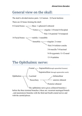
  • 1. Ahmad Amro Baradee General view on the skull: The skull is divided intotwo parts: 1) Cranium 2) Facial skeleton There are 22 bones forming the skull: 8 Cranial bones: Base: 1-sphenoid 2-ethmoid Vertex: singular: 3-Frontal 4-Occipital Pair: 5-6-parietal 7-8-temporal 14 Facial bones: mobile: 1-mandible Immobile: singular: 2-vomer Pair:3-4-inferior concha 5-6-maxilla 7-8-lacrimal 9-10-zygomatic 11-12-nasal 13-14-palatine The Ophthalmic nerve: Frontal Supraorbital(through supraorbital foramen) Supratrochlear (through supratrochler notch) Ophthalmic Lacrimal Infratochlear Nasociliary anterior ethmoid Posterior ethmoid The ophthalmic nerve gives collateral branches ( before the three terminal branches ) these are: recurrent meningeal branch , and anastomosis branches with the third and fourth cranial nerves and with the carotid plexus.
  • 2. Ahmad Amro Baradee The external carotid artery: Superior thyroid Anteroir lingual facial External medial: ascending pharyngeal Carotid Artery posterior occipital Posterior auricular terminal Maxillary Superficial temporal Transverse facial Middle temporal Zygomatico-orbital Anterior auricular 2 terminal br.: frontal parietal The Maxillary Artery: Classification "1": We say the maxillary artery gives 14 collateral branches , and they are classified among their directions into 4 categories: A. Three ascending branches heading to the cranium: 1) Tympanic artery 2) Middle meningeal artery 3) Accessory meningeal artery B. Three lateral branches heading to the muscles: 1) Masseteric artery 2) deep posterior temporal artery 3) deep anterior temporal artery C. Four descending branches heading to masticating system: 1) inferior dental artery 2) pterygoid artery 3) orobuccal artery (oro=mouth ) 4) alveolar artery D. Four deep branches evolve in the pterygopalatine fossa: 1) Infraorbital artery
  • 3. Ahmad Amro Baradee 2) Vidian artery ( the artery of pterygoid canal ) 3) Descending palatine artery 4) Pterygopalatine artery Classification "2": ( I, myself, prefer this way of classification) We say the maxillary artery gives 16 branches , and they are classified into 3 categories: A. First category: 1) middle meningeal 2) accessory meningeal 3)inferior alveolar 4) deep auricular 5) anterior tympanic B. Second category: 1) lateral pterygoid 2) medial pterygoid 3)masseter 4) temporal 5) buccinator C. Third category: 1) posterior superior alveolar 2) Vidian (artery of pterygoid canal) 3)greater palatine 4)lesser palatine 5)sphenopalatine 6)infraorbital Temoral bone's fissures: Squamotympanic f. Tympanic Squamos part part Petrotympanic f. petrosal squamopetrosal f. prominence Only the tympanomastoid fissure is left to be pointed out
  • 4. Ahmad Amro Baradee The paranasal sinuses (air sinuses): These sinuses: 1) Reduce the weight of the skull 2) Give the ability to make sounds 3) Give the shape of the face Opens into:The paranasal sinus Semilunar hiatus , middle meatusMaxillary Semilunar hiatus , middle meatusFrontal Semilunar hiatus , middle meatusAnterior and middle ethmoid Superior meatusPosterior ethmoid Sphenoethmoidal recess,above the superior meatusSphenoid Don't open..Mastoid air cells Remember: the nasolacrimal duct carries tears into the inferior meatus Note: "meatus" refers to the space under the concha , means: the inferior meatus is the space under the inferior concha The venouses sinuses: Singular:  Superior sagittal  Inferior sagittal  Straight  Occipital  intercavernous Pair:  superior petrosal  inferior petrosal  transverse  sigmoid  cavernous
  • 5. Ahmad Amro Baradee EndsVenouses sinus Right transverseSuperior sagittal StraightInferior sagittal Left transverseStraight Continues as: the sigmoidTransverse (right or left) Jugular bulbSigmoid SigmoidSuperior petrosal Jugular bulbInferior petrosal The Sphenoid bone:  The anterior aspect of the sphenoid bone we find the sphenoidal crest , and on each side we see the apertute of the sphenoidal sinus, and lateral to that aperture we see semi air cells which present the place of articulation between the ethmoid labyrinth and the sphenoid bone.  The anterior part of the sphenoidal concha articulates with the orbital process of the palatine bone.  The posterior part of the orbital process of the palatine bone gives an opening to connect with the sphenoidal sinus , whereas the margins of this opening articulates with the sphenoidal concha.  The inferior surface of the sphenoidal concha articulates with the superior surface of the sphenoidal process of the palatine bone.  We can recognise the following landmarks in an intercranial aspect of the sphenoid bone: (in this exact order postero-anteriorly): Dorsum sellae Pituitary gland's fossa Groove for intercavernous sinus Tuberculum sellae (on each of its sides we find the middle clinoid process ) Chiasmatic groove Sphenoidal limbus Sphenoidal jugum ethmoidal process
  • 6. Ahmad Amro Baradee The pterygopalatine fossa: It’s a pyramidal fossa 1) It's boundaries:  Lateral: the internal surface of the ramus of mandible  Anterior: maxillary tuber  Medial: the perpendicular plate of palatine bone (at it's top end we see the sphenopalatine foramen )  Posterior: the anterior surface of the pterygoid process  Superior: the inferior surface of the greater wing of the sphenoid bone (the maxillary surface) and the apex of this pyramidal fossa is at the inferior end (at the place of meeting: the pterygoid process with the pyramidal process of palatine bone together with the tuber of maxillary bone) 2) the foramina that open into the fossa:  Foramen rotundom  Foramen ovale  The aperture of the pterygoid canal  The aperture of the Palatovaginal canal  the sphenopalatine foramen (connects the fossa with the nasal cavity )  the inferior orbital fissure (connects it with the orbit)  the pterygomaxillary (pterygopalatine) fissure (connects it with the infratemporal fossa)  it connects with the oral cavity through the greater palatine Canal (the pterygopalatine canal) which has the (Mercedes sign shape: the posterior part of the canal is formed by the pterygoid process , the anterio-medial is formed by the perpendicular plate of palatine bone (with making a groove on it) , the anterio-lateral part is formed by the posterior part of the nasal surface of the maxillary bone (also with making a groove on it )
  • 7. Ahmad Amro Baradee Note: the term (pterygopalatine canal) can refer to one of two canals: 1) the greater palatine canal 2) OR:the palatovaginal canal 3) It's contents:  The two pterygoid muscles  The maxillary artery and it's branches  The maxillary nerve and it's branches  The mandibular nerve and it's branches  The pterygoid venouses plexus  The pterygopalatine ganglion: lies at the bottom of the fossa 4) The branches of the pterygopalatine ganglion:  Orbital branches(passes through the inferior orbital fissure)  nasopalatine nerve  posterior superior nasal branches  greater palatine nerve  lesser palatine nerve  Bock's nerve ( the pterygopalatine nerve) (passes through the palatovaginal canal) o The vidian nerve (the nerve of pterygoid canal) , and two sturdy branches of the maxillary nerve: end up at the ganglion. I hope this was helpful  By: Ahmad Amro Baradee