Avoidance of stochastic RNA interactions can be harnessed to control protein ...
PIIS2211124714009115
1. Report
An Active Role for the Ribosome in Determining the
Fate of Oxidized mRNA
Graphical Abstract
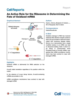
Highlights
Oxidized mRNA is detrimental for tRNA selection on the
ribosome
8-oxoG stalls translation regardless of its position within the
codon
In the absence of no-go decay factors, 8-oxoG-containing
mRNAs accumulate in vivo
mRNA surveillance mechanisms have evolved to deal with
damaged mRNA
Authors
Carrie L. Simms, Benjamin H. Hudson, ...,
Ali S. Rangwala, Hani S. Zaher
Correspondence
hzaher@wustl.edu
In Brief
Oxidative damage to RNA has received
relatively little attention despite evidence
that it can accumulate in cells and is asso-ciated
with numerous disease states.
Simms et al. demonstrate that a single
modified residue in an mRNA can lead
to ribosomal stalling. Cells in which no-go
decay is compromised show
increased levels of 8-oxoG mRNA, sug-gesting
that mRNA surveillance mecha-nisms
may have evolved to cope with
damaged mRNA.
Simms et al., 2014, Cell Reports 9, 1256–1264
November 20, 2014 ª2014 The Authors
http://dx.doi.org/10.1016/j.celrep.2014.10.042
2. Cell Reports
Report
An Active Role for the Ribosome
in Determining the Fate of Oxidized mRNA
Carrie L. Simms,1 Benjamin H. Hudson,1 John W. Mosior,1 Ali S. Rangwala,1 and Hani S. Zaher1,*
1Department of Biology, Washington University in St. Louis, St. Louis, MO 63130, USA
*Correspondence: hzaher@wustl.edu
http://dx.doi.org/10.1016/j.celrep.2014.10.042
This is an open access article under the CC BY-NC-ND license (http://creativecommons.org/licenses/by-nc-nd/3.0/).
SUMMARY
Chemical damage to RNA affects its functional prop-erties
and thus may pose a significant hurdle to
the translational apparatus; however, the effects of
damaged mRNA on the speed and accuracy of the
decoding process and their interplay with quality-control
processes are not known. Here, we system-atically
explore the effects of oxidative damage on
the decoding process using a well-defined bacterial
in vitro translation system. We find that the oxidative
lesion 8-oxoguanosine (8-oxoG) reduces the rate of
peptide-bond formation by more than three orders
of magnitude independent of its position within the
codon. Interestingly, 8-oxoG had little effect on the
fidelity of the selection process, suggesting that
the modification stalls the translational machinery.
Consistent with these findings, 8-oxoG mRNAs
were observed to accumulate and associate with
polyribosomes in yeast strains in which no-go decay
is compromised. Our data provide compelling evi-dence
that mRNA-surveillance mechanisms have
evolved to cope with damaged mRNA.
INTRODUCTION
The accurate translation of cellular mRNAs is an integral feature
of the ribosome and translation factors. On intact mRNAs, the
selection of aminoacylated tRNA substrates by the ribosome
has evolved to utilize thermodynamic and kinetic differences be-tween
cognate and noncognate interactions (Zaher and Green,
2009a). On aberrant mRNAs, such as truncated ones and those
either containing premature or lacking stop codons, a number of
ribosome-based-quality-control processes ensure that these
RNAs are not translated and instead are targeted for degradation
(Graille and Se´ raphin, 2012; Kervestin and Jacobson, 2012;
Shoemaker and Green, 2012). In addition to defects in its
sequence, mRNA is susceptible to chemically induced defects.
Nucleic acids are constantly under assault from both endoge-nous
and exogenous agents that can affect their chemical prop-erties
and hence their function (Wurtmann and Wolin, 2009).
These assaults include reactive oxygen species (ROS), UV light,
and alkylating agents. In contrast to DNA, damaged RNA has
received little attention, presumably due to its transient nature
(Li et al., 2006), even though damaged RNA is known to accumu-late
and has been linked to a number of neurodegenerative dis-eases
(Nunomura et al., 1999; Shan et al., 2003, 2007; Shan and
Lin, 2006; Tanaka et al., 2007). Indeed, in human fibroblast cells,
the median half-life of mRNAs is 10 hr (Yang et al., 2003), indi-cating
that damaged mRNAs may persist and are likely to pose
a challenge to the cell if not corrected. Furthermore, a number
of mRNA-surveillance mechanisms exist to handle aberrant
mRNAs, suggesting that high turnover of RNA alone is not a suf-ficient
means to cope with damaged RNAs.
ROS are present under normal conditions as byproducts of
metabolic reactions (Finkel and Holbrook, 2000), and their levels
increase significantly under stress conditions. The reaction of
ROS with nucleic acids results in a myriad of modifications (Bar-ciszewski
et al., 1999). Among these lesions, modification to
the guanine base resulting in the 8-oxo-7,8-dihydro-guanine
(8-oxoGua) is the quintessential archetype of oxidation adducts
due to its ability to pair with adenosine during DNA replication
(Figure 1A). In agreement with this altered base-pairing property,
RNA damage appears to affect decoding, as crudely oxidized
mRNA has been linked to reduced translational efficiency
and accuracy (Shan et al., 2003, 2007; Tanaka et al., 2007).
To date, mechanistic understanding of this inefficiency (i.e.,
whether it is due to miscoding, stalling, or degradation of the
damaged mRNA) is lacking. This is mainly due to the difficulty
of targeting a specific transcript at an exact location for
oxidation.
Oxidized RNA is known to accumulate in vivo, for which 8-
oxoG is present at 1 in 105 residues in total RNA (the levels
could potentially be higher in the mRNA pool) under normal con-ditions
and increases as much as 10-fold under oxidative stress
(Hofer et al., 2005; Shen et al., 2000), highlighting the potential
risk to cellular homeostasis. Additionally, oxidized RNA appears
to turn over at a faster rate (Hofer et al., 2005), suggesting that it
is selectively degraded; however, the exact details of such a
mechanism are currently unknown. It is conceivable that factors
that recognize lesions on RNA exist and target it for degrada-tion,
but a more appealing model would involve ribosomal ac-tion.
What is attractive about this model is the existence of a
number of ribosome-based mRNA-surveillance mechanisms
(Shoemaker and Green, 2012). Second, as the ribosome is the
only machine that sees all mRNAs, it is not difficult to imagine
how a damaged RNA quality-control mechanism can be built
into the ribosome. The signal for any degradation process,
1256 Cell Reports 9, 1256–1264, November 20, 2014 ª2014 The Authors
3. Figure 1. 8-oxoG Is Detrimental to the Decoding Process
(A) Chemical structures of G:C, 8-oxoG:C, and 8-oxoG:A base pairs. 8-oxoG adopts a syn conformation, allowing it to base pair with adenosine.
(B) Schematic representation of guanosine and 8-oxoG initiation complexes encoding for the dipeptide Met-Arg. Both complexes carry the initiator fMet-tRNAfMet
in the P site; the G complex displays a CGC codon in the A site, whereas the 8-oxoG complex displays a C8-oxoGC codon in the A site.
(C) Schematic representation of the cognate Arg-tRNAArg and near-cognate Leu-tRNALeu (G:A mismatch at the second position) ternary complexes.
(D) Phosphorimager scan of an electrophoretic TLC showing the reactivities of the initiation complexes with the indicated ternary complexes and release factors.
Green asterisk represents the cognate dipeptide, whereas the red one represents the near-cognate dipeptide resulting from a G:A mismatch.
(E) Quantification of the dipeptide yield in (D); the predicted codon-anticodon interaction is shown below the x axis, and the corresponding dipeptide is shown
above the bars.
(F and G) Time courses of peptide-bond formation between the initiation complexes and the cognate Arg-tRNAArg and near-cognate Leu-tRNALeu ternary
complexes, respectively.
See also Figures S1 and S2.
however, would have to initiate within the decoding center of the
ribosome as it selects an incoming aminoacyl-tRNA (aa-tRNA)
against the oxidized A-site codon. With this in mind, here we
examine the effect of 8-oxoG on decoding using a high-resolu-tion
approach, providing a comprehensive look at the ribosomal
response to oxidative damage.
RESULTS
8-oxoG Is Detrimental to the Decoding Process
To explore the effects of RNA damage on the decoding process,
we took advantage of our high-resolution reconstituted bacterial
system (Zaher and Green, 2009b), which can monitor the
Cell Reports 9, 1256–1264, November 20, 2014 ª2014 The Authors 1257
4. efficiency of incorporation of every single amino acid (Figure S1).
Weproduced ribosomal initiation complexes carrying the initiator
tRNA f-[35S]-Met-tRNAfMet in the P site combined with a variety
of intact or site-specifically damaged codons in the A site. We
started our studies with a pair of complexes displaying either
the intact CGC or C8-oxoGC codon in the A site (Figure 1B). Under
normal conditions, this codon is decoded by Arg-tRNAArg
ICG with
canonical Watson-Crick base pairs at the three positions. We
reasoned that the oxidized complex, if 8-oxoG base pairs with
A, should be decoded by Leu-tRNALeu
GAG (Figure 1C). The com-plexes
were reacted with different aa-tRNAs isoacceptors as well
as the two release factors (RF1 and RF2) for 5 s, which should be
sufficient to reach the endpoint of a normal peptidyl-transfer re-action
(Figure 1D). The production of dipeptides or RF-mediated
hydrolysis was visualized using an electrophoretic thin-layer
chromatography (TLC) system (Youngman et al., 2004). At first
glance, the survey appears to agree well with our prediction.
The intact complex efficiently reacts with Arg-tRNAArg and not
with Leu-tRNALeu; the oxidized complex reacts poorly with Arg-tRNA
Arg but slightly better with Leu-tRNALeu, relative to the native
one. Furthermore, the data suggest that the adduct affects the
decoding process in a more complicated fashion than we initially
anticipated; there are a set of aa-tRNAs that decode the intact
codon but appear not to decode the modified codon (Figure 1D).
Nonetheless, the reactivity of the damaged complex with the
near-cognate aa-tRNA appears to proceed with an overall poor
efficiency relative to normal peptide-bond formation (Figure 1E),
indicating that the oxidized base may have a substantial effect on
the speed of the ribosome.
Having established the crude reactivity of the complex
harboring the damaged mRNA, we next determined the effects
of the adduct on the rate of peptide-bond formation with cognate
and near-cognate aa-tRNAs. A comparison of rates for the reac-tion
between theCGC and C8-oxoGCcomplexes and the cognate
Arg-tRNAArg showed that the complex displaying the modified
nucleotides reacted much slower than the intact one; the
apparent rate was determined to be more than 1,000-fold slower
(Figure 1F). These findings suggest that 8-oxoG is likely to stall
the ribosome, unless the rate with the near-cognate ternary com-plexes
is greatly accelerated. In agreement with our endpoint
analysis (Figure 1D), the apparent rate for the 8-oxoG complex
with the near-cognate Leu-tRNALeu ternary complex (where G
mispairs with A in the second position), albeit faster than the
immeasurably slow rate for the intact complex, was very slow
(0.03 s1)—likely too slow to support efficient protein synthesis
in vivo (Figure 1G). This was unexpected, as our initial prediction
was that 8-oxoGwould lead to incorporation of the near-cognate
aa-tRNA and produce some level of miscoding.
The Effect on Decoding Is Insensitive to the Position of
the Lesion
The first two positions of the codon and anticodon minihelix
require strict Watson-Crick base pairing, whereas the third posi-tion
allows some wobble base pairing. Additionally, the decoding
center of the ribosome utilizes different interactions to recognize
the correct geometry for the different positions (Ogle et al., 2001).
As a result, we reasoned that the position of the modification
within the codon might have differential effects on tRNA selec-tion
by the ribosome. In particular, we expected the decoding
process to be normal in the presence of damage at the third po-sition
of the codon. To address this hypothesis, we generated
three new sets of complexes, placing the 8-oxoG at the first, sec-ond,
or third position of the codon (see Figure S2). In each case,
the 8-oxoG complex reacted much less efficiently with the
cognate aa-tRNA than its intact counterpart. The complexes
with 8-oxoG in the first or second position instead reacted
more efficiently with the predicted (if 8-oxoG pairs with A, or G
at the wobble position) near-cognate aa-tRNA, but again with
an overall poor yield. Lastly, the third set of complexes, carrying
the 8-oxoG at the wobble position, reacted poorly with its
cognate aa-tRNA, in direct contrast to our prediction that the
third position is insensitive to damage. Furthermore, the complex
reactivity with near-cognate aa-tRNAs displayed a distinct pro-file
relative to its intact counterpart, highlighting the detrimental
effect of the adduct on the decoding process even at the third
position of the codon (Figure S2). Taken together, the data reveal
a comprehensive overview of the potential havoc caused by
oxidative damage to mRNA on the translation machinery.
We next tested if modification of other positions would elicit
a less profound effect on the rate of peptide-bond formation.
We measured the rate of peptide-bond formation for the same
set of complexes described earlier with their respective cognate
and near-cognate aa-tRNAs. Cognate aa-tRNA incorporation
was three to four orders of magnitude slower for 8-oxoG codons
than for undamaged controls (Figure 2A). Conversely, 8-oxoG
codons incorporated near-cognate aa-tRNAs nearly 10-fold
faster than intact codons, although the observed rates (0.002–
0.03 s1) remained dramatically slower than that measured for
normal peptide-bond formation (20–40 s1) (Figure 2A). These
observations collectively argue that 8-oxoG likely stalls the elon-gation
phase of translation regardless of its location within the
anticodon. Furthermore the drastic inhibitory effect of 8-oxoG
on the decoding process was confirmed under competitive con-ditions.
In particular, in contrast to the native GGC complex,
which produced the expected full-length peptide when incu-bated
in the presence of the full complement of aa-tRNAs,
elongation factors and release factors (PURE system, NEB),
the oxidized G8-oxoGC complex failed to produce any detectable
peptide products (Figure 2B).
To provide mechanistic insight into the deleterious effects of
oxidized mRNA on the decoding process, we set out to explore
if the effects of 8-oxoG on peptidyl transfer result from inhibition
of conformational changes known to be important for the
decoding process (Ogle et al., 2002). In particular, we added
the aminoglycoside paromomycin to our reaction, which binds
the decoding center and induces a conformation in the 30S sub-unit
similar to that observed when cognate tRNA is bound; in do-ing
so, it allows the ribosome to accept near-cognate tRNAs as if
they were cognate ones (Carter et al., 2000). As expected (Pape
et al., 2000), the addition of paromomycin to a reaction between
the GGC complex and its cognate Gly-tRNAGly ternary complex
had no effect on the observed rate of peptide-bond formation
(Figure 2C, left panel). In contrast, addition of the antibiotic to
the oxidized G8oxoGC complex accelerated the rate of peptide-bond
formation more than 100-fold (Figure 2C, right panel).
These observations strongly argue that 8-oxoG prevents the
1258 Cell Reports 9, 1256–1264, November 20, 2014 ª2014 The Authors
5. Figure 2. 8-oxoG Inhibits Peptide-Bond Formation
(A) Observed rates for peptidyl transfer (kPT) measured on native (GUU, GGC, CGC, GAG) and 8-oxoG complexes (8-oxoGUU, G8-oxoGC, C8-oxoGC, GA8-oxoG) with
cognate (left two bars in each graph) and near-cognate (A:G mismatches at the 8-oxoG position) (right two bars in each graph) tRNAs. The codon-anticodon
interaction is shown below the x axis, and the corresponding dipeptide is shown above the bars. Clear bars represent rates observed with native complexes;
black ones represent those observed with 8-oxoG complexes. Reactions were carried out at least in duplicate ±SEM
(B) Phosphorimager scan of an electrophoretic TLC of reactions between native complex (GGC) or 8-oxoG complex (G8-oxoGC), which encode MGLYK peptide,
and the full complement of aa-tRNAs, elongation and release factors (PURE system, NEB). Reactions were allowed to proceed for 30 s before the addition of
100 mM KOH.
(C) Time-courses of peptide-bond formation between the native GGC complex and Gly-tRNAGly (left) or oxidized G8oxoGC complex and Gly-tRNAGly (right) in the
absence (blue circles) and presence of paromomycin (red squares).
(D) Time courses of RF2-mediated release on native UGA (blue circles) and U8-oxoGA (red squares) initiation complexes.
small subunit from adopting the active conformation required for
proper tRNA selection.
Peptide Release Is Only Marginally Affected in the
Presence of 8-oxoG
Two of the three stop codons contain a guanosine: UAG and
UGA. In E. coli, these are recognized by RF1 and RF2, respec-tively.
Stop-codon recognition by RFs is inherently different
than sense-codon recognition by aa-tRNA due to the intrinsic dif-ferences
in the polymer (protein versus RNA) carrying out the
recognition (Zaher and Green, 2009a). Unlike aa-tRNA selection,
which requires many residues of the decoding center to adopt a
distinct conformation from the ground state, RF selection in-volves
fewer and dissimilar conformational changes within the
decoding center (Korostelev et al., 2008; Laurberg et al., 2008;
Weixlbaumer et al., 2008). As a result, we reasoned that the ef-fects
of 8-oxoG on sense-codon recognition could be different
than those determined for RF-recognition on stop codons. Since
weexpected 8-oxoG to have the greatest effect at the second po-sition,
we programmed ribosomes with f-[35S]-Met-tRNAfMet in
the P site and either a UGAorU8-oxoGA in the A site and assessed
their reactivity with RF2. In direct contrast to peptide bond forma-tion,
the presence of 8-oxoG only marginally affected the rate
of peptide release (0.7 s1 and 0.1 s1 for control and 8-oxoG,
respectively) (Figure 2D). Together, these results suggest
that 8-oxoG prevents conformational changes associated with
RNA-RNA interactions (i.e., codon-anticodon), but not protein-
RNA interactions (i.e., RF-stop codon), in the decoding center.
8-oxoG Inhibits Protein Synthesis in Cell Extracts
So far, our characterization of the effects of 8-oxoG on the de-coding
process has focused on initiation complexes in a bacte-rial
reconstituted system, and the effects of adducts on authentic
elongation during protein synthesis were not discerned. Never-theless,
a number of studies have looked at the consequences
of oxidative RNA damage on its function, and all hinted at a pro-found
correlation between oxidized mRNA and reduced transla-tional
efficiency and accuracy (Shan et al., 2003, 2007; Tanaka
et al., 2007). However, in all of these studies, the identities of
the adducts formed from the crude hydrogen peroxide treatment
and their relative abundance were not quantified. Furthermore,
the effect of the treatment on the yield of full-length protein prod-ucts
was only marginal at best (Tanaka et al., 2007), presumably
due to the inefficiency of the hydrogen peroxide treatment.
To improve upon this and provide a better understanding of
the effects of the adduct on translation, we generated synthetic
reporter mRNAs containing the lesion at a specific location
within the coding sequence. We used in vitro transcription to
synthesize a 300 nt transcript and ligated this to an 8-oxoG-containing
synthetic RNA oligonucleotide (Figure S3). We note
that the final construct has the modification on the first position
of a GAC codon. We also constructed three control reporters
(one with the equivalent native codon, a second with a stop
codon and a third shorter transcript with no stop codon) (Figures
3A and S3). Using these reporters, we could assess how detri-mental
the 8-oxoG adduct is to translational speed and whether
our reconstituted system recapitulates what happens during
Cell Reports 9, 1256–1264, November 20, 2014 ª2014 The Authors 1259
6. bona fide translation in bacterial and eukaryotic extracts. We
initially analyzed the translation of these reporters in an S30 bac-terial
extract. In agreement with the reconstituted system, the
8-oxoG transcript produced a protein product of a size similar
to that of the stop transcript (Figure 3B). A priori, one would
expect the incubation of 8-oxoG and no-stop transcripts in the
S30 extract to result in the accumulation of peptidyl-tRNA, as
these transcripts are expected to stall the ribosome, unless a
ribosomal rescue system is operational in these extracts. Inter-estingly,
we observe no peptidyl-tRNA accumulation with either
reporter, even though the products were resolved on bis-Tris
gels (Figure 3B), which are known to maintain the integrity of
peptidyl-tRNA.
We next asked whether the inhibitory effects of 8-oxoG
on translation were evident in eukaryotic extracts. For efficient
translation in eukaryotes, the transcripts were capped using
vaccinia capping enzyme and poly(A)denylated (except for the
no stop reporter) using E. coli poly(A) polymerase. Consistent
with our bacterial studies, 8-oxoG prevented the translation of
full-length protein. The 8-oxoG reporter, when incubated in
wheat germ extracts or rabbit reticulocyte lysates, failed to pro-duce
the full-length protein product and instead generated a
product of similar length to that of the stop codon reporter (Fig-ures
3C, 3D, and S3). However, in contrast to the bacterial
extract results, we could detect accumulation of peptidyl tRNA
in the presence of the 8-oxoG and no-stop transcripts, suggest-ing
that the ribosomal rescue system in the wheat germ extract is
not efficient at clearing a stalled ribosome on the oxidized RNA.
Figure 3. 8-oxoG Stalls Translation in Cell
Extracts
(A) Schematic of the mRNA reporters used in
eukaryotic extracts. The full-length mRNA en-codes
a peptide that has a molecular weight of
9 kDa, whereas the stop mRNA, which has a
stop codon at the position of the 8-oxoG codon in
the 8-oxoG mRNA, encodes a peptide that has a
molecular weight of 8.4 kDa. Reporters used in
S30 reactions were identical minus the cap and
polyadenylation.
(B) Autoradiograph of a bis-Tris gel of translation
assays using bacterial S30 extracts. The 8-oxoG
transcript yields a peptide of a size similar to the
stop reporter. Asterisk (*) indicates a nonspecific
band.
(C and D) Autoradiograph of bis-Tris and Tris-
Tricine gels of in vitro translation assays in wheat
germ extracts. Proteins were labeled by the
addition of [35S]-Methionine to the reactions. The
8-oxoG mRNA and no-stop mRNA both accu-mulate
peptidyl-tRNA, visible on the bis-Tris gel,
which disappear upon the addition of RNase A
(C). Reactions separated on Tris-Tricine were
incubated with and without MG132. The 8-oxoG
mRNA produces truncated protein products
(marked by arrows); the largest of these has a size
similar to that observed in the presence of stop
mRNA (D).
See also Figure S3.
Additionally, the yield of the truncated protein products is much
less than those produced in the presence of the control and stop
mRNA, suggesting that 8-oxoG indeed inhibits the progress of
translation. To gain an improved resolution of the products being
produced from these reactions, we utilized Tris-Tricine gels to
resolve the peptide products. With this gel system, we noted
the appearance of several smaller protein bands in the presence
of 8-oxoG mRNA (Figure 3D), likely resulting from ribosomes
stacked behind the ribosome that stalls on the adduct. Further-more,
we observed no improvement in the yield of the truncated
protein products upon addition of the proteasome inhibitor
MG132 (Figure 3D), suggesting that the diminished yield of the
truncated product is likely due to slowed translation and not to
turnover of the protein products. We also note that incubation
of labeled RNA in extracts did not reveal significant degradation
of the RNA during the course of the reaction (Figure S3). These
observations strongly suggest that, similar to what we observe
in the bacterial reconstituted system, 8-oxoG halts translational
elongation in eukaryotes.
8-oxoG mRNAs Accumulate In Vivo in the Absence of
No-Go Decay
Three mRNA quality-control processes are known to exist in eu-karyotes:
nonsense-mediated decay, no-go decay (NGD), and
nonstop decay. In NGD, transcripts containing secondary struc-tures
such as hairpins or stretches of rare codons are cleaved
just upstream of the ribosome (Doma and Parker, 2006; Tsuboi
et al., 2012). In yeast, the process involves the proteins
1260 Cell Reports 9, 1256–1264, November 20, 2014 ª2014 The Authors
7. Dom34, Hbs1, and Rli1 that together are responsible for recog-nizing
and subsequent recycling of the stalled ribosome (Pisar-eva
et al., 2011; Shoemaker et al., 2010; Shoemaker and Green,
2011). The process results in an endonucleolytic cleavage of the
mRNA; the 30 end piece is then degraded by the 50-30 exonu-clease
Xrn1, and the 50 end piece is degraded by the exosome
(Tsuboi et al., 2012). The exact role of these various quality-con-trol
processes in recognizing damaged RNA (e.g., oxidized RNA)
has not been studied. Intriguingly, however, quality-control pro-cesses
have been linked to damaged RNA where enzymatic
depurination at specific sites within a transcript was found to stall
the ribosome and is likely to elicit NGD (Gandhi et al., 2008).
Our observation that the 8-oxoG modification stalls protein
synthesis suggests that 8-oxoG-containing mRNAs might be
subject to degradation through NGD. To address this possibility,
we quantified 8-oxoG levels in total and poly(A)-enriched RNA
from different yeast strains using a competitive ELISA protocol
(Yin et al., 1995) that specifically recognizes the oxidized base
(Figure S4). Interestingly, the levels of the adduct in wild-type
cells under normal conditions were approximately 5-fold higher
in poly(A)-enriched RNA than in total RNA (0.03 ng 8-oxoG/
mg total RNA versus 0.15 8-oxoG/mg poly(A)-enriched RNA),
suggesting that mRNAs are more susceptible to oxidative stress,
perhaps due to their naked nature and reduced secondary
structure relative to rRNA or tRNA. Furthermore, we observe little
to no change in the level of 8-oxoG adducts in total RNA in
the absence of quality-control or mRNA-decay pathways. In
contrast, the levels of 8-oxoG in poly(A)-enriched RNA increase
significantly by almost 2-fold (1.85-fold ± 0.17, p = 0.028) in the
absence of Dom34 (Figure 4A), suggesting that 8-oxoG-contain-ing
mRNA is subject to NGD and is stabilized in the absence of
Dom34. Additionally, Xrn1-deficient cells show a significant
accumulation of 8-oxoG (2.18-fold ± 0.25, p = 0.011), whereas
abrogating exosome function had no effect (Figure S4). We
next sought to confirm that the changes in 8-oxoG levels are
not the result of strain to strain variation by complementing the
dom34D and xrn1D strains and measuring the levels of 8-oxoG
in these rescued strains (dom34D comp. and xrn1D comp.,
respectively). These strains were complemented by integrating
the native genes at the his3 locus of chromosome XV (under
the control of the endogenous promoter and carrying the endog-enous
UTR), resulting in transcript levels that were similar to wild-type
(Figure S4). Consistent with our proposal, 8-oxoG levels
were restored to wild-type upon reintroduction of Dom34 and
Xrn1 (Figure 4A). It is worth noting that our measurements of
8-oxoG levels are likely an underrepresentation, since our
poly(A)-enriched samples still contain some rRNA (Figure S4).
Furthermore, the 2-fold increase in the steady-state levels of 8-
oxoG in the absence of Dom34 and Xrn1 is consistent with the
effects of these factors on the steady-state levels of typical
NGD reporters (Tsuboi et al., 2012).
These observations are in full agreement with our current un-derstanding
of the NGD process, wherein (1) translation arrest
results in an endonucleolytic cleavage upstream of the stall
sequence (in this case, 8-oxoG); (2) ribosomes are recycled by
Dom34:Hbs1; and (3) the 30 fragment, i.e., 8-oxoG containing,
is degraded by Xrn1, whereas the 50 fragment is degraded by
the exosome (Doma and Parker, 2006; Tsuboi et al., 2012). It is
worth noting that while endonucleolytic cleavage is not directly
mediated by Dom34:Hbs1, it is significantly stimulated in the
presence of these factors (Tsuboi et al., 2012). Two key predic-tions
emerge from this model: (1) in the absence of Dom34, 8-
oxoG-containing mRNA is more likely to associate with the ribo-somes;
(2) in the absence of Xrn1, 8-oxoG-containing mRNA is
stabilized but would not be associated with the ribosomes. We
set out to test these predictions by performing polysome anal-ysis
of the different yeast strains and quantifying the distribution
of 8-oxoG among the different fractions of ribonucleoproteins
(Figures 4B–4E). xrn1D strains have been shown to accumulate
poly(A) mRNAs in the light fraction of the polysome due to stabi-lization
of uncapped mRNA that cannot be translated (Hu et al.,
2009). To correct for this, we normalized the levels of 8-oxoG
to the total amount of RNA during our polysomes analysis
of poly(A)-enriched RNA, which should adjust for variation in
poly(A)-RNA occupancy across the sucrose gradient. As ex-pected,
for all strains, we observed very similar distributions of
8-oxoG in total RNA (Figure 4D), consistent with our measure-ments
from whole cells (Figure S4). However, in the absence of
Dom34, 8-oxoG levels in poly(A)-enriched RNA are significantly
higher in the 80S and polysome fractions (Figure 4E), demon-strating
that Dom34p plays a role in clearing stalled ribosomes
on damaged mRNA. Additionally, in the absence of Xrn1,
8-oxoG levels were elevated in the light fractions, containing
non-ribosome-bound RNAs, and unaltered in the 80S and poly-some
fractions. Supporting the model that Dom34 acts up-stream
of Xrn1 in NGD, Xrn1/Dom34 double-mutant 8-oxoG
levels were similar to those of Dom34 (Figure 4E).
We next hypothesized that if NGD is important in rescuing ri-bosomes
stalled on damaged mRNA, then deleting dom34 and
xrn1 should make yeast cells sensitive to nucleic-acid-damaging
agents. In particular, we were interested in determining the
sensitivity of the different yeast strains to the carcinogen 4-nitro-quinoline-
1-oxide (4NQO), which is known to react with RNA to
form the 8-oxoguanine lesion (Tada and Kohda, 1993). We uti-lized
a zone of inhibition (halo) assay to compare the growth of
the deletion strains in the presence of 4NQO (Figure 4F). Consis-tent
with a role for NGD in clearing damaged mRNA, deletion of
dom34 and xrn1 rendered yeast cells significantly more sensitive
to 4NQO (1.5-fold ± 0.05, p 0.0001 and 1.8-fold ± 0.06, p
0.0001, respectively), and reintroduction of the genes restored
normal growth relative to the wild-type strain (Figure 4G). Finally,
the dom34D xrn1D double mutant was found to exhibit dramatic
sensitivity to 4NQO compared to the single mutants (3.0-fold ±
0.18, p 0.0001) (Figure 4G). These observations strongly sug-gest
that Dom34 and Xrn1 protect the cell from the effects of
RNA-damaging agents.
DISCUSSION
Here, we demonstrated that mRNA containing the oxidized
nucleotide 8-oxoG stalls the ribosome and that NGD may have
evolved to clear these and other aberrant mRNAs. A priori,
we expected the adduct, based on its chemical properties,
to increase miscoding by allowing G:A mismatches between
the codon and anticodon. In contrast to this prediction, 8-
oxoG was found to inhibit tRNA selection in a well-defined
Cell Reports 9, 1256–1264, November 20, 2014 ª2014 The Authors 1261
8. Figure 4. 8-OxoG mRNA Is Stabilized in Yeast in the Absence of No-Go-Decay Factors and Associates with Polysomes in the Absence of
Dom34
(A) Bar graph showing the levels of 8-oxoG, relative to WT, in poly(A)-enriched RNAs isolated from the indicated yeast strains. The values shown are the means of
at least three measurements ± SEM. *p 0.05
(B) Polysomes profiles of the indicated strains.
(C) Denaturing agarose electrophoresis of the RNA samples isolated from the different fractions in the sucrose gradient.
(D and E) Distribution of 8-oxoG levels across the gradients in total RNA and poly(A)-enriched RNA samples, respectively. Levels of 8-oxoG in (D) were normalized
to the total amount of 8-oxoG across the gradient, whereas in (E), they were divided by the amount of poly(A)-enriched RNA.
(F) Representative images of zone of inhibition assays on the indicated yeast strains using 4-nitroquinoline-1-oxide (4NQO).
(G) Quantification of 4NQO sensitivity for the indicated strains, relative to WT. Data are mean ± SEM, from results of at least three biological replicates.
***p 0.001, ****p 0.0001.
See also Figure S4.
1262 Cell Reports 9, 1256–1264, November 20, 2014 ª2014 The Authors
9. reconstituted system, with rates of peptide-bond formation
that are three to four orders of magnitude slower than those
measured on intact complexes. Remarkably, the effects of the
modifications on the accuracy of tRNA selection appear to be
minimal; the rates with near-cognate ternary complexes in the
presence of 8-oxoG are slightly faster (10-fold) relative to intact
complexes. Furthermore, the position of the adduct, even at the
wobble position, resulted in a similar effect on the decoding pro-cess,
highlighting the severity of mRNA oxidation on the pro-tein
synthesis machinery. In agreement with observations from
the reconstituted systems, we find near-complete inhibition of
protein synthesis in the presence of 8-oxoG during authentic
elongation in eukaryotic extracts. The effects of 8-oxoG on the
decoding process appear to be a direct result of impaired
RNA-RNA interaction between the codon and anticodon, as
recognition of stop codons by protein factors was only margin-ally
affected.
How can a small change to the nucleobase result in such
drastic effects on the decoding process? One could imagine
that the addition of the carbonyl oxygen to the non-Watson-Crick
face of the base introduces a polar group that needs to be hy-drated.
The introduction of this energetic penalty is likely to be
inhibitory to the decoding process. On the other hand, in solu-tion,
8-oxoG exists in equilibrium between the anti and syn
conformation, but on the ribosome, the adduct might favor the
anti conformation, which is detrimental to proper A-minor inter-action
in the decoding center. Regardless of the conformation
of 8-oxoG in the decoding center, the modification appears to
prevent the decoding center from adopting an active conforma-tion.
This is supported by our data showing that the addition of
paromomycin alleviates most of the inhibitory effects observed
in the presence of 8-oxoG. These observations highlight the
sensitive nature of the decoding center, which is reinforced by
recent reports demonstrating a uridine to pseudouridine change
(a small change that does not affect the base-pairing properties)
has a dramatic affect on the decoding process and increases
stop-codon readthrough (Ferna´ ndez et al., 2013; Karijolich and
Yu, 2011).
Our in vitro studies suggested that oxidized RNA is subject
to NGD. NGD was initially characterized by Doma and Parker
(Doma and Parker, 2006), who showed that mRNAs with strong
secondary structure or stretches of rare codons are targeted for
degradation in a process that is dependent on translation. While
mRNAs with hairpins for the most part are not biologically
relevant, this study established a direct relationship between
translational speed and rapid degradation of the corresponding
mRNA. Consistent with this, we find 8-oxoG mRNA to be stabi-lized
in the absence of Dom34 (a key component in NGD) and
associated with polysomes. These observations strongly sug-gest
oxidized mRNA is a primary target of ribosome-based
mRNA quality-control processes. We note that NGD and in
particular Dom34 has been implicated in a number of processes;
for example, Inada and colleagues have shown the factor to be
important in rescuing ribosomes trapped on truncated mRNA
(Tsuboi et al., 2012). Recently, Guydosh and Green used ribo-somal
profiling to show that in the absence of the factor, ribo-somes
venture into the 30 UTR (Guydosh and Green, 2014).
Furthermore, the factor is critical for nonfunctional rRNA decay
(LaRiviere et al., 2006; Soudet et al., 2010) and ribosomal assem-bly
(Strunk et al., 2012). Interestingly, all of these processes
share a common feature whereby the target for the factors is a
stalled ribosome with an empty A site (van den Elzen et al.,
2014). It is highly likely that the list of NGD targets is long and
may include other modified mRNAs such as those resulting
from alkylative and UV damage.
Oxidative damage to RNA appears to be correlated with a
number of age-related neurodegenerative diseases and is likely
to be a contributing factor to their pathogenicity. As an example,
Shan et al. showed that in Alzheimer patients, greater than 50%
of mRNA isolated from cerebral cortex were oxidized (Shan and
Lin, 2006). Our results indicate that RNA damage represents a
real and serious threat to the cell and RNA surveillance pathways
such as NGD may have evolved to provide a mechanism to cope
with these damages. RNA metabolism could be a useful target
for delaying or eliminating the onset of age-related neurodegen-erative
diseases.
EXPERIMENTAL PROCEDURES
Dipeptide Formation Survey and Rates of Peptide-Bond Formation
For survey reactions, equal volume of purified initiation complexes (2 mM), pre-incubated
at 37C, was added to ternary complex. The mixture was incubated
at 37C for 5 s before the reaction was quenched by the addition of 100 mM
KOH. Peptide release was carried out in a similar manner, except initiation
complexes were combined with RF1 or RF2 at 10 mMand the reaction stopped
using 20 mM EDTA (pH 6.0). To determine rates of peptide-bond formation,
equal volumes of initiation and ternary complexes were mixed on a quench-flow
instrument (RQF-3 quench-flow, KinTek Corporation). For all reactions,
dipeptides were resolved from unreacted fMet using cellulose TLCs electro-phoresis
in pyridine-acetate buffer (pH 2.7) (Youngman et al., 2004).
To determine the observed rates of peptide-bond formation or peptide
release, dipeptide yield (PT rates) or fraction of released fMet (peptide release)
was plotted against time and the curve fit to a first-order rate equation using
GraphPad prism software. Observed rates reported were calculated by multi-plying
the fit rate with the endpoint and then divided by the maximal endpoint
(observed with the native complexes).
ELISA
8-oxoG was quantified using a competitive ELISA assay. Each experiment
included a set of standards (8-oxoG in 0.1% BSA), enabling quantification
by a standard curve (Abs = (Absmax)/(1 + ([8-OHG]/IC50)).
Zone of Inhibition Assay
Yeast cultures were grown to mid-log phase and plated in triplicate at 0.5
optical density per plate. After drying thoroughly, 25 mg of 4-nitroquinoline-
1-oxide in DMSO was spotted on a 0.5 cm filter paper disc and placed in
the center of the plate. Plates were incubated at 30C for 48 hr before imag-ing.
The area of inhibition was measured using ImageJ. Data were normalized
to wild-type and analyzed using the Student’s t test.
Detailed methods are provided in Supplemental Experimental Procedures.
SUPPLEMENTAL INFORMATION
Supplemental Information includes Supplemental Experimental Procedures
and four figures and can be found with this article online at http://dx.doi.org/
10.1016/j.celrep.2014.10.042.
AUTHOR CONTRIBUTIONS
C.L.S, B.H.H., and H.S.Z. designed the experiments and wrote the manu-script.
C.L.S, B.H.H., J.W.M., A.S.R., and H.S.Z. performed the experiments.
Cell Reports 9, 1256–1264, November 20, 2014 ª2014 The Authors 1263
10. ACKNOWLEDGMENTS
This work was supported by an NIH grant (R00GM094210) and funding from
the Searle Scholars Program to H.S.Z. We thank Allen Buskirk and Joseph
Jez for helpful comments on the manuscript and members of the laboratory
for useful discussions.
Received: June 25, 2014
Revised: September 23, 2014
Accepted: October 15, 2014
Published: November 13, 2014
REFERENCES
Barciszewski, J., Barciszewska, M.Z., Siboska, G., Rattan, S.I., and Clark, B.F.
(1999). Some unusual nucleic acid bases are products of hydroxyl radical
oxidation of DNA and RNA. Mol. Biol. Rep. 26, 231–238.
Carter, A.P., Clemons, W.M., Brodersen, D.E., Morgan-Warren, R.J., Wimberly,
B.T., and Ramakrishnan, V. (2000). Functional insights from the structure of the
30S ribosomal subunit and its interactions with antibiotics. Nature 407, 340–348.
Doma, M.K., and Parker, R. (2006). Endonucleolytic cleavage of eukaryotic
mRNAs with stalls in translation elongation. Nature 440, 561–564.
Ferna´ ndez, I.S., Ng, C.L., Kelley, A.C., Wu, G., Yu, Y.T., and Ramakrishnan, V.
(2013). Unusual base pairing during the decoding of a stop codon by the ribo-some.
Nature 500, 107–110.
Finkel, T., and Holbrook, N.J. (2000). Oxidants, oxidative stress and the
biology of ageing. Nature 408, 239–247.
Gandhi, R., Manzoor, M., and Hudak, K.A. (2008). Depurination of Brome
mosaic virus RNA3 in vivo results in translation-dependent accelerated degra-dation
of the viral RNA. J. Biol. Chem. 283, 32218–32228.
Graille, M., and Se´ raphin, B. (2012). Surveillance pathways rescuing eukary-otic
ribosomes lost in translation. Nat. Rev. Mol. Cell Biol. 13, 727–735.
Guydosh, N.R., and Green, R. (2014). Dom34 rescues ribosomes in 30 untrans-lated
regions. Cell 156, 950–962.
Hofer, T., Badouard, C., Bajak, E., Ravanat, J.L., Mattsson, A., and Cotgreave,
I.A. (2005). Hydrogen peroxide causes greater oxidation in cellular RNA than in
DNA. Biol. Chem. 386, 333–337.
Hu, W., Sweet, T.J., Chamnongpol, S., Baker, K.E., and Coller, J. (2009). Co-translational
mRNA decay in Saccharomyces cerevisiae. Nature 461, 225–229.
Karijolich, J., and Yu, Y.T. (2011). Converting nonsense codons into sense
codons by targeted pseudouridylation. Nature 474, 395–398.
Kervestin, S., and Jacobson, A. (2012). NMD: a multifaceted response to pre-mature
translational termination. Nat. Rev. Mol. Cell Biol. 13, 700–712.
Korostelev, A., Asahara, H., Lancaster, L., Laurberg, M., Hirschi, A., Zhu, J.,
Trakhanov, S., Scott, W.G., and Noller, H.F. (2008). Crystal structure of a trans-lation
termination complex formed with release factor RF2. Proc. Natl. Acad.
Sci. USA 105, 19684–19689.
LaRiviere, F.J., Cole, S.E., Ferullo, D.J., and Moore, M.J. (2006). A late-acting
quality control process for mature eukaryotic rRNAs. Mol. Cell 24, 619–626.
Laurberg, M., Asahara, H., Korostelev, A., Zhu, J., Trakhanov, S., and Noller,
H.F. (2008). Structural basis for translation termination on the 70S ribosome.
Nature 454, 852–857.
Li, Z., Wu, J., and Deleo, C.J. (2006). RNA damage and surveillance under
oxidative stress. IUBMB Life 58, 581–588.
Nunomura, A., Perry, G., Pappolla, M.A., Wade, R., Hirai, K., Chiba, S., and
Smith, M.A. (1999). RNA oxidation is a prominent feature of vulnerable neurons
in Alzheimer’s disease. J. Neurosci. 19, 1959–1964.
Ogle, J.M., Brodersen, D.E., Clemons, W.M., Jr., Tarry, M.J., Carter, A.P., and
Ramakrishnan, V. (2001). Recognition of cognate transfer RNA by the 30S
ribosomal subunit. Science 292, 897–902.
Ogle, J.M., Murphy, F.V., Tarry, M.J., and Ramakrishnan, V. (2002). Selection
of tRNA by the ribosome requires a transition from an open to a closed form.
Cell 111, 721–732.
Pape, T., Wintermeyer, W., and Rodnina, M.V. (2000). Conformational switch in
the decoding region of 16S rRNA during aminoacyl-tRNA selection on the ribo-some.
Nat. Struct. Biol. 7, 104–107.
Pisareva, V.P., Skabkin, M.A., Hellen, C.U., Pestova, T.V., and Pisarev, A.V.
(2011). Dissociation by Pelota, Hbs1 and ABCE1 of mammalian vacant 80S
ribosomes and stalled elongation complexes. EMBO J. 30, 1804–1817.
Shan, X., and Lin, C.L. (2006). Quantification of oxidized RNAs in Alzheimer’s
disease. Neurobiol. Aging 27, 657–662.
Shan, X., Tashiro, H., and Lin, C.L. (2003). The identification and characteriza-tion
of oxidized RNAs in Alzheimer’s disease. J. Neurosci. 23, 4913–4921.
Shan, X., Chang, Y., and Lin, C.L. (2007). Messenger RNA oxidation is an early
event preceding cell death and causes reduced protein expression. FASEB J.
21, 2753–2764.
Shen, Z., Wu, W., and Hazen, S.L. (2000). Activated leukocytes oxidatively
damage DNA, RNA, and the nucleotide pool through halide-dependent forma-tion
of hydroxyl radical. Biochemistry 39, 5474–5482.
Shoemaker, C.J., and Green, R. (2011). Kinetic analysis reveals the ordered
coupling of translation termination and ribosome recycling in yeast. Proc.
Natl. Acad. Sci. USA 108, E1392–E1398.
Shoemaker, C.J., and Green, R. (2012). Translation drives mRNA quality con-trol.
Nat. Struct. Mol. Biol. 19, 594–601.
Shoemaker, C.J., Eyler, D.E., and Green, R. (2010). Dom34:Hbs1 promotes
subunit dissociation and peptidyl-tRNA drop-off to initiate no-go decay. Sci-ence
330, 369–372.
Soudet, J., Ge´ lugne, J.P., Belhabich-Baumas, K., Caizergues-Ferrer, M., and
Mougin, A. (2010). Immature small ribosomal subunits can engage in transla-tion
initiation in Saccharomyces cerevisiae. EMBO J. 29, 80–92.
Strunk, B.S., Novak, M.N., Young, C.L., and Karbstein, K. (2012). A translation-like
cycle is a quality control checkpoint for maturing 40S ribosome subunits.
Cell 150, 111–121.
Tada, M., and Kohda, K. (1993). Identification of N4-(guanosin-7-yl)-4-amino-quinoline
1-oxide and its possible role in guanine C8 adduction. Nucleic Acids
Symp. Ser. 29, 25–26.
Tanaka, M., Chock, P.B., and Stadtman, E.R. (2007). Oxidized messenger
RNA induces translation errors. Proc. Natl. Acad. Sci. USA 104, 66–71.
Tsuboi, T., Kuroha, K., Kudo, K., Makino, S., Inoue, E., Kashima, I., and Inada,
T. (2012). Dom34:hbs1 plays a general role in quality-control systems by
dissociation of a stalled ribosome at the 30 end of aberrant mRNA. Mol. Cell
46, 518–529.
van den Elzen, A.M., Schuller, A., Green, R., and Se´ raphin, B. (2014). Dom34-
Hbs1 mediated dissociation of inactive 80S ribosomes promotes restart of
translation after stress. EMBO J. 33, 265–276.
Weixlbaumer, A., Jin, H., Neubauer, C., Voorhees, R.M., Petry, S., Kelley, A.C.,
and Ramakrishnan, V. (2008). Insights into translational termination from the
structure of RF2 bound to the ribosome. Science 322, 953–956.
Wurtmann, E.J., and Wolin, S.L. (2009). RNA under attack: cellular handling of
RNA damage. Crit. Rev. Biochem. Mol. Biol. 44, 34–49.
Yang, E., van Nimwegen, E., Zavolan, M., Rajewsky, N., Schroeder, M., Mag-nasco,
M., and Darnell, J.E., Jr. (2003). Decay rates of human mRNAs: corre-lation
with functional characteristics and sequence attributes. Genome Res.
13, 1863–1872.
Yin, B., Whyatt, R.M., Perera, F.P., Randall, M.C., Cooper, T.B., and Santella,
R.M. (1995). Determination of 8-hydroxydeoxyguanosine by an immunoaffinity
chromatography-monoclonal antibody-based ELISA. Free Radic. Biol. Med.
18, 1023–1032.
Youngman, E.M., Brunelle, J.L.,Kochaniak, A.B.,andGreen, R. (2004). The active
site of the ribosome is composed of two layers of conserved nucleotides with
distinct roles in peptide bond formation and peptide release. Cell 117, 589–599.
Zaher, H.S., and Green, R. (2009a). Fidelity at the molecular level: lessons from
protein synthesis. Cell 136, 746–762.
Zaher, H.S., and Green, R. (2009b). Quality control by the ribosome following
peptide bond formation. Nature 457, 161–166.
1264 Cell Reports 9, 1256–1264, November 20, 2014 ª2014 The Authors