Recommended

Recommended
More Related Content
What's hot
What's hot (20)
Similar to poster isev
Similar to poster isev (20)
poster isev
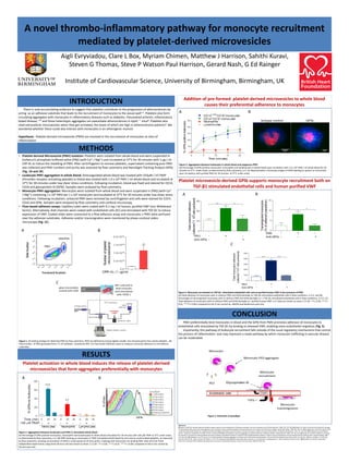
- 1. A novel thrombo-inflammatory pathway for monocyte recruitment mediated by platelet-derived microvesicles INTRODUCTION Aigli Evryviadou, Clare L Box, Myriam Chimen, Matthew J Harrison, Sahithi Kuravi, Steven G Thomas, Steve P Watson Paul Harrison, Gerard Nash, G Ed Rainger Institute of Cardiovascular Science, University of Birmingham, Birmingham, UK • Platelet-derived Microvesicle (PMV) Isolation: Platelets were isolated from whole blood and were suspended in Dulbecco’s phosphate buffered saline (PBS) (with Ca2+ / Mg2+) and incubated at 37°C for 30 minutes with 1 μg / ml CRP-XL to induce the shedding of PMV. After centrifugation to remove platelets, supernatant containing pure PMV was collected and PMV numbers and purity was assessed by flow cytometry and NanoSight Tracking Analysis (NTA) (Fig. 1A and 1B). • Leukocyte-PMV aggregation in whole blood: Anticoagulated whole blood was treated with 333μM / ml TRAP (thrombin receptor activating peptide) or blood was treated with 1.0 x 10⁹ PMV / ml whole blood and incubated at 37°C for 30 minutes under low shear stress conditions. Following incubation, blood was fixed and stained for CD14, CD16 and glycoprotein Ib (GPIb). Samples were analysed by flow cytometry. • Monocyte-PMV aggregation: Monocytes were isolated from whole blood and were suspended in (PBS) (with Ca2+ / Mg2+) containing 1 x 10⁹ PMV per 1 x 10⁶ monocytes and incubated at 37°C for 30 minutes under low shear stress conditions. Following incubation, unbound PMV were removed by centrifugation and cells were stained for CD14, CD16 and GPIb. Samples were analysed by flow cytometry and confocal microscopy. • Flow-based adhesion assays: Capillary tubes were coated with 0.1 mg / ml human, purified VWF (von Willebrand factor). Alternatively, ibidi channels were coated with endothelial cells (EC) and stimulated with TGF-β1 to induce expression of VWF. Coated slides were connected to a flow adhesion assay and monocytes ± PMV were perfused over the adhesive substrates. Adhesion and/or transmigration were monitored by phase-contrast video- microscopy (Fig. 1C). There is now accumulating evidence to suggest that platelets contribute to the progression of atherosclerosis by acting as an adhesive substrate that leads to the recruitment of monocytes to the vessel wall1-3. Platelets also form circulating aggregates with monocytes in inflammatory diseases such as diabetes, rheumatoid arthritis, inflammatory bowel disease, 4-6 and these heterotypic aggregates can exacerbate atherosclerosis in ApoE -/- mice8. Platelets also shed extracellular microvesicles when they get activated, the levels of which are high in atherosclerosis patients9. We wondered whether these could also interact with monocytes in an atherogenic manner. Hypothesis: Platelet-derived microvesicles (PMV) are involved in the recruitment of monocytes at sites of inflammation. METHODS RESULTS Platelet activation in whole blood induces the release of platelet-derived microvesicles that form aggregates preferentially with monocytes Figure 2. Aggregation between leukocytes and PMV in stimulated whole blood (A) Percentage of GPIb-positive monocytes, neutrophils and lymphocytes in whole blood stimulated for 30 minutes with 100 μM TRAP at 37°C under shear, as determined by flow cytometry, n=3. (B) GPIb staining on monocytes in TRAP-stimulated whole blood by time and on unstimulated platelets, as measured by flow cytometry, showing accumulation of GPIb in small quanta at all time points, implying that monocytes are binding PMV. Data are from three independent experiments using three donours and are shown as mean ± S.E.M. * P ≤ 0.05, ** P ≤ 0.01, *** P ≤ 0.001 compared to the 0 min control by Dunnet post-test. Addition of pre-formed platelet-derived microvesicles to whole blood causes their preferential adherence to monocytes Platelet microvesicle-derived GPIb supports monocyte recruitment both on TGF-β1 stimulated endothelial cells and human purified VWF CONCLUSION Figure 3. Aggregation between leukocytes in whole blood and exogenous PMV (A) Percentage of GPIb-positive monocytes, neutrophils and lymphocytes in whole blood upon incubation with 1.0 x 10⁹ PMV / ml whole blood for 20 minutes at 37°C under shear, as determined by flow cytometry, n=3. (C) Representative microscopy images of GPIb labelling (in green) on monocytes upon incubation with purified PMV for 30 minutes at 37°C under shear. Figure 4. Monocyte recruitment on TGF-β1- stimulated endothelial cells and on purified human VWF in the presence of PMV (A) Total adhesion of monocytes with or without PMV and GPIb blockade on TGF-β1-stimulated endothelial cells in flow conditions, n=3-5. and (B) Percentage of transmigrated monocytes with or without PMV and GPIb blockade on n TGF-β1-stimulated endothelial cells in flow conditions, n=3-5. (C) Total adhesion of monocytes with or without PMV and GPIb blockade on purified human VWF, n=3. Data are shown as mean ± S.E.M. * P ≤ 0.05, ** P ≤ 0.01, *** P ≤ 0.001 compared to the 0 min control by ANOVA and Bonferroni post-test. References (1) Bahra P, Nash GB. Sparsely adherent platelets support capture and immobilization of flowing neutrophils. Journal of Laboratory and Clinical Medicine. 1998; 132: 223-228. (2) Diakovo TG, Roth SJ, Buccola JM, Bainton DF, Springer TA. Neutrophil rolling, arrest and transmigration across activated, surface-adherent platelets via sequential action of p-selectin and the beta 2-integrin cd11b/cd18. Blood. 1996; 88: 146-157. (3) Kuckleburg CJ, Yates CM, Kalia N, Zhao Y, Nash GB, Watson SP, Rainger GE. Endothelial cell-borne bridges selectively recruit monocytes in human and mouse models of vascular inflammation. Cardiovascular Research. 2011; 91: 134-141. (4) Harding SA, Sommerfield AJ, Sarma J, Twomey PJ, Newby DE, Frier BM, Fox KA. Increased cd40 ligand and platelet-monocyte aggregates in patients with type 1 diabetes mellitus. Atherosclerosis. 2004; 176: 321-325. (5) Joseph JE, Harrison P, Mackie IJ, Isenberg DA, Machin SJ. Increased circulating platelet-leucocyte complexes and platelet activation in patients with antiphospholipid syndrome, systemic lupus erythematosus and rheumatoid arthritis. British Journal of Haematology. 2001; 115: 451-459. (6) Tekelioglu Y, Uzun H, Gucer H. Circulating platelet-leukocyte aggregates in patients with inflammatory bowel disease. Journal of Chinese Medical Association. 2013; 76: 182-185. (7) Huo Y, Schober A, Forlow SB, Smith DF, Hyman MC, Jung S, Littman DR, Weber C, Ley K. Circulating activated platelets exacerbate atherosclerosis in mice deficient in apolipoprotein e. Nature Medicine. 2003; 9: 61-67. (8) Michelsen AE, Brodin E, Brosstad F, Hansen JB. Increased level of platelet microparticles in survivors of myocardial infarction. Scand J Clin Lab Invest. 2008; 68: 386–392. Figure 5. Schematic of paradigm PMV preferentially bind monocytes in blood and the GPIb from PMV promotes adhesion of monocytes to endothelial cells stimulated by TGF-β1 by binding to released VWF, enabling trans-endothelial migration (Fig. 5). Importantly, this pathway of leukocyte recruitment falls outside of the usual regulatory mechanisms that control the process of inflammation and may represent a novel pathway by which monocyte trafficking in vascular disease can be moderated. Figure 1. (A) Gating strategy for detecting PMV by flow cytometry. PMV are defined by having slightly smaller size and granularity than whole platelets. (B) Total number of PMV generated from 3 x 10⁸ platelets, counted by NTA. (C) Flow-based Adhesion assay to measure monocyte adhesion to immobilised substrates. A B A C B A B C A B