More Related Content
Similar to Pain, Heat, & Emotion. NEJM
Similar to Pain, Heat, & Emotion. NEJM (20)
More from Paul Coelho, MD
More from Paul Coelho, MD (20)
Pain, Heat, & Emotion. NEJM
- 1. editorials
n engl j med 368;15 nejm.org april 11, 2013 1447
reduce mortality in patients undergoing primary
PCI.7 Second-generation drug-eluting stents have
increased the durability of primary PCI and may
even have lowered rates of stent thrombosis, as
compared with first-generation drug-eluting stents
or bare-metal stents.8 Thus, since the start of
the STREAM trial, the results of primary PCI
have gotten better and safer, creating an even
higher bar for prehospital fibrinolysis.
The findings of this trial could have a major
effect on clinical practice and further highlight
the prominence of timely PCI as the treatment
of choice for STEMI (Fig. 1). Health care systems
can be reconfigured to provide such care, but
there are a variety of practical barriers.9 When
primary PCI cannot be performed, prompt fibri-
nolysis should be administered, with transfer to
a PCI-capable center in the next several hours,
especially in high-risk patients.10 A pharmaco
invasive approach, including initial half-dose fi-
brinolysis in the elderly, may be an option in
selected circumstances, though it does not repre-
sent optimal care as compared with timely pri-
mary PCI. The STREAM trial shows us that the
best therapy for STEMI remains rapid mechani-
cal restoration of coronary flow with a stent.
Disclosure forms provided by the author are available with the
full text of this article at NEJM.org.
From the VA Boston Healthcare System, Brigham and Wom-
en’s Hospital, and Harvard Medical School — all in Boston.
This article was published on March 10, 2013, at NEJM.org.
1. Keeley EC, Boura JA, Grines CL. Primary angioplasty versus
intravenous thrombolytic therapy for acute myocardial infarc-
tion: a quantitative review of 23 randomised trials. Lancet 2003;
361:13-20.
2. Andersen HR, Nielsen TT, Rasmussen K, et al. A compari-
son of coronary angioplasty with fibrinolytic therapy in acute
myocardial infarction. N Engl J Med 2003;349:733-42.
3. Pinto DS, Kirtane AJ, Nallamothu BK, et al. Hospital delays
in reperfusion for ST-elevation myocardial infarction: implica-
tions when selecting a reperfusion strategy. Circulation 2006;
114:2019-25.
4. Ellis SG, Tendera M, de Belder MA, et al. Facilitated PCI in
patients with ST-elevation myocardial infarction. N Engl J Med
2008;358:2205-17.
5. Armstrong PW, Gershlick AH, Goldstein P, et al. Fibrinolysis
or primary PCI in ST-segment elevation myocardial infarction.
N Engl J Med 2013;368:1379-87.
6. Desai NR, Bhatt DL. The state of periprocedural antiplatelet
therapy after recent trials. JACC Cardiovasc Interv 2010;3:571-83.
7. Bavry AA, Kumbhani DJ, Bhatt DL. Role of adjunctive throm-
bectomy and embolic protection devices in acute myocardial
infarction: a comprehensive meta-analysis of randomized trials.
Eur Heart J 2008;29:2989-3001.
8. Bhatt DL. EXAMINATION of new drug-eluting stents — top
of the class! Lancet 2012;380:1453-5.
9. Pottenger BC, Diercks DB, Bhatt DL. Regionalization of care
for ST-segment elevation myocardial infarction: is it too soon?
Ann Emerg Med 2008;52:677-85.
10. Cantor WJ, Fitchett D, Borgundvaag B, et al. Routine early
angioplasty after fibrinolysis for acute myocardial infarction.
N Engl J Med 2009;360:2705-18.
DOI: 10.1056/NEJMe1302670
Copyright © 2013 Massachusetts Medical Society.
Is a PCI-capable
hospital nearby?
Perform primary PCI
Acute STEMI diagnosed
in the field?
Bring to closest hospital
for further chest-pain
evaluation
No
Yes
Transfer to PCI-capable
hospital, especially
if high risk
Stabilize at receiving
hospital; transfer for
primary PCI
Yes
Yes
Is hospital part of a
STEMI network?
No
Administer full-dose
fibrinolysis, if no contra-
indications; consider
half-dose agent if
≥75 yr of age
No
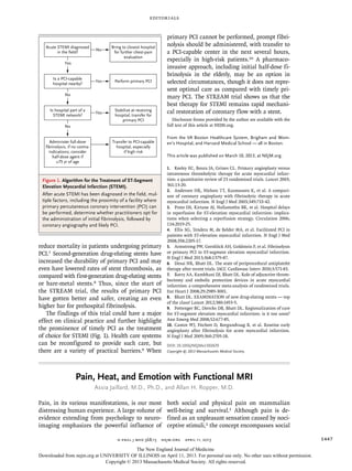
Figure 1. Algorithm for the Treatment of ST-Segment
Elevation Myocardial Infarction (STEMI).
After acute STEMI has been diagnosed in the field, mul-
tiple factors, including the proximity of a facility where
primary percutaneous coronary intervention (PCI) can
be performed, determine whether practitioners opt for
the administration of initial fibrinolysis, followed by
coronary angiography and likely PCI.
Pain, Heat, and Emotion with Functional MRI
Assia Jaillard, M.D., Ph.D., and Allan H. Ropper, M.D.
Pain, in its various manifestations, is our most
distressing human experience. A large volume of
evidence extending from psychology to neuro-
imaging emphasizes the powerful influence of
both social and physical pain on mammalian
well-being and survival.1 Although pain is de-
fined as an unpleasant sensation caused by noci-
ceptive stimuli,2 the concept encompasses social
The New England Journal of Medicine
Downloaded from nejm.org at UNIVERSITY OF ILLINOIS on April 11, 2013. For personal use only. No other uses without permission.
Copyright © 2013 Massachusetts Medical Society. All rights reserved.
- 2. The new engl and jour nal of medicine
n engl j med 368;15 nejm.org april 11, 20131448
as well as physical pain. Painful emotional feel-
ings are associated with the loss of social con-
nection owing to rejection, exclusion from a
group, personal failure, or the death of a loved
one. We often describe the experience of social
pain by using terms for physical pain,1 making
reference to broken hearts, hurt feelings, heart-
ache, or being crushed or wounded to the quick.
Moreover, social pain activates neural circuitry
that is related to somatosensory pain, and an-
algesic agents have ameliorating effects on both
physical pain and pain caused by social rejection,
providing mechanistic links between them.1,3
However, a dissociation between the sensory
and affective components of physical pain has
long been known from clinical work, in which
lesions of the lateral thalamus render a person
insensate on the opposite side of the body while
still permitting a display of grimacing, restless-
ness, and autonomic responses to pain. Func-
tional magnetic resonance imaging (fMRI) stud-
ies have confirmed this separation by showing a
neural circuitry for physical pain that has two
disparate ensembles: first, a sensory system in
the primary and secondary somatosensory cor-
texes and posterior insula that codes for the
qualitative and quantitative characteristics of a
stimulus, and second, an affective system in the
dorsal anterior cingulate cortex, anterior insula,
and the limbic system that signals aversive
states.1,4-6 The insula, which is embedded in both
systems, is a pivotal hub of a salience network that
identifies the most relevant internal and exter-
nal stimuli, including pain, from moment to mo-
ment, in order to guide attention and behavior.6,7
The question of whether particular regions of
the brain are specific for physical pain and
whether activity in these regions can be quanti-
fied are the main issues addressed by Wager
and colleagues in this issue of the Journal.8 The
investigators, using fMRI and machine-learning
methods, identified a widely distributed, multi-
regional pattern (or signature response) that was
activated by physical pain applied in the form of
heat to the forearm of healthy volunteers. The
pattern that Wager and colleagues detected had
high sensitivity and specificity in discriminating
painful heat from nonpainful warmth, pain an-
ticipation, pain recall, and provocatively, social
pain. In addition, activity in these regions in
response to pain was reduced by an opioid an-
algesic agent.
These results may be of great practical impor-
tance, because physical pain is the most com-
mon reason for consultation with a physician.
We comprehend our own pain only as a subjec-
tive phenomenon and recognize that the experi-
ence and affective display of pain differ from
person to person and from culture to culture.
Physicians are flummoxed by pain because of a
paucity of objective manifestations and are re-
duced to using clinical instruments, such as the
visual-analogue scale to quantitate pain. Imag-
ine how all fields of medicine would be altered
if pain could be objectified by a measure that
did not require direct patient reporting. For ex-
ample, what would be seen in patients with fi-
bromyalgia, depression, or narcotic addiction,
who have both physical and emotional pain?
Wager and colleagues describe potential appli-
cations of their method, including detecting and
quantifying pain in persons who cannot com-
municate and in those for whom the self-report
of the intensity of pain is suspect.
The results, however, require cautious evalua-
tion for several reasons. First, the authors make
it clear that they have studied only cutaneous
pain and not pain in the context of disease, so
their findings may not apply to clinical circum-
stances. They also do not shed light on the issue
of chronic pain, one of the most vexing prob-
lems in general medicine. Second, their assess-
ment of social pain, in which participants re-
called a recent romantic breakup while viewing
a photograph of their ex-partner, used an uncer-
tain stimulus with respect to the neural processes
that are engaged. Participants in these studies
may have experienced many feelings, including
social rejection, love, or attachment, which led
to changes in the activity of reward centers in
the brain.9 Finally, the spatial resolution used in
this study was limited, reflecting the low sensi-
tivity of the 1.5-T fMRI system that was used for
most of the testing, and this may have led to the
misidentification of small deep-brain structures
that contributed to the neurologic signature re-
sponse for pain. Therefore, further studies in
diverse clinical circumstances with the use of
more-sensitive MRI acquisition techniques will
be necessary to validate any pain biomarker.
The studies conducted by Wager and col-
leagues serve as an example of how functional
neuroimaging may help clinicians assess clini-
cal symptoms, such as somatic and emotional
The New England Journal of Medicine
Downloaded from nejm.org at UNIVERSITY OF ILLINOIS on April 11, 2013. For personal use only. No other uses without permission.
Copyright © 2013 Massachusetts Medical Society. All rights reserved.
- 3. editorials
n engl j med 368;15 nejm.org april 11, 2013 1449
pain, that were previously thought to be impen-
etrable. Being doctors, though, we may ultimate-
ly have to acknowledge that “pain is pain” and
can be reported only by the patient.
Disclosure forms provided by the authors are available with
the full text of this article at NEJM.org.
From the Unité d’Imagerie par Résonance Magnétique, Struc-
ture Federative de Recherche 1, Pôle de Recherche, Centre Hos-
pitalier Universitaire de Grenoble, Grenoble, France (A.J.); and
the Department of Neurology, Brigham and Women’s Hospi-
tal, Boston (A.H.R.).
1. Eisenberger NI. The pain of social disconnection: examin-
ing the shared neural underpinnings of physical and social pain.
Nat Rev Neurosci 2012;13:421-34.
2. Price DD. Psychological and neural mechanisms of the af-
fective dimension of pain. Science 2000;288:1769-72.
3. Dewall CN, Macdonald G, Webster GD, et al. Acetamino-
phen reduces social pain: behavioral and neural evidence. Psy-
chol Sci 2010;21:931-7.
4. Kross E, Berman MG, Mischel W, Smith EE, Wager TD. So-
cial rejection shares somatosensory representations with physi-
cal pain. Proc Natl Acad Sci U S A 2011;108:6270-5.
5. Cauda F, D’Agata F, Sacco K, Duca S, Geminiani G, Vercelli
A. Functional connectivity of the insula in the resting brain.
Neuroimage 2011;55:8-23.
6. Cauda F, Torta DM, Sacco K, et al. Functional anatomy of
cortical areas characterized by Von Economo neurons. Brain
Struct Funct 2012 January 29 (Epub ahead of print).
7. Menon V, Uddin LQ. Saliency, switching, attention and con-
trol: a network model of insula function. Brain Struct Funct
2010;214:655-67.
8. Wager TD, Atlas LY, Lindquist MA, Roy M, Woo C-W, Kross E.
An fMRI-based neurologic signature of physical pain. N Engl J
Med 2013;368:1388-97.
9. O’Connor MF, Wellisch DK, Stanton AL, Eisenberger NI, Ir-
win MR, Lieberman MD. Craving love? Enduring grief activates
brain’s reward center. Neuroimage 2008;42:969-72.
DOI: 10.1056/NEJMe1213074
Copyright © 2013 Massachusetts Medical Society.
The New England Journal of Medicine
Downloaded from nejm.org at UNIVERSITY OF ILLINOIS on April 11, 2013. For personal use only. No other uses without permission.
Copyright © 2013 Massachusetts Medical Society. All rights reserved.