Short case...Spinal multiple sclerosis
•
4 gefällt mir•813 views

Short case...Spinal multiple sclerosis

Empfohlen
Empfohlen
Weitere ähnliche Inhalte
Ähnlich wie Short case...Spinal multiple sclerosis
Ähnlich wie Short case...Spinal multiple sclerosis (20)
Mehr von Professor Yasser Metwally
Mehr von Professor Yasser Metwally (20)
Kürzlich hochgeladen
Kürzlich hochgeladen (20)
Short case...Spinal multiple sclerosis
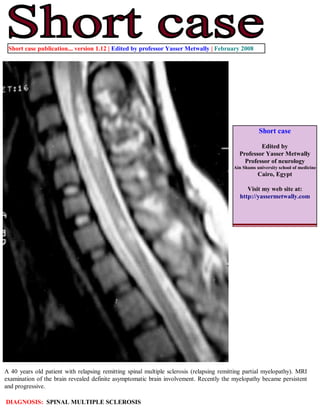
- 1. Short case publication... version 1.12 | Edited by professor Yasser Metwally | February 2008 Short case Edited by Professor Yasser Metwally Professor of neurology Ain Shams university school of medicine Cairo, Egypt Visit my web site at: http://yassermetwally.com A 40 years old patient with relapsing remitting spinal multiple sclerosis (relapsing remitting partial myelopathy). MRI examination of the brain revealed definite asymptomatic brain involvement. Recently the myelopathy became persistent and progressive. DIAGNOSIS: SPINAL MULTIPLE SCLEROSIS
- 2. Figure 1. A case of spinal multiple sclerosis. Precontrast MRI T1 images showing peripherally located, pencil shaped multiple sclerosis plaques that occupy 2-3 spinal segments. The plaques are well demarcated and hypointense. No evidence of spinal cord atrophy and postcontrast studies did not show evidence of contrast enhancement. Figure 2. A case of spinal multiple sclerosis. MRI T2 images showing peripherally located, pencil shaped multiple sclerosis plaques that occupy 2-3 spinal segments The plaques are orientated along the longitudinal axis of the spinal cord on sagittal sections. The plaques are short (less than two spinal segments), pencil shaped, multiple and well demarcated. No evidence of spinal cord atrophy. The spinal cord parenchyma is asymmetrically involved. Diffuse abnormalities seen as poorly demarcated areas of increased signal intensity on MRI T2 images are also seen in this study. Diffuse abnormalities are more common in primary progressive MS and secondary progressive MS.
- 3. Figure 3. A case of spinal multiple sclerosis. MRI T2 and precontrast T1 cross section images showing diffuse abnormalities seen as poorly demarcated areas of increased signal intensity on MRI T2 images and low signal on the MRI T1 images. Diffuse abnormalities are more common in primary progressive MS and secondary progressive MS. Figure 4. Atrophy affecting the upper cervical spinal segments. Over the past decade, researchers and clinicians have gained new insights into the core of demyelinating diseases of the spinal cord, and much progress has been made in the management of these diseases. Although we are starting to uncover some of the structural and physiologic substrates of demyelination of the CNS, we are far from understanding what causes many of these demyelinating disorders and how to prevent their progression. With further development of new techniques, such as DTI and more potent MR units, spinal cord diseases may be distinguished from each other, and effective therapeutic strategies may be initiated before any cord damage occurs. In particular MRI is very helpful in differentiation between Spinal multiple sclerosis and transverse myelitis In the series reported by Choi et al, [2] the centrally located MRI T2 high signal intensity occupied more than two thirds
- 4. of the cross-sectional area of the cord in transverse myelitis. In multiple sclerosis, plaques are usually located peripherally and occupy less than half the cross-sectional area of the cord. The central isointensity, or dot (commonly seen in transverse myelitis), represents central gray matter squeezed by the uniform, evenly distributed oedematous changes of the cord. Choi and colleagues [2] have demonstrated the role of contrast media in differentiating transverse myelitis from multiple sclerosis. In transverse myelitis, enhancement is in the periphery of a centrally located area of high T2 weighted images. In multiple sclerosis, the lesions show enhancement in the central zone of peripherally located high signal intensity on T2 weighted images. In conclusion, certain MRI characteristics help in differentiating acute transverse myelitis from spinal form of multiple sclerosis. These include: 1) centrally located high intensity signal extending over 3 to 4 segments and occupying more than two thirds of the cord cross-sectional area and 2) peripheral contrast enhancement of high intensity signal. Figure 5. Differential diagnoses of intramedullary lesions based on their location at the cross-sectional area of the cord. (A) MS: Dorsally located wedge-shaped lesion involving less then two thirds of the cross-sectional area of the spinal cord seen on axial T2-Wi MR image. (B) Poliomyelitis: Bilateral enhancing anterior nerve roots demonstrated on postcontrast T1-Wi MR image. (C) Vacuolar myelopathy: Bilateral, symmetrical, high-signal-intensity MS Poliomyelitis Sbacute combined degeneration abnormality located dorsally in the spinal cord in an HIV-positive patient. DD: Subacute combined degeneration. (D) ATM: On axial T2-Wi, a high-signal-intensity lesion involving more than two thirds of cross- sectional area of the spinal cord is observed. (E) Herpes-simplex-virus myelitis: Postcontrast T1-Wi axial MR image showing nodular enhancing lesion located in the lateral part of the cervical spinal cord. DD: active MS plaque. (F) Spinal cord infarction: Swelling of the anterior parts of the spinal cord is shown on axial T2-Wi MR images, indicating vulnerability of the anterior portions of the spinal cord to ischemia. Transverse myelitis HIV myelitis Infarction Addendum A new version of short case is uploaded in my web site every week (every Saturday and remains available till Friday.) To download the current version follow the link quot;http://pdf.yassermetwally.com/short.pdfquot;. You can download the long case version of this short case during the same week from: http://pdf.yassermetwally.com/case.pdf or visit web site: http://pdf.yassermetwally.com To download the software version of the publication (crow.exe) follow the link: http://neurology.yassermetwally.com/crow.zip At the end of each year, all the publications are compiled on a single CD-ROM, please contact the author to know more details. Screen resolution is better set at 1024*768 pixel screen area for optimum display For an archive of the previously reported cases go to www.yassermetwally.net, then under pages in the right panel, scroll down and click on the text entry quot;downloadable short cases in PDF formatquot;
- 5. References 1. Metwally, MYM: Textbook of neurimaging, A CD-ROM publication, (Metwally, MYM editor) WEB-CD agency for electronic publishing, version 9.1a January 2008 2. Choi KH, Lee KS, Chung SO, et al.. Idiopathic transverse myelitis: MR characteristics. AJNR Am J Neuroradiol. 1996;17:1151–1160.