Short case...Postinfectious transverse myelitis
•
2 gefällt mir•1,204 views

Short case...Postinfectious transverse myelitis

Empfohlen
Weitere ähnliche Inhalte
Andere mochten auch
Andere mochten auch (14)
Ähnlich wie Short case...Postinfectious transverse myelitis
Ähnlich wie Short case...Postinfectious transverse myelitis (20)
Mehr von Professor Yasser Metwally
Mehr von Professor Yasser Metwally (20)
Kürzlich hochgeladen
Kürzlich hochgeladen (20)
Short case...Postinfectious transverse myelitis
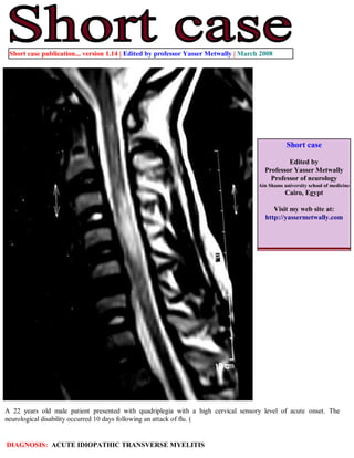
- 1. Short case publication... version 1.14 | Edited by professor Yasser Metwally | March 2008 Short case Edited by Professor Yasser Metwally Professor of neurology Ain Shams university school of medicine Cairo, Egypt Visit my web site at: http://yassermetwally.com A 22 years old male patient presented with quadriplegia with a high cervical sensory level of acute onset. The neurological disability occurred 10 days following an attack of flu. ( DIAGNOSIS: ACUTE IDIOPATHIC TRANSVERSE MYELITIS
- 2. Figure 1. A case of transverse myelitis. A, precontrast MRI T1 image and B,C,D postcontrast MRI T1 images. showing mild dilatation of C2,C3,C4,C5 cervical spinal segments with central intramedullary, multisegmental hypointensity and with peripheral contrast enhancement. Figure 2. A case of transverse myelitis. MRI T2 images showing multisegmental (about five spinal segments from C2-C6) intramedullary hyperintensity with mild cord dilatation.
- 3. Figure 3. A case of transverse myelitis. Cross-sectional MRI T2 images showing mild dilatation of the spinal cord with central hyperintensities occupying more than 2/3 of the cross-sectional area of the spinal cord with absence of the central dot sign. Figure 4. MRI T2 showing a case of acute idiopathic transverse myelitis. Notice cord swelling and the multisegmental, central increased cord signal intensity at the cervicodorsal region Figure 5. Notice cord swelling in the cervico dorsal region with patchy irregular and peripheral contrast enhancement. Also notice the central T2 hyperintensity. Peripheral contrast enhancement is outside and peripheral to the central T2 hyperintensity.
- 4. The MRI picture characteristic of idiopathic transverse myelitis A centrally located multisegmental (3 to 8 spinal segments) MRI T2 hyperintensity that occupies more than two thirds of the cross-sectional area of the cord is characteristic of transverse myelitis. The MRI T2 hyperintensity commonly shows a slow regression with clinical improvement. The central spinal cord MRI T2 hyperintensity represents evenly distributed central cord edema. MRI T1 Hypointensity might be present in the same spinal segments that show T2 hyperintensity although to a lesser extent. The MRI T2 hyperintensity is central, bilateral, more or less symmetrical and multisegmental. MRI T2 central isointensity, or dot (within and in the core of the MRI T2 hyperintensity) might be present and is believed to represent central gray matter squeezed by the uniform, evenly distributed edematous changes of the cord. (central dot sign). It might not be of any clinical significance. Contrast enhancement is commonly focal or peripheral and maximal at or near the segmental MRI T2 hyperintensity. In idiopathic transverse myelitis enhancement is peripheral to the centrally located area of high T2 signal intensity rather than in the very same area. The prevalence of cord enhancement is significantly higher in patients with cord expansion. Spinal cord expansion might or might not be present and when present is usually multisegmental and better appreciated on the sagittal MRI T1 images. Spinal cord expansion tapers smoothly to the normal cord, and is of lesser extent than the high T2 signal abnormality. Multiple sclerosis plaques (and subsequent T2 hyperintensity) are located peripherally, are less than 2 vertebral segments in length, and occupies less than half the cross-sectional area of the cord. In contrast to transverse myelitis, enhancement in MS occurs in the same location of high-signal-intensity lesions seen on T2-weighted images. Figure 6. Differential diagnoses of intramedullary lesions based on their location at the cross-sectional area of the cord. (A) MS: Dorsally located wedge- shaped lesion involving less then two thirds of the cross-sectional area of the spinal cord seen on axial T2-Wi MR image. (B) Poliomyelitis: Bilateral enhancing anterior nerve roots demonstrated on postcontrast T1-Wi MR image. (C) Vacuolar myelopathy: Bilateral, symmetrical, high- signal-intensity abnormality located dorsally in the spinal cord in an HIV- MS Poliomyelitis Sbacute combined degeneration positive patient. DD: Subacute combined degeneration. (D) ATM: On axial T2-Wi, a high-signal-intensity lesion involving more than two thirds of cross-sectional area of the spinal cord is observed. (E) Herpes- simplex-virus myelitis: Postcontrast T1-Wi axial MR image showing nodular enhancing lesion located in the lateral part of the cervical spinal cord. DD: active MS plaque. (F) Spinal cord infarction: Swelling of the anterior parts of the spinal cord is shown on axial T2-Wi MR images, indicating vulnerability of the anterior portions of the spinal cord to ischemia. Transverse myelitis HIV myelitis Infarction
- 5. Addendum A new version of short case is uploaded in my web site every week (every Saturday and remains available till Friday.) To download the current version follow the link quot;http://pdf.yassermetwally.com/short.pdfquot;. You can download the long case version of this short case during the same week from: http://pdf.yassermetwally.com/case.pdf or visit web site: http://pdf.yassermetwally.com To download the software version of the publication (crow.exe) follow the link: http://neurology.yassermetwally.com/crow.zip At the end of each year, all the publications are compiled on a single CD-ROM, please contact the author to know more details. Screen resolution is better set at 1024*768 pixel screen area for optimum display For an archive of the previously reported cases go to www.yassermetwally.net, then under pages in the right panel, scroll down and click on the text entry quot;downloadable short cases in PDF formatquot; References 1. Metwally, MYM: Textbook of neurimaging, A CD-ROM publication, (Metwally, MYM editor) WEB-CD agency for electronic publishing, version 9.1a January 2008