Short case...Occipital lobe cavernoma
•
1 gefällt mir•518 views

Short case...Occipital lobe cavernoma

Empfohlen
Empfohlen
Weitere ähnliche Inhalte
Andere mochten auch
Andere mochten auch (15)
Ähnlich wie Short case...Occipital lobe cavernoma
Ähnlich wie Short case...Occipital lobe cavernoma (20)
Mehr von Professor Yasser Metwally
Mehr von Professor Yasser Metwally (20)
Kürzlich hochgeladen
Kürzlich hochgeladen (20)
Short case...Occipital lobe cavernoma
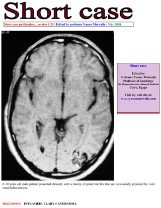
- 1. Short case publication... version 1.21 | Edited by professor Yasser Metwally | May 2008 Short case Edited by Professor Yasser Metwally Professor of neurology Ain Shams university school of medicine Cairo, Egypt Visit my web site at: http://yassermetwally.com A 30 years old male patient presented clinically with a history of grand mal fits that are occasionally preceded by vivid visual hallucinations. DIAGNOSIS: INTRAMEDULLARY CAVERNOMA
- 2. Figure 1. Type II left occipital cavernoma. Precontrast MRI T1 images. The characteristic MR imaging appearance of cavernomas is a well-defined, lobulated lesion with a reticulated core of heterogenous signal intensity on both T1 and T2 weighted sequences resulting from thrombosis, fibrosis, calcification, and hemorrhage. Extracellular and intracellular methemoglobin and thrombosis are responsible for the high intensity signal within the lesion, while calcifications, fibrosis, and acute and subacute blood are responsible for the low signal areas. The cavernoma in the current case has the classic quot;popcornquot; appearance characteristic of type II cavernomas. Figure 2. Type II left occipital cavernoma. Precontrast MRI T1 images. The characteristic MR imaging appearance of cavernomas is a well-defined, lobulated lesion with a reticulated core of heterogenous signal intensity on both T1 and T2 weighted sequences resulting from thrombosis, fibrosis, calcification, and hemorrhage. Extracellular and intracellular methemoglobin and thrombosis are responsible for the high intensity signal within the lesion, while calcifications, fibrosis, and acute and subacute blood are responsible for the low signal areas. The cavernoma in the current case has the classic quot;popcornquot; appearance characteristic of type II cavernomas.
- 3. Figure 3. Type II left occipital cavernoma. Precontrast MRI T1 images. The characteristic MR imaging appearance of cavernomas is a well-defined, lobulated lesion with a reticulated core of heterogenous signal intensity on both T1 and T2 weighted sequences resulting from thrombosis, fibrosis, calcification, and hemorrhage. Extracellular and intracellular methemoglobin and thrombosis are responsible for the high intensity signal within the lesion, while calcifications, fibrosis, and acute and subacute blood are responsible for the low signal areas. The cavernoma in the current case has the classic quot;popcornquot; appearance characteristic of type II cavernomas. Conclusion MRI picture of occipital cavernoma. The MRI mixed signal intensity of the precontrast MRI T1 images and the MRI T2 images is due to the presence of altered blood of different ages. The MRI T1 precontrast hyperintensity is due to methemoglobin The MRI T2 hypointensity is due to hemosiderin pigments. Addendum A new version of short case is uploaded in my web site every week (every Saturday and remains available till Friday.) To download the current version follow the link quot;http://pdf.yassermetwally.com/short.pdfquot;. You can download the long case version of this short case during the same week from: http://pdf.yassermetwally.com/case.pdf or visit web site: http://pdf.yassermetwally.com To download the software version of the publication (crow.exe) follow the link: http://neurology.yassermetwally.com/crow.zip At the end of each year, all the publications are compiled on a single CD-ROM, please contact the author to know more details. Screen resolution is better set at 1024*768 pixel screen area for optimum display For an archive of the previously reported cases go to www.yassermetwally.net, then under pages in the right panel, scroll down and click on the text entry quot;downloadable short cases in PDF formatquot; References 1. Metwally, MYM: Textbook of neurimaging, A CD-ROM publication, (Metwally, MYM editor) WEB-CD agency for electronic publishing, version 9.1a January 2008