Short case...Myxopapillary ependymoma
•
2 likes•253 views

Short case...Myxopapillary ependymoma http://yassermetwally.com http://yassermetwally.net

Recommended
Recommended
More Related Content
Similar to Short case...Myxopapillary ependymoma
Similar to Short case...Myxopapillary ependymoma (20)
More from Professor Yasser Metwally
More from Professor Yasser Metwally (20)
Recently uploaded
Recently uploaded (20)
Short case...Myxopapillary ependymoma
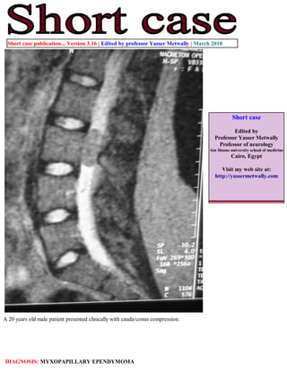
- 1. Short case publication... Version 3.16 | Edited by professor Yasser Metwally | March 2010 Short case Edited by Professor Yasser Metwally Professor of neurology Ain Shams university school of medicine Cairo, Egypt Visit my web site at: http://yassermetwally.com A 20 years old male patient presented clinically with cauda/conus compression. DIAGNOSIS: MYXOPAPILLARY EPENDYMOMA
- 2. Figure 1. Myxopapillary ependymoma: Precontrast MRI T1 image (A) and postcontrast MRI T1 image (B) showing a well defined, well circumscribed oval (sausage-shaped) mass at L1 lumbar region. The mass is hyperintense on precontrast scan and showed dense contrast enhancement. The precontrast T1 hyperintensity could be due to presence of abundant mucin and hemorrhagic products. Figure 2. MRI T2 image showing the lumbar myxopapillary ependymoma as a well defined, well circumscribed oval (sausage-shaped) mass at L1 lumbar region. The ependymoma is hypointense relative to the CSF. References 1. Metwally, MYM: Textbook of neurimaging, A CD-ROM publication, (Metwally, MYM editor) WEB-CD agency for electronic publishing, version 11.1a December 2010
- 3. Addendum A new version of short case is uploaded in my web site every week (every Saturday and remains available till Friday.) To download the current version follow the link "http://pdf.yassermetwally.com/short.pdf". You can download the long case version of this short case during the same week from: http://pdf.yassermetwally.com/case.pdf or visit web site: http://pdf.yassermetwally.com To download the software version of the publication (crow.exe) follow the link: http://neurology.yassermetwally.com/crow.zip At the end of each year, all the publications are compiled on a single CD-ROM, please contact the author to know more details. Also to view a list of the previously published case records follow the following link (http://wordpress.com/tag/case- record/) or click on it if it appears as a link in your PDF reader To inspect the patient's full radiological study, click on the attachment icon (the paper clip icon in the left pane) of the acrobat reader then double click on the attached file Click here to download the long case version of this short case in PDF format