Short case...Dysembryoplastic neuroepithelial tumor
•
2 likes•742 views

Short case...Dysembryoplastic neuroepithelial tumor http://yassermetwally.com http://yassermetwally.net

Recommended
More Related Content
Similar to Short case...Dysembryoplastic neuroepithelial tumor
Similar to Short case...Dysembryoplastic neuroepithelial tumor (20)
More from Professor Yasser Metwally
More from Professor Yasser Metwally (20)
Recently uploaded
Recently uploaded (20)
Short case...Dysembryoplastic neuroepithelial tumor
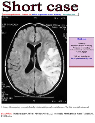
- 1. Short case publication... Version 3.4| Edited by professor Yasser Metwally | November 2009 Short case Edited by Professor Yasser Metwally Professor of neurology Ain Shams university school of medicine Cairo, Egypt Visit my web site at: http://yassermetwally.com A 6 years old male patient presented clinically with intractable complex partial seizure. The child is mentally subnormal. DIAGNOSIS: DYSEMBRYOPLASTIC NEUROEPITHELIAL TUMOUR ASSOCIATED WITH CORTICAL DYSPLASIA
- 2. Figure 1. Precontrast MRI T1 image (A) and postcontrast MRI T1 images (B,C) showing evidence of lissencephaly and pachygyria. Also observed an intracortical mass in the left posterior parieto-occipital; area. The mass is hypointense on precontrast scans with no observable postcontrast enhancement. The mass has a very minimal mas effect. No evidence of perilesional edema is observed. Figure 2. MRI T2 images showing showing similar finding observed in figure 1. The mass is hyperintense on the MRI T2 images, purely intracortical with minimal mass effect. Also noted lissencephaly, pachygyria with probable subependymal heterotopia. No evidence of perilesional edema is observed.
- 3. Figure 3. MRI FLAIR images showing bilateral hyperintense zones scattered in the cerebral white matter and the immediate periventricular area, especially involving the genu of the corpus callosum. The hyperintense lesions apparently have no mass effect. Notice the moderate hydrocephalic changes. Figure 4. Photomicrograph (original magnification, x160; hematoxylineosin stain) of a dysembryoplastic neuroepithelial tumor with cystic degeneration shows a trabecular pattern (long arrows) of glial elements, including astrocytes and oligodendrocytes. Oligodendroglial cells (arrowhead) contain small dark nuclei, whereas the astrocytic cells (short arrow) are somewhat larger with pink cytoplasm. Dysembryoplastic neuroepithelial tumor [DNET] is a relatively newly described benign tumor arising within the supratentorial cortex and almost always associated with partial complex seizures. These lesions may occasionally appear cystic and show one of the three characteristics which include: a) specific glioneuronal element, b) nodular component, or c) association with cortical dysplasia.
- 4. MR scan usually demonstrates a focal cortical lesion most commonly in the temporal lobe that is hypodense on T1 and hyperintense on T2 weighted studies. It is not uncommon for a small subset of these tumors to resemble benign cysts with slightly increased signal on proton density-weighted and FLAIR sequences. Post-contrast enhancement and calcification may also occur occasionally. Imaging Findings for Dysembryoplastic neuroepithelial tumor Imaging characteristics of dysembryoplastic neuroepithelial tumor is similar to those of other low-grade glial tumors and may not be possible to distinguish this tumor from diffuse astrocytoma, ganglioglioma, oligodendroglioma, or other low- grade neoplasms. At CT, the tumor manifests as a hypoattenuating mass. Calcification may be seen. Remodeling of the adjacent inner table of the skull may also be seen. At MR imaging, dysembryoplastic neuroepithelial tumors most commonly manifest as cortical masses that are hypointense on T1-weighted images and hyperintense on T2-weighted images without surrounding vasogenic edema. Some lesions may appear as an enlarged gyrus, producing a soap bubble appearance at the cortical margin. Approximately one-third of dysembryoplastic neuroepithelial tumors enhance following intravenous administration of contrast material. References 1. Metwally, MYM: Textbook of neurimaging, A CD-ROM publication, (Metwally, MYM editor) WEB-CD agency for electronic publishing, version 10.4a October 2009 Addendum A new version of short case is uploaded in my web site every week (every Saturday and remains available till Friday.) To download the current version follow the link "http://pdf.yassermetwally.com/short.pdf". You can download the long case version of this short case during the same week from: http://pdf.yassermetwally.com/case.pdf or visit web site: http://pdf.yassermetwally.com To download the software version of the publication (crow.exe) follow the link: http://neurology.yassermetwally.com/crow.zip At the end of each year, all the publications are compiled on a single CD-ROM, please contact the author to know more details. Screen resolution is better set at 1024*768 pixel screen area for optimum display For an archive of the previously reported cases go to www.yassermetwally.net, then under pages in the right panel, scroll down and click on the text entry "downloadable short cases in PDF format" Also to view a list of the previously published case records follow the following link (http://wordpress.com/tag/case- record/) or click on it if it appears as a link in your PDF reader