SlideShare ist ein Scribd-Unternehmen logo
1 von 6
Downloaden Sie, um offline zu lesen
Version 12                    A Monthly Publication presented by Professor Yasser Metwally                    April 2009

EEG EVALUATION OF FOCAL CEREBRAL LESIONS

    Role of Electroencephalography in the Era of Computed Tomography

The role of electroencephalography (EEG) in detecting focal cerebral disturbances has undergone a significant change in the last decade
owing to the development and availability of computerized imaging techniques. EEG is generally complementary to these as, with the
exception of positron emission tomography (PET) and single photon emission tomography (SPECT), only EEG evaluates changes in brain
physiology. Furthermore, EEG provides the only continuous measure of cerebral function over time. Computerized transaxial tomography
(CT) and magnetic resonance imaging (MRI) scanning are clearly the procedures of choice for delineating structural lesions. However, they
do not currently reveal abnormalities unless anatomic alterations in brain tissue have occurred.

In this edition, we will review the major electroencephalographic changes that occur with focal cerebral lesions, describe how they relate to
CT findings, and indicate the relevance of focal physiologic dysfunction in the absence of structural pathology. Although this article deals
with conventional electroencephalographic techniques, newer developments such as topographic EEG mapping and magnetoencephalography
suggest that the monitoring of spontaneous and evoked electrical brain activity will continue to play an important role in neurologic diagnosis.

ELECTROENCEPHALOGRAPHIC ABNORMALITIES INDICATING FOCAL DYSFUNCTION

    Focal Delta Activity

Focal delta activity is the classic electroencephalographic sign of a local disturbance in cerebral function. A structural lesion is most strongly
suggested if the delta activity is continuously present, shows variability in waveform amplitude, duration, and morphology (so-called
quot;polymorphicquot; or quot;arrhythmicquot; activity), and persists during changes in physiologic state. Delta waves that attenuate with eye opening (or
other alerting maneuvers), or fail to persist into sleep, are less indicative of structural pathology.


                                                                                                              Figure 1. A, Continuous left parietal-
                                                                                                              occipital polymorphic delta activity. Note
                                                                                                              associated loss of alpha rhythm and
                                                                                                              attenuation of faster frequencies over the
                                                                                                              occipital region. The responsible lesion was
                                                                                                              a malignant glioma of the left parietal lobe
                                                                                                              (B).

                                                                                                   The localizing value of focal delta is
                                                                                                   increased when it is topographically
                                                                                                   discrete or associated with depression of
                                                                                                   superimposed         faster       background
                                                                                                   frequencies. 3,19,34 Superficial lesions
                                                                                                   tend to produce more restricted EEG
changes, whereas deep cerebral lesions may result in hemispheric, or even bilateral, delta. Lesions involving the central and parietal areas are
less likely to present with a circumscribed delta focus, and are also correspondingly more apt to produce delta activity falsely localized to the
temporal areas.

Focal delta is often, but by no means always, maximal over the actual lesion. If sufficient destruction of cortex has occurred, the voltage of
delta activity may actually be reduced over the area of maximal cortical involvement and thus be higher in the areas bordering the lesion. 19 If
two or more delta foci are present, the one that is most persistent and least rhythmic indicates the site of the major lesion, regardless of voltage.
Few studies have correlated focal delta with CT abnormalities. Gilmore and Brenner, 17 examined 100 consecutive EEGs containing focal
polymorphic delta activity and reviewed the CT findings in these patients. Sixty-eight patients had focal CT lesions, 10 had nonfocal
abnormalities, and 22 had normal scans. Although peak delta voltage was not always directly over the lesion, laterality was invariably correct.
Normal CT scans occurred in patients with seizure disorders (12), concussion or contusion (5), ischemic strokes (3), viral encephalitis (1), and a
progressive undiagnosed neurologic syndrome (1). Twelve patients with focal neurologic examinations had normal CT scans.

Weisberg and associated 57 studied 50 consecutive patients with a unilateral temporal delta pattern and neurologic signs. CT in these patients
showed tumor (40 per cent), vascular lesions (20 per cent), diffuse atrophy (16 per cent), or hydrocephalus (4 per cent). Twenty per cent had
normal CT scans, a figure similar to that of Gilmore and Brenner. 17 Half of the patients with normal CT had probable epilepsy; the remainder
had no further evolution of their neurologic findings over a 4-year follow-up.

In another study, Weisberg and colleagues, 57 reviewed CT findings in 20 patients who had a quot;unilateral delta patternquot; but normal neurologic
examination, cerebrospinal fluid (CSF), and isotope brain scan. Six had abnormal CT scans: three showing diffuse atrophy and three with
infarcts. The three patients with atrophy developed Alzheimer's disease within 1 year. The authors did not indicate why the remaining 14
patients with normal CT had been referred to the EEG laboratory.

Reports such as these demonstrate that although focal polymorphic delta is strongly correlated with localized anatomic pathology, EEG findings
may occur in the absence of a demonstrable CT lesion. When focal delta is found without a corresponding CT abnormality, it is usually in the
setting of seizures, nonhemorrhagic infarction, or trauma. 17, 57

Clinical, 46 and experimental, 18 observations indicate that polymorphic delta results primarily from lesions affecting cerebral white matter.
Involvement of superficial cortex is not essential, and, indeed, lesions restricted to the cortical mantle do not generally produce significant focal
delta. 18,46 It is likely that functional deafferentation of cortex, rather than a change in cortical metabolic rate, is critical. 18 Cerebral edema
does not appear to make a substantial contribution to the production of delta waves. 16,18,46

Persistent polymorphic delta activity may not precisely match the true location of the lesion, particularly since it presumably arises from
                                                                                     physiological deranged neurons often lying on the
                                                                                     margin of the destructive lesion. Persistent
                                                                                     polymorphic delta activity is aetiologically nonspecific
                                                                                     and is seen in a variety of subcortical (while matter)
                                                                                     destructive lesions including neoplasms, infarctions,
                                                                                     abscesses, trauma, and haemorrhage. It can also be
                                                                                     seen in reversible processes such as focal ischemia in
                                                                                     transient ischemic attacks or focal depression from a
                                                                                     recent seizure.

                                                                                            Because the likelihood of a demonstrable structural
                                                                                            change is strongly correlated with the degree of
                                                                                            slowing, the clinical associations of focal theta activity
                                                                                            are less striking, especially for acute or subacute
                                                                                            lesions. Focal theta may be seen in the early stages of a
                                                                                            slowly growing neoplasm or in the resolution of acute
                                                                                            lesions caused by stroke or trauma.

Figure 2. Polymorphic slow wave activity in a patient with subcortical glioma, notice the marked variability in wave shape morphology,
frequency and amplitude.

    Beta Activity

Abnormalities in beta activity are usually limited to voltage asymmetries. To be considered unequivocally abnormal, there should be a persistent
amplitude difference of 35 per cent or greater (expressed as a percentage of the higher voltage). 32

Diminished beta activity results either from cortical dysfunction or from an increase in resistance of the medium separating cortex from scalp
recording electrodes. Thus, local attenuation of beta may occur with a cortical infarction, for example, or in the presence of a subdural or
epidural fluid collection. A beta asymmetry may also result from localized scalp edema caused by head injury or infiltration from an intravenous
line. Similar considerations apply as well to the localized absence or attenuation of background rhythms other than beta.

Focally increased beta activity is usually associated with a skull defect. 7,25 Occasionally, localized enhancement of beta may occur over a tumor
or as the manifestation of an epileptogenic focus. 20

    Epileptiform Activity

Focal epileptiform activity (spikes or sharp waves) may antedate the appearance of focal EEG-slowing or other clues to a tumor by months or
years. 21 In a multicenter study of 1396 patients with epilepsy, 10 per cent had tumors detected by CT. 15 However, the incidence of tumor rose
to 22 per cent when only patients with partial seizures were considered. Brain tumor did not occur in patients with primary generalized epilepsy
and was found in only 5 per cent of those with secondary generalized seizures.
Periodic lateralized epileptiform discharges (PLEDS) usually occur in the setting of an acute or subacute destructive process. Impaired
                                                                                            consciousness is virtually always present,
                                                                                            and seizures are evident nearly 80 per cent of
                                                                                            the time. The complexes are most often
                                                                                            composed of di- or triphasic spikes or sharp
                                                                                            waves recurring at approximately regular I to
                                                                                            2-second      intervals.     However,      the
                                                                                            distribution, morphology, voltage, and rate of
                                                                                            repetition vary substantially among patients.
                                                                                            Schwartz and coworkers, 53 studied 52
                                                                                            patients and found a typical electrographic
                                                                                            evolution for PLEDS. Gradual simplification
                                                                                            in morphology and progressive prolongation
                                                                                            of the interval between discharges usually
                                                                                            occurred within 4 weeks. In a few patients,
                                                                                            however, clinical relapses were accompanied
                                                                                            by reappearance of PLEDS.

Figure 3. A, Left-sided PLEDS, maximally involving the left parietal-occipital region. Background rhythms are slowed bilaterally, and there is
a slight reduction in faster frequencies on the left. The patient had an intracerebral tuberculoma on that side.

PLEDs may also occur independently over both hemispheres, a situation referred to as BIPLEDs. 11 In patients exhibiting BIPLEDs, diffuse
diseases, rather than focal lesions, are the rule. Thus, BIPLEDs are most often seen with infections (particularly herpes simplex encephalitis),
anoxic encephalopathy, epilepsy, and sickle cell anemia. 01,11


LATERALIZED AND GENERALIZED ELECTROENCEPHALOGRAPHIC FINDINGS

    Lateralised EEG changes

The character and distribution of the electroencephalographic changes produced by a focal lesion depend on size of lesion, its distance from the
cortical surface, and the specific structures involved. A small lesion critically located in the thalamus, for example, may produce widespread
hemispheric slowing and alteration in sleep spindles and alpha rhythm regulation. The same discrete lesion, however, located at the cortical
surface, may produce few, if any, electroencephalographic findings. Indeed, cortical lesions must involve relatively large areas to produce
                                                                                             attenuation of background rhythms in the relative
                                                                                             absence of slowing. Examples include subdural
                                                                                             hematomas and meningiomas. Large infarcts (due to
                                                                                             middle cerebral or carotid artery occlusions, for
                                                                                             instance) involve extensive areas of cortex as well as
                                                                                             adjacent white matter, thus producing both
                                                                                             hemispheric polymorphic delta and loss of
                                                                                             overriding faster frequencies. Lesions that produce
                                                                                             hemispheric depression of background rhythms
                                                                                             affect both normal and abnormal patterns, as
                                                                                             illustrated by the case of a subdural hematoma
                                                                                             causing an interhemispheric asymmetry of triphasic
                                                                                             waves in an alcoholic with hepatic failure.

                                                                                          Figure 4. A, Failure of alpha rhythm to attenuate
                                                                                          normally with eye-opening on the left. The patient
had a giant aneurysm of the left internal carotid-middle cerebral artery bifurcation with compression of the frontal and temporal lobes from
below (B).

Focal lesions may slow or attenuate the alpha rhythm unilaterally. A particularly striking abnormality of the alpha rhythm is unilateral failure
to attenuate normally with eye opening (Bancaud's phenomenon) or other alerting maneuvers. These changes are reliable indicators of an
ipsilateral, usually posterior, cerebral lesion, but they do not provide more specific localizing information.

The photic driving response to repetitive flash stimulation may be consistently lateralized in normal individuals. 9 When it is the only finding
in an otherwise normal record, an asymmetry of photic driving may usually be ignored. It is clear that a cortical lesion may depress the photic
response unilaterally, but under these circumstances, the asymmetric photic response occurs in conjunction with other indications of focal
dysfunction. Occasionally, focal lesions (especially subcortical or epileptogenic ones) may enhance the photic response on one side. 9
Hyperventilation will often enhance localized, low-amplitude polymorphic delta or convert intermittent slowing into a continuous focal
abnormality. Focal spikes, or even seizures, sometimes appear only during hyperventilation. 42 A consistently asymmetric response to
hyperventilation is always abnormal.
Generalized EEG changes

Generalized electroencephalographic abnormalities do not contribute to localization of a focal lesion or by themselves even suggest the presence
of localized structural pathology. They do, however, provide information about the extent of dysfunction resulting from a focal lesion or about a
coexisting abnormality (metabolic encephalopathy, for example).

Subfrontal, diencephalic, or infratentorial lesions may produce generalized electroencephalographic changes, usually a combination of
intermittent bursts of rhythmic delta waves and continuous, widespread polymorphic theta and delta slowing. In the absence of obstructive
hydrocephalus, electroencephalographic abnormalities are more frequent with rostral than caudal brain-stem lesions. Schaul and coworkers, 51
reviewed the EEGs of 154 patients with diencephalic or posterior fossa lesions. Only 12 per cent of patients with diencephalic lesions had
normal EEGS, whereas 60 per cent and 73 per cent of patients with lower brain-stem or cerebellar pathology respectively had normal EEGS. If
the EEG abnormality was clearly lateralized, an infratentorial lesion was unlikely.
Paroxysmal bursts of rhythmic delta waves with frontal or occipital predominance (the latter especially common in children) have been
associated with subfrontal, deep midline, or posterior fossa lesions. In fact, however, intermittent rhythmic delta activity (IRDA) is nonspecific
and is seen much more often in the setting of metabolic disorders or other encephalopathies affecting the brain diffusely than with focal lesions,
regardless of location.

IRDA may appear against an otherwise normal background. In contrast to polymorphic delta, IRDA is usually reactive to alerting maneuvers,
disappears in sleep, and is augmented by hyperventilation or drowsiness. Correlative studies using CT and PET 16,29,41,46 have failed to
demonstrate a particular anatomic structure responsible for generating IRDA. Physiologic investigations, 18,46 implicate dysfunction of
thalamocortical interactions.

Rhythmic delta activity consists of sinusoidal waveforms of approximately 2.5 Hz that occur intermittently in the EEG recording. It is most often
symmetric but can be lateralized. In adults, the delta activity has a frontal predominance (frontal intermittent rhythmic delta activity [FIRDA]).
In children, it is maximal posteriorly (occipital intermittent rhythmic delta activity [OIRDA]). Intermittent rhythmic delta activity is associated
with structural lesions, most commonly diencephalic, infratentorial or intraventricular tumors, or with diffuse encephalopathies. FIRDA
occurring in patients with a normal EEG background suggests that the pattern is due to a structural lesion; when associated with EEG
background abnormalities, it is likely to be due to encephalopathy. In cases of encephalopathy with FIRDA, the pathophysiologic processes are
believed to involve cortical and subcortical gray matter. OIRDA is associated with absence epilepsy in children aged 6-10 years.


References

1. Annegers, J. F., Grabow, J. D., Groover, R. V., et al.: Seizures after head trauma: a population study. Neurology (N.Y.), 30:683-689, 1980.
2. Aoki, Y., Hiraga, H., and Ichijo, S.: EEG of moyamoya disease. Electroencephalogr. Clin. Neurophysiol., 43:490, 1977.
3. Arfel, G., and Fischgold, H.; EEG signs in tumors of the brain. Electroencephalogr. Clin. Neurophysiol., 19(Suppl.);36-50, 1961.
4. Brodtkorb, E., Lindqvist, M., jonsson, M., et al.: Diagnosis of herpes simplex encephalitis, a comparison between electroencephalography
     and computed tomography findings. Acta Neurol. Scand., 66:462-471, 1982.
5. Chatrian, G. E., Shaw, C., and Leffman, H.: The significance of periodic lateralized epileptiform discharges in EEG: An electrographic,
     clinical, and pathological study. Electroencephalogr. Clin. Neurophysiol., 17:177-193, 1964.
6. Chien, L. T., Boehm, R. M., Robinson, H., et al.: Characteristic early electroencephalographic changes in herpes simplex encephalitis,
     clinical and virologic studies. Arch. Neurol., 34:361-364, 1977.
7. Cobb, W. A., Guiloff, R. J., and Cast, J.: Breach rhythm: The EEG related to skull defects. Electroencephalogr. Clin. Neurophysiol., 47:251-
     271, 1979.
8. Cohen, D., and Cuffin, B. N.: Demonstration of useful differences between magnetoencephalogram and electroencephalogram.
     Electroencephalogr. Clin. Neurophysiol., 56:38-51, 1983.
9. Coull, B. M., and Pedley, T. A.: Intermittent photic stimulation: Clinical usefulness of nonconvulsive responses. Electroencephalogr. Clin.
     Neurophysiol., 44:353-363, 1978.
10. Daly, D. D.: Use of EEG for diagnosis and evaluation of epileptic seizures and nonepileptic episodic disorders. In Klass, D., and Daly, D.
     (eds.): Current Practice of Clinical Electroencephalography. New York, Raven Press, 1979.
11. de la Paz, D., and Brenner, R. P.: Bilateral independent periodic lateralized epileptiform discharges. Arch. Neurol., 38:713-715, 1981.
12. Dorfman, L. J., Marshall, W. H., and Enzmann, D. R.: Cerebral infarction and migraine: Clinical and radiologic correlations. Neurology
     (N.Y.), 29:317-322, 1979.
13. Elian, M.: Herpes simplex encephalitis: Prognosis and long-term follow-up. Arch. Neurol., 32:39-43, 1975.
14. Enzmann, D. R., Ransom, B., Norman, D., et al.: Computed tomography of herpes simplex encephalitis. Radiology, 129:419-425, 1978.
15. Gastaut, H.: Conclusions: Computerized transverse axial tomography in epilepsy. Epilepsia, 17:337-338, 1976.
16. Gastaut, J. L., Michel, B., Hassan, S., et al.: Electroencephalography in brain edema (127 cases of brain tumor investigated by cranial
     computerized tomography). Electroencephalogr. Clin. Neurophysiol., 46:239-255, 1979.
17. Gilmore, P. C., and Brenner, R. P.: Correlation of EEG, computerized tomography, and clinical findings: Study of 100 patients with focal
     delta activity. Arch. Neurol., 38:371-372, 1981.
18. Gloor, P., Ball, G., and Schaul, N.: Brain lesions that produce delta waves in the EEG. Neurology (Minneap.), 27:326-333, 1977.
19. Goldensohn, E. S.: Use of the EEG for evaluation of focal intracranial lesions. In Klass, D., and Daly, D. (eds.): Current Practice of
Clinical Electroencephalography. New York, Raven Press, 1979.
20. Green, R. L. and Wilson, W. P.: Asymmetries of beta activity in epilepsy, brain tumor, and cerebrovascular disease. Electroencephalogr.
     Clin. Neurophysiol., 13:75-78, 1961.
21. Hess, R.: Brain tumors and other space occupying processes. In Remorid, A., and Hess, R. (eds): Handbook of Electroencephalography and
     Clinical Neurophysiology. Vol. 14, Part C. Amsterdam, Elsevier Scientific Publishing Co., 1975.
22. Hockaday, J. M., and Whitty, C. W. M.: Factors determining the electroencephalogram in migraine: A study of 560 patients according to
     clinical type of migraine. Brain, 92:769-788, 1969.
23. Hungerford, G. D., du Boulay, G. H., and Zilka, K. J.: Computerized axial tomography in patients with severe migraine: A preliminary
     report. J. Neurol. Neurosurg. Psychiatry, 39:990-994, 1976.
24. jabbari, B., Maulsby, R. L., Holtzapple, P. A., et a].; Prognostic value of EEG in acute vascular aphasia: A long-term clinical-EEG study of
     53 patients. Clin. Electroencephalogr., 10:190-197, 1979.
25. Jaffe, R. and Jacobs, L.: The beta focus: Its nature and significance. Acta Neurol. Scand., 48:191 203, 1972.
26. jasper, H. H., and van Buren, J.: Interrelationship between cortex and subcortical structures: Clinical electroencephalographic studies.
     Electroencephalogr. Clin. Neurophysiol. 4(Suppl.):168-188, 1953.
27. jennctt, B.: Epilepsy after Nonmissile Head Injuries. Ed. 2. Chicago, Year Book Medical Publishers, 1975.
28. Johnson, B. T.: Viral Infections of the Nervous System. New York, Raven Press, 1982.
29. jonkman, E. J., van der Holst, M. J. C., and Posen, L.: Relation between EEG, CAT, and clinical data. Electroencephalogr. Clin.
     Neurophysiol., 43:547, 1977.
30. Kaufman, D. M., Zimmerman, R. D., and Leeds, N. E.: Computed tomography in herpes simplex encephalitis. Neurology (N.Y.), 29:1392-
     1396, 1979.
31. Kayser-Gatchalian, M. C., and Neundorfer, B.: The prognostic value of EEG in ischemic cerebral insults. Electroencephalogr. Clin.
     Neurophysiol., 49:608-617, 1980.
32. Kellaway, P.: An orderly approach to visual analysis: The parameters of the normal EEG in adults and children. In Klass, D., and Daly, D.
     (eds.): Current Practice of Clinical Electroencephalography, New York, Raven Press, 1979.
33. Ketz, E.: Brain tumors and epilepsy. In Vinken, P. J., and Bruyn, G. W. (eds): Handbook of Clinical Neurology. Vol. 16. Amsterdam, North
     Holland Publishing Co., 1974.
34. Kooi, K. A., Tucker, R. P., and Marshall, R. E.: Fundamentals of Electroencephalography. Ed. 2. Hagerstown, New York, Harper and Row,
     1978.
35. Krenkel, W.: The electroencephalogram in tumors of the brain. In Vinken, P. J., and Bruyn, G. W. (eds.): Handbook of Clinical Neurology.
     Vol. 16. Amsterdam, North Holland Publishing Co., 1974.
36. Legg, N. J., Gupta, P. C., and Scott, D. F.: Epilepsy following cerebral abscess: A clinical and EEG study of 70 patients. Brain, 96:259-268,
     1973.
37. Lenzi, G. L., Gracowiak, R. S. J., and Jones, T.: Cerebral oxygen metabolism and blood flow in human cerebral ischemic infarction. J.
     Cereb. Blood Flow Metab., 2:321-335, 1982.
38. Lombroso, C. T., and Duffy, F. H.: Brain electrical activity mapping as an adjunct to CT scanning. In Canger, R., Angeleri, F., and Penry, J.
     K. (eds.): Advances in Epileptology: Xlth Epilepsy International Symposium. New York, Raven Press, 1980.
39. Markand, 0. N., and Daly, D. D.: Pseudoperiodic lateralized paroxysmal discharges in electroencephalogram. Neurology (Minneap.), 21:976-
     981, 1971.
40. Masdeu, J. C., Azar-Kia, B., and Rubino, F.: Evaluation of recent cerebral infarction by computerized tomography. Arch. Neurol., 34:417-
     421, 1977.
41. Michel, B., Gastaut, J. L., and Bianchi, L.: Electroencephalographic cranial computerized tomographic correlations in brain abscess.
     Electroencephalogr. Clin. Neurophysiol., 46:256-273, 1979.
42. Miley, C. E., and Forster, F. M.: Activation of partial complex seizures by hyperventilation. Arch. Neurol., 34:371-373, 1977.
43. Mizrahi, E. M., and Tharp, B. R.: A characteristic EEG pattern in neonatal herpes simplex encephalitis. Neurology (N.Y.), 32:1215-1220,
     1982.
44. Morawetz, R. B., Whittey, R. J., and Murphy, D. M.: Experience with brain biopsy for suspected herpes encephalitis: A review of forty
     consecutive cases. Neurosurgery, 12:654-657, 1983.
45. Niedermeyer, E.: Cerebrovascular disorders and EEG. In Niedermeyer, E., and Lopes da Silva, F. (eds.): Electroencephalography: Basic
     Principles, Clinical Applications, and Related Fields. Baltimore, Urban and Schwarzenberg, 1982.
46. Newmark, M. E., Theodore, W. H., Sato, S., et al.: EEG, transmission computed tomography, and positron emission tomography with
     fluorodeoxyglucose 18F: Their use in adults with gliomas. Arch. Neurol., 40:607-610, 1983.
47. Paulson, 0. B., and Sharbrough, F. W.: Physiologic and Pathophysiologic relationship between the EEG and the regional cerebral blood flow.
     Acta Neurol. Scand., 50:194-220, 1974.
48. Roseman, E., Schmidt, R., and Foltz, E.: Serial electroencephalography in vascular lesions of the brain. Neurology (Minneap.), 2:311-331,
     1952.
49. Rosenberg, C. E., Anderson, D. C., Mahowald, M. W., et al.: Computerized tomography and EEG in patients without focal neurological
     findings. Arch. Neurol., 39:291-292, 1982.
50. Scarf, J. E., and Rahm, W. E.: The human electrocorticogram: A report of spontaneous electrical potentials obtained from the exposed brain.
     J. Neurophysiol., 4:418-426, 1941.
51. Schaul, N., Gloor, P., and Gotman, J.: The EEG in deep midline lesions. Neurology (N.Y.), 31;157-167, 1981.
52. Schracder, P. L., and Navjeet, S.: Seizure disorders following periodic lateralized epileptiform discharges. Epilepsia, 21:647-653, 1980.
53. Schwartz, M. S., Prior, P. F., and Scott, D. F.: The occurrence and evolution in the EEG of a lateralized periodic phenomenon. Brain,
     92:613-622, 1973.
54. Sunder, T. R., Erwin, C. W., and Dubois, P. J.: Hyperventilation induced abnormalities in the electroencephalogram of children with
moyamoya disease. Electroencephalogr. Clin. Neurophysiol., 49:414-420, 1980.
55. Tiikel, K., and jasper, H.: The electroencephalogram in parasagittal lesions. Electroencephalogr. Clin. Neurophysiol., 4:481-494, 1952.
56. Upton, A., and Gumpert, J.: Electroencephalography in diagnosis of herpes simplex encephalitis. Lancet, 1:650-652, 1970.
57. Weisberg, L., Nice, C., and Katz, M.: Cerebral Computed Tomography: A Text Atlas. Ed. 2. Philadelphia, W. B. Saunders Co., 1984.
58. Westmoreland, B. F.: EEG in the evaluation of headaches. In Klass, D., and Daly, D. (eds.): Current Practice of Clinical
    Electroencephalography. New York, Raven Press, 1979.
59. Yanigahara, T., Houser, D. W., and Klass, D. W.: Computed tomography and EEG in cerebrovascular disease. Arch. Neurol., 38:597-600,
    1981.


                                                          The author,
                                                   Professor Yasser Metwally
                             Professor of clinical neurology, Ain Shams university, Cairo, Egypt.
                                                    www.yassermetwally.com

                           A new version of this publication is uploaded in my web site every month
                                 Follow the following link to download the current version:
                                    http://brainmapping.yassermetwally.com/map.pdf

                                              © Yasser Metwally, all rights reserved

Weitere ähnliche Inhalte

Andere mochten auch

Topic of the month...Neuroimaging of smelling
Topic of the month...Neuroimaging of smellingTopic of the month...Neuroimaging of smelling
Topic of the month...Neuroimaging of smellingProfessor Yasser Metwally
 
Topic of the month.... The role of serotonin in attention deficit/hyperactivi...
Topic of the month.... The role of serotonin in attention deficit/hyperactivi...Topic of the month.... The role of serotonin in attention deficit/hyperactivi...
Topic of the month.... The role of serotonin in attention deficit/hyperactivi...Professor Yasser Metwally
 
Talking psychiatry...The neurology of schizophrenia
Talking psychiatry...The neurology of schizophreniaTalking psychiatry...The neurology of schizophrenia
Talking psychiatry...The neurology of schizophreniaProfessor Yasser Metwally
 
case record...Heavily calcified dorsal disc herniation at D11,D2 disc level
case record...Heavily calcified dorsal disc herniation at D11,D2 disc levelcase record...Heavily calcified dorsal disc herniation at D11,D2 disc level
case record...Heavily calcified dorsal disc herniation at D11,D2 disc levelProfessor Yasser Metwally
 
Undergraduate downloads...Clinical neurological examination
Undergraduate downloads...Clinical neurological examinationUndergraduate downloads...Clinical neurological examination
Undergraduate downloads...Clinical neurological examinationProfessor Yasser Metwally
 
Topic of the month.... Antiangiogenic Therapy in Brain Tumors
Topic of the month.... Antiangiogenic Therapy in Brain TumorsTopic of the month.... Antiangiogenic Therapy in Brain Tumors
Topic of the month.... Antiangiogenic Therapy in Brain TumorsProfessor Yasser Metwally
 
Talking psychiatry...Functional neuroimaging of schizophrenia
Talking psychiatry...Functional neuroimaging of schizophreniaTalking psychiatry...Functional neuroimaging of schizophrenia
Talking psychiatry...Functional neuroimaging of schizophreniaProfessor Yasser Metwally
 
Case record...Epidural secondary CNS lymphoma
Case record...Epidural secondary CNS lymphomaCase record...Epidural secondary CNS lymphoma
Case record...Epidural secondary CNS lymphomaProfessor Yasser Metwally
 
Topic of the month.... Pathology of primary CNS lymphoma
Topic of the month.... Pathology of primary CNS lymphomaTopic of the month.... Pathology of primary CNS lymphoma
Topic of the month.... Pathology of primary CNS lymphomaProfessor Yasser Metwally
 
Neurological differential diagnosis...Differential diagnosis of parkinsonian ...
Neurological differential diagnosis...Differential diagnosis of parkinsonian ...Neurological differential diagnosis...Differential diagnosis of parkinsonian ...
Neurological differential diagnosis...Differential diagnosis of parkinsonian ...Professor Yasser Metwally
 
Topic of the month.... Hereditary subcortical vascular dementia (CADASIL)
Topic of the month.... Hereditary subcortical vascular dementia (CADASIL)Topic of the month.... Hereditary subcortical vascular dementia (CADASIL)
Topic of the month.... Hereditary subcortical vascular dementia (CADASIL)Professor Yasser Metwally
 
Presentation...Non-surgical management of brain tumors
Presentation...Non-surgical management of brain tumorsPresentation...Non-surgical management of brain tumors
Presentation...Non-surgical management of brain tumorsProfessor Yasser Metwally
 
Short case...Dysembryoplastic neuroepithelial tumor
Short case...Dysembryoplastic neuroepithelial tumorShort case...Dysembryoplastic neuroepithelial tumor
Short case...Dysembryoplastic neuroepithelial tumorProfessor Yasser Metwally
 

Andere mochten auch (20)

Topic of the month...Neuroimaging of smelling
Topic of the month...Neuroimaging of smellingTopic of the month...Neuroimaging of smelling
Topic of the month...Neuroimaging of smelling
 
Topic of the month.... The role of serotonin in attention deficit/hyperactivi...
Topic of the month.... The role of serotonin in attention deficit/hyperactivi...Topic of the month.... The role of serotonin in attention deficit/hyperactivi...
Topic of the month.... The role of serotonin in attention deficit/hyperactivi...
 
Talking psychiatry...The neurology of schizophrenia
Talking psychiatry...The neurology of schizophreniaTalking psychiatry...The neurology of schizophrenia
Talking psychiatry...The neurology of schizophrenia
 
Short case...Neuromyelitis optica
Short case...Neuromyelitis opticaShort case...Neuromyelitis optica
Short case...Neuromyelitis optica
 
Short case...Spinal metastasis
Short case...Spinal metastasisShort case...Spinal metastasis
Short case...Spinal metastasis
 
Short case...Cerebral palsy
Short case...Cerebral palsyShort case...Cerebral palsy
Short case...Cerebral palsy
 
Issues in brainmapping...Neonatal EEG
Issues in brainmapping...Neonatal EEGIssues in brainmapping...Neonatal EEG
Issues in brainmapping...Neonatal EEG
 
case record...Heavily calcified dorsal disc herniation at D11,D2 disc level
case record...Heavily calcified dorsal disc herniation at D11,D2 disc levelcase record...Heavily calcified dorsal disc herniation at D11,D2 disc level
case record...Heavily calcified dorsal disc herniation at D11,D2 disc level
 
Short case...Myxopapillary ependymoma
Short case...Myxopapillary ependymomaShort case...Myxopapillary ependymoma
Short case...Myxopapillary ependymoma
 
Short case...Neuro-Behcet
Short case...Neuro-BehcetShort case...Neuro-Behcet
Short case...Neuro-Behcet
 
Undergraduate downloads...Clinical neurological examination
Undergraduate downloads...Clinical neurological examinationUndergraduate downloads...Clinical neurological examination
Undergraduate downloads...Clinical neurological examination
 
Topic of the month.... Antiangiogenic Therapy in Brain Tumors
Topic of the month.... Antiangiogenic Therapy in Brain TumorsTopic of the month.... Antiangiogenic Therapy in Brain Tumors
Topic of the month.... Antiangiogenic Therapy in Brain Tumors
 
Talking psychiatry...Functional neuroimaging of schizophrenia
Talking psychiatry...Functional neuroimaging of schizophreniaTalking psychiatry...Functional neuroimaging of schizophrenia
Talking psychiatry...Functional neuroimaging of schizophrenia
 
Case record...Epidural secondary CNS lymphoma
Case record...Epidural secondary CNS lymphomaCase record...Epidural secondary CNS lymphoma
Case record...Epidural secondary CNS lymphoma
 
Topic of the month.... Pathology of primary CNS lymphoma
Topic of the month.... Pathology of primary CNS lymphomaTopic of the month.... Pathology of primary CNS lymphoma
Topic of the month.... Pathology of primary CNS lymphoma
 
Neurological differential diagnosis...Differential diagnosis of parkinsonian ...
Neurological differential diagnosis...Differential diagnosis of parkinsonian ...Neurological differential diagnosis...Differential diagnosis of parkinsonian ...
Neurological differential diagnosis...Differential diagnosis of parkinsonian ...
 
Short case...Epidural empyema
Short case...Epidural empyemaShort case...Epidural empyema
Short case...Epidural empyema
 
Topic of the month.... Hereditary subcortical vascular dementia (CADASIL)
Topic of the month.... Hereditary subcortical vascular dementia (CADASIL)Topic of the month.... Hereditary subcortical vascular dementia (CADASIL)
Topic of the month.... Hereditary subcortical vascular dementia (CADASIL)
 
Presentation...Non-surgical management of brain tumors
Presentation...Non-surgical management of brain tumorsPresentation...Non-surgical management of brain tumors
Presentation...Non-surgical management of brain tumors
 
Short case...Dysembryoplastic neuroepithelial tumor
Short case...Dysembryoplastic neuroepithelial tumorShort case...Dysembryoplastic neuroepithelial tumor
Short case...Dysembryoplastic neuroepithelial tumor
 

Ähnlich wie Issues in brainmapping...EEG in the evaluation of focal cerebral dysfunction

Issues in brainmapping...Nonspecific EEG patterns
Issues in brainmapping...Nonspecific EEG patternsIssues in brainmapping...Nonspecific EEG patterns
Issues in brainmapping...Nonspecific EEG patternsProfessor Yasser Metwally
 
Issues in brainmapping...The secrets of conventional EEG
Issues in brainmapping...The secrets of conventional EEGIssues in brainmapping...The secrets of conventional EEG
Issues in brainmapping...The secrets of conventional EEGProfessor Yasser Metwally
 
Issues in brainmapping...EEG in brain tumors
Issues in brainmapping...EEG in brain tumorsIssues in brainmapping...EEG in brain tumors
Issues in brainmapping...EEG in brain tumorsProfessor Yasser Metwally
 
Issues in brainmapping...EEG and brainmap spectral profiles in cortical lesio...
Issues in brainmapping...EEG and brainmap spectral profiles in cortical lesio...Issues in brainmapping...EEG and brainmap spectral profiles in cortical lesio...
Issues in brainmapping...EEG and brainmap spectral profiles in cortical lesio...Professor Yasser Metwally
 

Ähnlich wie Issues in brainmapping...EEG in the evaluation of focal cerebral dysfunction (7)

Issues in brainmapping...Nonspecific EEG patterns
Issues in brainmapping...Nonspecific EEG patternsIssues in brainmapping...Nonspecific EEG patterns
Issues in brainmapping...Nonspecific EEG patterns
 
Issues in brainmapping...The secrets of conventional EEG
Issues in brainmapping...The secrets of conventional EEGIssues in brainmapping...The secrets of conventional EEG
Issues in brainmapping...The secrets of conventional EEG
 
Issues in brainmapping...EEG in brain tumors
Issues in brainmapping...EEG in brain tumorsIssues in brainmapping...EEG in brain tumors
Issues in brainmapping...EEG in brain tumors
 
Electroencephalography
ElectroencephalographyElectroencephalography
Electroencephalography
 
Issues in brainmapping...EEG and brainmap spectral profiles in cortical lesio...
Issues in brainmapping...EEG and brainmap spectral profiles in cortical lesio...Issues in brainmapping...EEG and brainmap spectral profiles in cortical lesio...
Issues in brainmapping...EEG and brainmap spectral profiles in cortical lesio...
 
Presentación1
Presentación1Presentación1
Presentación1
 
Recognition of abnormal EEG.
Recognition of abnormal EEG.Recognition of abnormal EEG.
Recognition of abnormal EEG.
 

Mehr von Professor Yasser Metwally

The Snake, the Scorpion, the turtle in Egypt
The Snake, the Scorpion, the turtle in EgyptThe Snake, the Scorpion, the turtle in Egypt
The Snake, the Scorpion, the turtle in EgyptProfessor Yasser Metwally
 
Radiological pathology of epileptic disorders
Radiological pathology of epileptic disordersRadiological pathology of epileptic disorders
Radiological pathology of epileptic disordersProfessor Yasser Metwally
 
Radiological pathology of cerebrovascular disorders
Radiological pathology of cerebrovascular disordersRadiological pathology of cerebrovascular disorders
Radiological pathology of cerebrovascular disordersProfessor Yasser Metwally
 
Radiological pathology of spontaneous cerebral hemorrhage
Radiological pathology of spontaneous cerebral hemorrhageRadiological pathology of spontaneous cerebral hemorrhage
Radiological pathology of spontaneous cerebral hemorrhageProfessor Yasser Metwally
 
Radiological pathology of cerebral amyloid angiography
Radiological pathology of cerebral amyloid angiographyRadiological pathology of cerebral amyloid angiography
Radiological pathology of cerebral amyloid angiographyProfessor Yasser Metwally
 
Radiological pathology of cerebral microbleeds
Radiological pathology of cerebral microbleedsRadiological pathology of cerebral microbleeds
Radiological pathology of cerebral microbleedsProfessor Yasser Metwally
 
Issues in radiological pathology: Radiological pathology of watershed infarct...
Issues in radiological pathology: Radiological pathology of watershed infarct...Issues in radiological pathology: Radiological pathology of watershed infarct...
Issues in radiological pathology: Radiological pathology of watershed infarct...Professor Yasser Metwally
 
Radiological pathology of cortical laminar necrosis
Radiological pathology of cortical laminar necrosisRadiological pathology of cortical laminar necrosis
Radiological pathology of cortical laminar necrosisProfessor Yasser Metwally
 

Mehr von Professor Yasser Metwally (20)

The Egyptian Zoo in Cairo 2015
The Egyptian Zoo in Cairo 2015The Egyptian Zoo in Cairo 2015
The Egyptian Zoo in Cairo 2015
 
End of the great nile river in Ras Elbar
End of the great nile river in Ras ElbarEnd of the great nile river in Ras Elbar
End of the great nile river in Ras Elbar
 
The Lion and The tiger in Egypt
The Lion and The tiger in EgyptThe Lion and The tiger in Egypt
The Lion and The tiger in Egypt
 
The monkeys in Egypt
The monkeys in EgyptThe monkeys in Egypt
The monkeys in Egypt
 
The Snake, the Scorpion, the turtle in Egypt
The Snake, the Scorpion, the turtle in EgyptThe Snake, the Scorpion, the turtle in Egypt
The Snake, the Scorpion, the turtle in Egypt
 
The Egyptian Parrot
The Egyptian ParrotThe Egyptian Parrot
The Egyptian Parrot
 
The Egyptian Deer
The Egyptian DeerThe Egyptian Deer
The Egyptian Deer
 
The Egyptian Pelican
The Egyptian PelicanThe Egyptian Pelican
The Egyptian Pelican
 
The Flamingo bird in Egypt
The Flamingo bird in EgyptThe Flamingo bird in Egypt
The Flamingo bird in Egypt
 
Egyptian Cats
Egyptian CatsEgyptian Cats
Egyptian Cats
 
Radiological pathology of epileptic disorders
Radiological pathology of epileptic disordersRadiological pathology of epileptic disorders
Radiological pathology of epileptic disorders
 
Radiological pathology of cerebrovascular disorders
Radiological pathology of cerebrovascular disordersRadiological pathology of cerebrovascular disorders
Radiological pathology of cerebrovascular disorders
 
Radiological pathology of spontaneous cerebral hemorrhage
Radiological pathology of spontaneous cerebral hemorrhageRadiological pathology of spontaneous cerebral hemorrhage
Radiological pathology of spontaneous cerebral hemorrhage
 
Radiological pathology of cerebral amyloid angiography
Radiological pathology of cerebral amyloid angiographyRadiological pathology of cerebral amyloid angiography
Radiological pathology of cerebral amyloid angiography
 
Radiological pathology of cerebral microbleeds
Radiological pathology of cerebral microbleedsRadiological pathology of cerebral microbleeds
Radiological pathology of cerebral microbleeds
 
The Egyptian Zoo in Cairo
The Egyptian Zoo in CairoThe Egyptian Zoo in Cairo
The Egyptian Zoo in Cairo
 
Progressive multifocal leukoencephalopathy
Progressive multifocal leukoencephalopathyProgressive multifocal leukoencephalopathy
Progressive multifocal leukoencephalopathy
 
Progressive multifocal leukoencephalopathy
Progressive multifocal leukoencephalopathyProgressive multifocal leukoencephalopathy
Progressive multifocal leukoencephalopathy
 
Issues in radiological pathology: Radiological pathology of watershed infarct...
Issues in radiological pathology: Radiological pathology of watershed infarct...Issues in radiological pathology: Radiological pathology of watershed infarct...
Issues in radiological pathology: Radiological pathology of watershed infarct...
 
Radiological pathology of cortical laminar necrosis
Radiological pathology of cortical laminar necrosisRadiological pathology of cortical laminar necrosis
Radiological pathology of cortical laminar necrosis
 

Kürzlich hochgeladen

Call Girls Frazer Town Just Call 7001305949 Top Class Call Girl Service Avail...
Call Girls Frazer Town Just Call 7001305949 Top Class Call Girl Service Avail...Call Girls Frazer Town Just Call 7001305949 Top Class Call Girl Service Avail...
Call Girls Frazer Town Just Call 7001305949 Top Class Call Girl Service Avail...narwatsonia7
 
Call Girl Surat Madhuri 7001305949 Independent Escort Service Surat
Call Girl Surat Madhuri 7001305949 Independent Escort Service SuratCall Girl Surat Madhuri 7001305949 Independent Escort Service Surat
Call Girl Surat Madhuri 7001305949 Independent Escort Service Suratnarwatsonia7
 
Call Girls Jp Nagar Just Call 7001305949 Top Class Call Girl Service Available
Call Girls Jp Nagar Just Call 7001305949 Top Class Call Girl Service AvailableCall Girls Jp Nagar Just Call 7001305949 Top Class Call Girl Service Available
Call Girls Jp Nagar Just Call 7001305949 Top Class Call Girl Service Availablenarwatsonia7
 
97111 47426 Call Girls In Delhi MUNIRKAA
97111 47426 Call Girls In Delhi MUNIRKAA97111 47426 Call Girls In Delhi MUNIRKAA
97111 47426 Call Girls In Delhi MUNIRKAAjennyeacort
 
Call Girl Service Bidadi - For 7001305949 Cheap & Best with original Photos
Call Girl Service Bidadi - For 7001305949 Cheap & Best with original PhotosCall Girl Service Bidadi - For 7001305949 Cheap & Best with original Photos
Call Girl Service Bidadi - For 7001305949 Cheap & Best with original Photosnarwatsonia7
 
Call Girls Whitefield Just Call 7001305949 Top Class Call Girl Service Available
Call Girls Whitefield Just Call 7001305949 Top Class Call Girl Service AvailableCall Girls Whitefield Just Call 7001305949 Top Class Call Girl Service Available
Call Girls Whitefield Just Call 7001305949 Top Class Call Girl Service Availablenarwatsonia7
 
Low Rate Call Girls Mumbai Suman 9910780858 Independent Escort Service Mumbai
Low Rate Call Girls Mumbai Suman 9910780858 Independent Escort Service MumbaiLow Rate Call Girls Mumbai Suman 9910780858 Independent Escort Service Mumbai
Low Rate Call Girls Mumbai Suman 9910780858 Independent Escort Service Mumbaisonalikaur4
 
call girls in munirka DELHI 🔝 >༒9540349809 🔝 genuine Escort Service 🔝✔️✔️
call girls in munirka  DELHI 🔝 >༒9540349809 🔝 genuine Escort Service 🔝✔️✔️call girls in munirka  DELHI 🔝 >༒9540349809 🔝 genuine Escort Service 🔝✔️✔️
call girls in munirka DELHI 🔝 >༒9540349809 🔝 genuine Escort Service 🔝✔️✔️saminamagar
 
Call Girls Service Chennai Jiya 7001305949 Independent Escort Service Chennai
Call Girls Service Chennai Jiya 7001305949 Independent Escort Service ChennaiCall Girls Service Chennai Jiya 7001305949 Independent Escort Service Chennai
Call Girls Service Chennai Jiya 7001305949 Independent Escort Service ChennaiNehru place Escorts
 
Book Call Girls in Yelahanka - For 7001305949 Cheap & Best with original Photos
Book Call Girls in Yelahanka - For 7001305949 Cheap & Best with original PhotosBook Call Girls in Yelahanka - For 7001305949 Cheap & Best with original Photos
Book Call Girls in Yelahanka - For 7001305949 Cheap & Best with original Photosnarwatsonia7
 
Call Girls Hebbal Just Call 7001305949 Top Class Call Girl Service Available
Call Girls Hebbal Just Call 7001305949 Top Class Call Girl Service AvailableCall Girls Hebbal Just Call 7001305949 Top Class Call Girl Service Available
Call Girls Hebbal Just Call 7001305949 Top Class Call Girl Service Availablenarwatsonia7
 
Call Girl Nagpur Sia 7001305949 Independent Escort Service Nagpur
Call Girl Nagpur Sia 7001305949 Independent Escort Service NagpurCall Girl Nagpur Sia 7001305949 Independent Escort Service Nagpur
Call Girl Nagpur Sia 7001305949 Independent Escort Service NagpurRiya Pathan
 
Russian Call Girls Chickpet - 7001305949 Booking and charges genuine rate for...
Russian Call Girls Chickpet - 7001305949 Booking and charges genuine rate for...Russian Call Girls Chickpet - 7001305949 Booking and charges genuine rate for...
Russian Call Girls Chickpet - 7001305949 Booking and charges genuine rate for...narwatsonia7
 
Call Girls In Andheri East Call 9920874524 Book Hot And Sexy Girls
Call Girls In Andheri East Call 9920874524 Book Hot And Sexy GirlsCall Girls In Andheri East Call 9920874524 Book Hot And Sexy Girls
Call Girls In Andheri East Call 9920874524 Book Hot And Sexy Girlsnehamumbai
 
Russian Call Girl Brookfield - 7001305949 Escorts Service 50% Off with Cash O...
Russian Call Girl Brookfield - 7001305949 Escorts Service 50% Off with Cash O...Russian Call Girl Brookfield - 7001305949 Escorts Service 50% Off with Cash O...
Russian Call Girl Brookfield - 7001305949 Escorts Service 50% Off with Cash O...narwatsonia7
 
Dwarka Sector 6 Call Girls ( 9873940964 ) Book Hot And Sexy Girls In A Few Cl...
Dwarka Sector 6 Call Girls ( 9873940964 ) Book Hot And Sexy Girls In A Few Cl...Dwarka Sector 6 Call Girls ( 9873940964 ) Book Hot And Sexy Girls In A Few Cl...
Dwarka Sector 6 Call Girls ( 9873940964 ) Book Hot And Sexy Girls In A Few Cl...rajnisinghkjn
 
call girls in Connaught Place DELHI 🔝 >༒9540349809 🔝 genuine Escort Service ...
call girls in Connaught Place  DELHI 🔝 >༒9540349809 🔝 genuine Escort Service ...call girls in Connaught Place  DELHI 🔝 >༒9540349809 🔝 genuine Escort Service ...
call girls in Connaught Place DELHI 🔝 >༒9540349809 🔝 genuine Escort Service ...saminamagar
 
Call Girls Service in Bommanahalli - 7001305949 with real photos and phone nu...
Call Girls Service in Bommanahalli - 7001305949 with real photos and phone nu...Call Girls Service in Bommanahalli - 7001305949 with real photos and phone nu...
Call Girls Service in Bommanahalli - 7001305949 with real photos and phone nu...narwatsonia7
 
Call Girls Jayanagar Just Call 7001305949 Top Class Call Girl Service Available
Call Girls Jayanagar Just Call 7001305949 Top Class Call Girl Service AvailableCall Girls Jayanagar Just Call 7001305949 Top Class Call Girl Service Available
Call Girls Jayanagar Just Call 7001305949 Top Class Call Girl Service Availablenarwatsonia7
 
Noida Sector 135 Call Girls ( 9873940964 ) Book Hot And Sexy Girls In A Few C...
Noida Sector 135 Call Girls ( 9873940964 ) Book Hot And Sexy Girls In A Few C...Noida Sector 135 Call Girls ( 9873940964 ) Book Hot And Sexy Girls In A Few C...
Noida Sector 135 Call Girls ( 9873940964 ) Book Hot And Sexy Girls In A Few C...rajnisinghkjn
 

Kürzlich hochgeladen (20)

Call Girls Frazer Town Just Call 7001305949 Top Class Call Girl Service Avail...
Call Girls Frazer Town Just Call 7001305949 Top Class Call Girl Service Avail...Call Girls Frazer Town Just Call 7001305949 Top Class Call Girl Service Avail...
Call Girls Frazer Town Just Call 7001305949 Top Class Call Girl Service Avail...
 
Call Girl Surat Madhuri 7001305949 Independent Escort Service Surat
Call Girl Surat Madhuri 7001305949 Independent Escort Service SuratCall Girl Surat Madhuri 7001305949 Independent Escort Service Surat
Call Girl Surat Madhuri 7001305949 Independent Escort Service Surat
 
Call Girls Jp Nagar Just Call 7001305949 Top Class Call Girl Service Available
Call Girls Jp Nagar Just Call 7001305949 Top Class Call Girl Service AvailableCall Girls Jp Nagar Just Call 7001305949 Top Class Call Girl Service Available
Call Girls Jp Nagar Just Call 7001305949 Top Class Call Girl Service Available
 
97111 47426 Call Girls In Delhi MUNIRKAA
97111 47426 Call Girls In Delhi MUNIRKAA97111 47426 Call Girls In Delhi MUNIRKAA
97111 47426 Call Girls In Delhi MUNIRKAA
 
Call Girl Service Bidadi - For 7001305949 Cheap & Best with original Photos
Call Girl Service Bidadi - For 7001305949 Cheap & Best with original PhotosCall Girl Service Bidadi - For 7001305949 Cheap & Best with original Photos
Call Girl Service Bidadi - For 7001305949 Cheap & Best with original Photos
 
Call Girls Whitefield Just Call 7001305949 Top Class Call Girl Service Available
Call Girls Whitefield Just Call 7001305949 Top Class Call Girl Service AvailableCall Girls Whitefield Just Call 7001305949 Top Class Call Girl Service Available
Call Girls Whitefield Just Call 7001305949 Top Class Call Girl Service Available
 
Low Rate Call Girls Mumbai Suman 9910780858 Independent Escort Service Mumbai
Low Rate Call Girls Mumbai Suman 9910780858 Independent Escort Service MumbaiLow Rate Call Girls Mumbai Suman 9910780858 Independent Escort Service Mumbai
Low Rate Call Girls Mumbai Suman 9910780858 Independent Escort Service Mumbai
 
call girls in munirka DELHI 🔝 >༒9540349809 🔝 genuine Escort Service 🔝✔️✔️
call girls in munirka  DELHI 🔝 >༒9540349809 🔝 genuine Escort Service 🔝✔️✔️call girls in munirka  DELHI 🔝 >༒9540349809 🔝 genuine Escort Service 🔝✔️✔️
call girls in munirka DELHI 🔝 >༒9540349809 🔝 genuine Escort Service 🔝✔️✔️
 
Call Girls Service Chennai Jiya 7001305949 Independent Escort Service Chennai
Call Girls Service Chennai Jiya 7001305949 Independent Escort Service ChennaiCall Girls Service Chennai Jiya 7001305949 Independent Escort Service Chennai
Call Girls Service Chennai Jiya 7001305949 Independent Escort Service Chennai
 
Book Call Girls in Yelahanka - For 7001305949 Cheap & Best with original Photos
Book Call Girls in Yelahanka - For 7001305949 Cheap & Best with original PhotosBook Call Girls in Yelahanka - For 7001305949 Cheap & Best with original Photos
Book Call Girls in Yelahanka - For 7001305949 Cheap & Best with original Photos
 
Call Girls Hebbal Just Call 7001305949 Top Class Call Girl Service Available
Call Girls Hebbal Just Call 7001305949 Top Class Call Girl Service AvailableCall Girls Hebbal Just Call 7001305949 Top Class Call Girl Service Available
Call Girls Hebbal Just Call 7001305949 Top Class Call Girl Service Available
 
Call Girl Nagpur Sia 7001305949 Independent Escort Service Nagpur
Call Girl Nagpur Sia 7001305949 Independent Escort Service NagpurCall Girl Nagpur Sia 7001305949 Independent Escort Service Nagpur
Call Girl Nagpur Sia 7001305949 Independent Escort Service Nagpur
 
Russian Call Girls Chickpet - 7001305949 Booking and charges genuine rate for...
Russian Call Girls Chickpet - 7001305949 Booking and charges genuine rate for...Russian Call Girls Chickpet - 7001305949 Booking and charges genuine rate for...
Russian Call Girls Chickpet - 7001305949 Booking and charges genuine rate for...
 
Call Girls In Andheri East Call 9920874524 Book Hot And Sexy Girls
Call Girls In Andheri East Call 9920874524 Book Hot And Sexy GirlsCall Girls In Andheri East Call 9920874524 Book Hot And Sexy Girls
Call Girls In Andheri East Call 9920874524 Book Hot And Sexy Girls
 
Russian Call Girl Brookfield - 7001305949 Escorts Service 50% Off with Cash O...
Russian Call Girl Brookfield - 7001305949 Escorts Service 50% Off with Cash O...Russian Call Girl Brookfield - 7001305949 Escorts Service 50% Off with Cash O...
Russian Call Girl Brookfield - 7001305949 Escorts Service 50% Off with Cash O...
 
Dwarka Sector 6 Call Girls ( 9873940964 ) Book Hot And Sexy Girls In A Few Cl...
Dwarka Sector 6 Call Girls ( 9873940964 ) Book Hot And Sexy Girls In A Few Cl...Dwarka Sector 6 Call Girls ( 9873940964 ) Book Hot And Sexy Girls In A Few Cl...
Dwarka Sector 6 Call Girls ( 9873940964 ) Book Hot And Sexy Girls In A Few Cl...
 
call girls in Connaught Place DELHI 🔝 >༒9540349809 🔝 genuine Escort Service ...
call girls in Connaught Place  DELHI 🔝 >༒9540349809 🔝 genuine Escort Service ...call girls in Connaught Place  DELHI 🔝 >༒9540349809 🔝 genuine Escort Service ...
call girls in Connaught Place DELHI 🔝 >༒9540349809 🔝 genuine Escort Service ...
 
Call Girls Service in Bommanahalli - 7001305949 with real photos and phone nu...
Call Girls Service in Bommanahalli - 7001305949 with real photos and phone nu...Call Girls Service in Bommanahalli - 7001305949 with real photos and phone nu...
Call Girls Service in Bommanahalli - 7001305949 with real photos and phone nu...
 
Call Girls Jayanagar Just Call 7001305949 Top Class Call Girl Service Available
Call Girls Jayanagar Just Call 7001305949 Top Class Call Girl Service AvailableCall Girls Jayanagar Just Call 7001305949 Top Class Call Girl Service Available
Call Girls Jayanagar Just Call 7001305949 Top Class Call Girl Service Available
 
Noida Sector 135 Call Girls ( 9873940964 ) Book Hot And Sexy Girls In A Few C...
Noida Sector 135 Call Girls ( 9873940964 ) Book Hot And Sexy Girls In A Few C...Noida Sector 135 Call Girls ( 9873940964 ) Book Hot And Sexy Girls In A Few C...
Noida Sector 135 Call Girls ( 9873940964 ) Book Hot And Sexy Girls In A Few C...
 

Issues in brainmapping...EEG in the evaluation of focal cerebral dysfunction

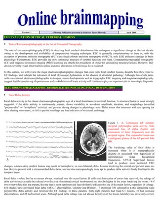
  • 1. Version 12 A Monthly Publication presented by Professor Yasser Metwally April 2009 EEG EVALUATION OF FOCAL CEREBRAL LESIONS Role of Electroencephalography in the Era of Computed Tomography The role of electroencephalography (EEG) in detecting focal cerebral disturbances has undergone a significant change in the last decade owing to the development and availability of computerized imaging techniques. EEG is generally complementary to these as, with the exception of positron emission tomography (PET) and single photon emission tomography (SPECT), only EEG evaluates changes in brain physiology. Furthermore, EEG provides the only continuous measure of cerebral function over time. Computerized transaxial tomography (CT) and magnetic resonance imaging (MRI) scanning are clearly the procedures of choice for delineating structural lesions. However, they do not currently reveal abnormalities unless anatomic alterations in brain tissue have occurred. In this edition, we will review the major electroencephalographic changes that occur with focal cerebral lesions, describe how they relate to CT findings, and indicate the relevance of focal physiologic dysfunction in the absence of structural pathology. Although this article deals with conventional electroencephalographic techniques, newer developments such as topographic EEG mapping and magnetoencephalography suggest that the monitoring of spontaneous and evoked electrical brain activity will continue to play an important role in neurologic diagnosis. ELECTROENCEPHALOGRAPHIC ABNORMALITIES INDICATING FOCAL DYSFUNCTION Focal Delta Activity Focal delta activity is the classic electroencephalographic sign of a local disturbance in cerebral function. A structural lesion is most strongly suggested if the delta activity is continuously present, shows variability in waveform amplitude, duration, and morphology (so-called quot;polymorphicquot; or quot;arrhythmicquot; activity), and persists during changes in physiologic state. Delta waves that attenuate with eye opening (or other alerting maneuvers), or fail to persist into sleep, are less indicative of structural pathology. Figure 1. A, Continuous left parietal- occipital polymorphic delta activity. Note associated loss of alpha rhythm and attenuation of faster frequencies over the occipital region. The responsible lesion was a malignant glioma of the left parietal lobe (B). The localizing value of focal delta is increased when it is topographically discrete or associated with depression of superimposed faster background frequencies. 3,19,34 Superficial lesions tend to produce more restricted EEG changes, whereas deep cerebral lesions may result in hemispheric, or even bilateral, delta. Lesions involving the central and parietal areas are less likely to present with a circumscribed delta focus, and are also correspondingly more apt to produce delta activity falsely localized to the temporal areas. Focal delta is often, but by no means always, maximal over the actual lesion. If sufficient destruction of cortex has occurred, the voltage of delta activity may actually be reduced over the area of maximal cortical involvement and thus be higher in the areas bordering the lesion. 19 If two or more delta foci are present, the one that is most persistent and least rhythmic indicates the site of the major lesion, regardless of voltage. Few studies have correlated focal delta with CT abnormalities. Gilmore and Brenner, 17 examined 100 consecutive EEGs containing focal polymorphic delta activity and reviewed the CT findings in these patients. Sixty-eight patients had focal CT lesions, 10 had nonfocal abnormalities, and 22 had normal scans. Although peak delta voltage was not always directly over the lesion, laterality was invariably correct.
  • 2. Normal CT scans occurred in patients with seizure disorders (12), concussion or contusion (5), ischemic strokes (3), viral encephalitis (1), and a progressive undiagnosed neurologic syndrome (1). Twelve patients with focal neurologic examinations had normal CT scans. Weisberg and associated 57 studied 50 consecutive patients with a unilateral temporal delta pattern and neurologic signs. CT in these patients showed tumor (40 per cent), vascular lesions (20 per cent), diffuse atrophy (16 per cent), or hydrocephalus (4 per cent). Twenty per cent had normal CT scans, a figure similar to that of Gilmore and Brenner. 17 Half of the patients with normal CT had probable epilepsy; the remainder had no further evolution of their neurologic findings over a 4-year follow-up. In another study, Weisberg and colleagues, 57 reviewed CT findings in 20 patients who had a quot;unilateral delta patternquot; but normal neurologic examination, cerebrospinal fluid (CSF), and isotope brain scan. Six had abnormal CT scans: three showing diffuse atrophy and three with infarcts. The three patients with atrophy developed Alzheimer's disease within 1 year. The authors did not indicate why the remaining 14 patients with normal CT had been referred to the EEG laboratory. Reports such as these demonstrate that although focal polymorphic delta is strongly correlated with localized anatomic pathology, EEG findings may occur in the absence of a demonstrable CT lesion. When focal delta is found without a corresponding CT abnormality, it is usually in the setting of seizures, nonhemorrhagic infarction, or trauma. 17, 57 Clinical, 46 and experimental, 18 observations indicate that polymorphic delta results primarily from lesions affecting cerebral white matter. Involvement of superficial cortex is not essential, and, indeed, lesions restricted to the cortical mantle do not generally produce significant focal delta. 18,46 It is likely that functional deafferentation of cortex, rather than a change in cortical metabolic rate, is critical. 18 Cerebral edema does not appear to make a substantial contribution to the production of delta waves. 16,18,46 Persistent polymorphic delta activity may not precisely match the true location of the lesion, particularly since it presumably arises from physiological deranged neurons often lying on the margin of the destructive lesion. Persistent polymorphic delta activity is aetiologically nonspecific and is seen in a variety of subcortical (while matter) destructive lesions including neoplasms, infarctions, abscesses, trauma, and haemorrhage. It can also be seen in reversible processes such as focal ischemia in transient ischemic attacks or focal depression from a recent seizure. Because the likelihood of a demonstrable structural change is strongly correlated with the degree of slowing, the clinical associations of focal theta activity are less striking, especially for acute or subacute lesions. Focal theta may be seen in the early stages of a slowly growing neoplasm or in the resolution of acute lesions caused by stroke or trauma. Figure 2. Polymorphic slow wave activity in a patient with subcortical glioma, notice the marked variability in wave shape morphology, frequency and amplitude. Beta Activity Abnormalities in beta activity are usually limited to voltage asymmetries. To be considered unequivocally abnormal, there should be a persistent amplitude difference of 35 per cent or greater (expressed as a percentage of the higher voltage). 32 Diminished beta activity results either from cortical dysfunction or from an increase in resistance of the medium separating cortex from scalp recording electrodes. Thus, local attenuation of beta may occur with a cortical infarction, for example, or in the presence of a subdural or epidural fluid collection. A beta asymmetry may also result from localized scalp edema caused by head injury or infiltration from an intravenous line. Similar considerations apply as well to the localized absence or attenuation of background rhythms other than beta. Focally increased beta activity is usually associated with a skull defect. 7,25 Occasionally, localized enhancement of beta may occur over a tumor or as the manifestation of an epileptogenic focus. 20 Epileptiform Activity Focal epileptiform activity (spikes or sharp waves) may antedate the appearance of focal EEG-slowing or other clues to a tumor by months or years. 21 In a multicenter study of 1396 patients with epilepsy, 10 per cent had tumors detected by CT. 15 However, the incidence of tumor rose to 22 per cent when only patients with partial seizures were considered. Brain tumor did not occur in patients with primary generalized epilepsy and was found in only 5 per cent of those with secondary generalized seizures.
  • 3. Periodic lateralized epileptiform discharges (PLEDS) usually occur in the setting of an acute or subacute destructive process. Impaired consciousness is virtually always present, and seizures are evident nearly 80 per cent of the time. The complexes are most often composed of di- or triphasic spikes or sharp waves recurring at approximately regular I to 2-second intervals. However, the distribution, morphology, voltage, and rate of repetition vary substantially among patients. Schwartz and coworkers, 53 studied 52 patients and found a typical electrographic evolution for PLEDS. Gradual simplification in morphology and progressive prolongation of the interval between discharges usually occurred within 4 weeks. In a few patients, however, clinical relapses were accompanied by reappearance of PLEDS. Figure 3. A, Left-sided PLEDS, maximally involving the left parietal-occipital region. Background rhythms are slowed bilaterally, and there is a slight reduction in faster frequencies on the left. The patient had an intracerebral tuberculoma on that side. PLEDs may also occur independently over both hemispheres, a situation referred to as BIPLEDs. 11 In patients exhibiting BIPLEDs, diffuse diseases, rather than focal lesions, are the rule. Thus, BIPLEDs are most often seen with infections (particularly herpes simplex encephalitis), anoxic encephalopathy, epilepsy, and sickle cell anemia. 01,11 LATERALIZED AND GENERALIZED ELECTROENCEPHALOGRAPHIC FINDINGS Lateralised EEG changes The character and distribution of the electroencephalographic changes produced by a focal lesion depend on size of lesion, its distance from the cortical surface, and the specific structures involved. A small lesion critically located in the thalamus, for example, may produce widespread hemispheric slowing and alteration in sleep spindles and alpha rhythm regulation. The same discrete lesion, however, located at the cortical surface, may produce few, if any, electroencephalographic findings. Indeed, cortical lesions must involve relatively large areas to produce attenuation of background rhythms in the relative absence of slowing. Examples include subdural hematomas and meningiomas. Large infarcts (due to middle cerebral or carotid artery occlusions, for instance) involve extensive areas of cortex as well as adjacent white matter, thus producing both hemispheric polymorphic delta and loss of overriding faster frequencies. Lesions that produce hemispheric depression of background rhythms affect both normal and abnormal patterns, as illustrated by the case of a subdural hematoma causing an interhemispheric asymmetry of triphasic waves in an alcoholic with hepatic failure. Figure 4. A, Failure of alpha rhythm to attenuate normally with eye-opening on the left. The patient had a giant aneurysm of the left internal carotid-middle cerebral artery bifurcation with compression of the frontal and temporal lobes from below (B). Focal lesions may slow or attenuate the alpha rhythm unilaterally. A particularly striking abnormality of the alpha rhythm is unilateral failure to attenuate normally with eye opening (Bancaud's phenomenon) or other alerting maneuvers. These changes are reliable indicators of an ipsilateral, usually posterior, cerebral lesion, but they do not provide more specific localizing information. The photic driving response to repetitive flash stimulation may be consistently lateralized in normal individuals. 9 When it is the only finding in an otherwise normal record, an asymmetry of photic driving may usually be ignored. It is clear that a cortical lesion may depress the photic response unilaterally, but under these circumstances, the asymmetric photic response occurs in conjunction with other indications of focal dysfunction. Occasionally, focal lesions (especially subcortical or epileptogenic ones) may enhance the photic response on one side. 9 Hyperventilation will often enhance localized, low-amplitude polymorphic delta or convert intermittent slowing into a continuous focal abnormality. Focal spikes, or even seizures, sometimes appear only during hyperventilation. 42 A consistently asymmetric response to hyperventilation is always abnormal.
  • 4. Generalized EEG changes Generalized electroencephalographic abnormalities do not contribute to localization of a focal lesion or by themselves even suggest the presence of localized structural pathology. They do, however, provide information about the extent of dysfunction resulting from a focal lesion or about a coexisting abnormality (metabolic encephalopathy, for example). Subfrontal, diencephalic, or infratentorial lesions may produce generalized electroencephalographic changes, usually a combination of intermittent bursts of rhythmic delta waves and continuous, widespread polymorphic theta and delta slowing. In the absence of obstructive hydrocephalus, electroencephalographic abnormalities are more frequent with rostral than caudal brain-stem lesions. Schaul and coworkers, 51 reviewed the EEGs of 154 patients with diencephalic or posterior fossa lesions. Only 12 per cent of patients with diencephalic lesions had normal EEGS, whereas 60 per cent and 73 per cent of patients with lower brain-stem or cerebellar pathology respectively had normal EEGS. If the EEG abnormality was clearly lateralized, an infratentorial lesion was unlikely. Paroxysmal bursts of rhythmic delta waves with frontal or occipital predominance (the latter especially common in children) have been associated with subfrontal, deep midline, or posterior fossa lesions. In fact, however, intermittent rhythmic delta activity (IRDA) is nonspecific and is seen much more often in the setting of metabolic disorders or other encephalopathies affecting the brain diffusely than with focal lesions, regardless of location. IRDA may appear against an otherwise normal background. In contrast to polymorphic delta, IRDA is usually reactive to alerting maneuvers, disappears in sleep, and is augmented by hyperventilation or drowsiness. Correlative studies using CT and PET 16,29,41,46 have failed to demonstrate a particular anatomic structure responsible for generating IRDA. Physiologic investigations, 18,46 implicate dysfunction of thalamocortical interactions. Rhythmic delta activity consists of sinusoidal waveforms of approximately 2.5 Hz that occur intermittently in the EEG recording. It is most often symmetric but can be lateralized. In adults, the delta activity has a frontal predominance (frontal intermittent rhythmic delta activity [FIRDA]). In children, it is maximal posteriorly (occipital intermittent rhythmic delta activity [OIRDA]). Intermittent rhythmic delta activity is associated with structural lesions, most commonly diencephalic, infratentorial or intraventricular tumors, or with diffuse encephalopathies. FIRDA occurring in patients with a normal EEG background suggests that the pattern is due to a structural lesion; when associated with EEG background abnormalities, it is likely to be due to encephalopathy. In cases of encephalopathy with FIRDA, the pathophysiologic processes are believed to involve cortical and subcortical gray matter. OIRDA is associated with absence epilepsy in children aged 6-10 years. References 1. Annegers, J. F., Grabow, J. D., Groover, R. V., et al.: Seizures after head trauma: a population study. Neurology (N.Y.), 30:683-689, 1980. 2. Aoki, Y., Hiraga, H., and Ichijo, S.: EEG of moyamoya disease. Electroencephalogr. Clin. Neurophysiol., 43:490, 1977. 3. Arfel, G., and Fischgold, H.; EEG signs in tumors of the brain. Electroencephalogr. Clin. Neurophysiol., 19(Suppl.);36-50, 1961. 4. Brodtkorb, E., Lindqvist, M., jonsson, M., et al.: Diagnosis of herpes simplex encephalitis, a comparison between electroencephalography and computed tomography findings. Acta Neurol. Scand., 66:462-471, 1982. 5. Chatrian, G. E., Shaw, C., and Leffman, H.: The significance of periodic lateralized epileptiform discharges in EEG: An electrographic, clinical, and pathological study. Electroencephalogr. Clin. Neurophysiol., 17:177-193, 1964. 6. Chien, L. T., Boehm, R. M., Robinson, H., et al.: Characteristic early electroencephalographic changes in herpes simplex encephalitis, clinical and virologic studies. Arch. Neurol., 34:361-364, 1977. 7. Cobb, W. A., Guiloff, R. J., and Cast, J.: Breach rhythm: The EEG related to skull defects. Electroencephalogr. Clin. Neurophysiol., 47:251- 271, 1979. 8. Cohen, D., and Cuffin, B. N.: Demonstration of useful differences between magnetoencephalogram and electroencephalogram. Electroencephalogr. Clin. Neurophysiol., 56:38-51, 1983. 9. Coull, B. M., and Pedley, T. A.: Intermittent photic stimulation: Clinical usefulness of nonconvulsive responses. Electroencephalogr. Clin. Neurophysiol., 44:353-363, 1978. 10. Daly, D. D.: Use of EEG for diagnosis and evaluation of epileptic seizures and nonepileptic episodic disorders. In Klass, D., and Daly, D. (eds.): Current Practice of Clinical Electroencephalography. New York, Raven Press, 1979. 11. de la Paz, D., and Brenner, R. P.: Bilateral independent periodic lateralized epileptiform discharges. Arch. Neurol., 38:713-715, 1981. 12. Dorfman, L. J., Marshall, W. H., and Enzmann, D. R.: Cerebral infarction and migraine: Clinical and radiologic correlations. Neurology (N.Y.), 29:317-322, 1979. 13. Elian, M.: Herpes simplex encephalitis: Prognosis and long-term follow-up. Arch. Neurol., 32:39-43, 1975. 14. Enzmann, D. R., Ransom, B., Norman, D., et al.: Computed tomography of herpes simplex encephalitis. Radiology, 129:419-425, 1978. 15. Gastaut, H.: Conclusions: Computerized transverse axial tomography in epilepsy. Epilepsia, 17:337-338, 1976. 16. Gastaut, J. L., Michel, B., Hassan, S., et al.: Electroencephalography in brain edema (127 cases of brain tumor investigated by cranial computerized tomography). Electroencephalogr. Clin. Neurophysiol., 46:239-255, 1979. 17. Gilmore, P. C., and Brenner, R. P.: Correlation of EEG, computerized tomography, and clinical findings: Study of 100 patients with focal delta activity. Arch. Neurol., 38:371-372, 1981. 18. Gloor, P., Ball, G., and Schaul, N.: Brain lesions that produce delta waves in the EEG. Neurology (Minneap.), 27:326-333, 1977. 19. Goldensohn, E. S.: Use of the EEG for evaluation of focal intracranial lesions. In Klass, D., and Daly, D. (eds.): Current Practice of
  • 5. Clinical Electroencephalography. New York, Raven Press, 1979. 20. Green, R. L. and Wilson, W. P.: Asymmetries of beta activity in epilepsy, brain tumor, and cerebrovascular disease. Electroencephalogr. Clin. Neurophysiol., 13:75-78, 1961. 21. Hess, R.: Brain tumors and other space occupying processes. In Remorid, A., and Hess, R. (eds): Handbook of Electroencephalography and Clinical Neurophysiology. Vol. 14, Part C. Amsterdam, Elsevier Scientific Publishing Co., 1975. 22. Hockaday, J. M., and Whitty, C. W. M.: Factors determining the electroencephalogram in migraine: A study of 560 patients according to clinical type of migraine. Brain, 92:769-788, 1969. 23. Hungerford, G. D., du Boulay, G. H., and Zilka, K. J.: Computerized axial tomography in patients with severe migraine: A preliminary report. J. Neurol. Neurosurg. Psychiatry, 39:990-994, 1976. 24. jabbari, B., Maulsby, R. L., Holtzapple, P. A., et a].; Prognostic value of EEG in acute vascular aphasia: A long-term clinical-EEG study of 53 patients. Clin. Electroencephalogr., 10:190-197, 1979. 25. Jaffe, R. and Jacobs, L.: The beta focus: Its nature and significance. Acta Neurol. Scand., 48:191 203, 1972. 26. jasper, H. H., and van Buren, J.: Interrelationship between cortex and subcortical structures: Clinical electroencephalographic studies. Electroencephalogr. Clin. Neurophysiol. 4(Suppl.):168-188, 1953. 27. jennctt, B.: Epilepsy after Nonmissile Head Injuries. Ed. 2. Chicago, Year Book Medical Publishers, 1975. 28. Johnson, B. T.: Viral Infections of the Nervous System. New York, Raven Press, 1982. 29. jonkman, E. J., van der Holst, M. J. C., and Posen, L.: Relation between EEG, CAT, and clinical data. Electroencephalogr. Clin. Neurophysiol., 43:547, 1977. 30. Kaufman, D. M., Zimmerman, R. D., and Leeds, N. E.: Computed tomography in herpes simplex encephalitis. Neurology (N.Y.), 29:1392- 1396, 1979. 31. Kayser-Gatchalian, M. C., and Neundorfer, B.: The prognostic value of EEG in ischemic cerebral insults. Electroencephalogr. Clin. Neurophysiol., 49:608-617, 1980. 32. Kellaway, P.: An orderly approach to visual analysis: The parameters of the normal EEG in adults and children. In Klass, D., and Daly, D. (eds.): Current Practice of Clinical Electroencephalography, New York, Raven Press, 1979. 33. Ketz, E.: Brain tumors and epilepsy. In Vinken, P. J., and Bruyn, G. W. (eds): Handbook of Clinical Neurology. Vol. 16. Amsterdam, North Holland Publishing Co., 1974. 34. Kooi, K. A., Tucker, R. P., and Marshall, R. E.: Fundamentals of Electroencephalography. Ed. 2. Hagerstown, New York, Harper and Row, 1978. 35. Krenkel, W.: The electroencephalogram in tumors of the brain. In Vinken, P. J., and Bruyn, G. W. (eds.): Handbook of Clinical Neurology. Vol. 16. Amsterdam, North Holland Publishing Co., 1974. 36. Legg, N. J., Gupta, P. C., and Scott, D. F.: Epilepsy following cerebral abscess: A clinical and EEG study of 70 patients. Brain, 96:259-268, 1973. 37. Lenzi, G. L., Gracowiak, R. S. J., and Jones, T.: Cerebral oxygen metabolism and blood flow in human cerebral ischemic infarction. J. Cereb. Blood Flow Metab., 2:321-335, 1982. 38. Lombroso, C. T., and Duffy, F. H.: Brain electrical activity mapping as an adjunct to CT scanning. In Canger, R., Angeleri, F., and Penry, J. K. (eds.): Advances in Epileptology: Xlth Epilepsy International Symposium. New York, Raven Press, 1980. 39. Markand, 0. N., and Daly, D. D.: Pseudoperiodic lateralized paroxysmal discharges in electroencephalogram. Neurology (Minneap.), 21:976- 981, 1971. 40. Masdeu, J. C., Azar-Kia, B., and Rubino, F.: Evaluation of recent cerebral infarction by computerized tomography. Arch. Neurol., 34:417- 421, 1977. 41. Michel, B., Gastaut, J. L., and Bianchi, L.: Electroencephalographic cranial computerized tomographic correlations in brain abscess. Electroencephalogr. Clin. Neurophysiol., 46:256-273, 1979. 42. Miley, C. E., and Forster, F. M.: Activation of partial complex seizures by hyperventilation. Arch. Neurol., 34:371-373, 1977. 43. Mizrahi, E. M., and Tharp, B. R.: A characteristic EEG pattern in neonatal herpes simplex encephalitis. Neurology (N.Y.), 32:1215-1220, 1982. 44. Morawetz, R. B., Whittey, R. J., and Murphy, D. M.: Experience with brain biopsy for suspected herpes encephalitis: A review of forty consecutive cases. Neurosurgery, 12:654-657, 1983. 45. Niedermeyer, E.: Cerebrovascular disorders and EEG. In Niedermeyer, E., and Lopes da Silva, F. (eds.): Electroencephalography: Basic Principles, Clinical Applications, and Related Fields. Baltimore, Urban and Schwarzenberg, 1982. 46. Newmark, M. E., Theodore, W. H., Sato, S., et al.: EEG, transmission computed tomography, and positron emission tomography with fluorodeoxyglucose 18F: Their use in adults with gliomas. Arch. Neurol., 40:607-610, 1983. 47. Paulson, 0. B., and Sharbrough, F. W.: Physiologic and Pathophysiologic relationship between the EEG and the regional cerebral blood flow. Acta Neurol. Scand., 50:194-220, 1974. 48. Roseman, E., Schmidt, R., and Foltz, E.: Serial electroencephalography in vascular lesions of the brain. Neurology (Minneap.), 2:311-331, 1952. 49. Rosenberg, C. E., Anderson, D. C., Mahowald, M. W., et al.: Computerized tomography and EEG in patients without focal neurological findings. Arch. Neurol., 39:291-292, 1982. 50. Scarf, J. E., and Rahm, W. E.: The human electrocorticogram: A report of spontaneous electrical potentials obtained from the exposed brain. J. Neurophysiol., 4:418-426, 1941. 51. Schaul, N., Gloor, P., and Gotman, J.: The EEG in deep midline lesions. Neurology (N.Y.), 31;157-167, 1981. 52. Schracder, P. L., and Navjeet, S.: Seizure disorders following periodic lateralized epileptiform discharges. Epilepsia, 21:647-653, 1980. 53. Schwartz, M. S., Prior, P. F., and Scott, D. F.: The occurrence and evolution in the EEG of a lateralized periodic phenomenon. Brain, 92:613-622, 1973. 54. Sunder, T. R., Erwin, C. W., and Dubois, P. J.: Hyperventilation induced abnormalities in the electroencephalogram of children with
  • 6. moyamoya disease. Electroencephalogr. Clin. Neurophysiol., 49:414-420, 1980. 55. Tiikel, K., and jasper, H.: The electroencephalogram in parasagittal lesions. Electroencephalogr. Clin. Neurophysiol., 4:481-494, 1952. 56. Upton, A., and Gumpert, J.: Electroencephalography in diagnosis of herpes simplex encephalitis. Lancet, 1:650-652, 1970. 57. Weisberg, L., Nice, C., and Katz, M.: Cerebral Computed Tomography: A Text Atlas. Ed. 2. Philadelphia, W. B. Saunders Co., 1984. 58. Westmoreland, B. F.: EEG in the evaluation of headaches. In Klass, D., and Daly, D. (eds.): Current Practice of Clinical Electroencephalography. New York, Raven Press, 1979. 59. Yanigahara, T., Houser, D. W., and Klass, D. W.: Computed tomography and EEG in cerebrovascular disease. Arch. Neurol., 38:597-600, 1981. The author, Professor Yasser Metwally Professor of clinical neurology, Ain Shams university, Cairo, Egypt. www.yassermetwally.com A new version of this publication is uploaded in my web site every month Follow the following link to download the current version: http://brainmapping.yassermetwally.com/map.pdf © Yasser Metwally, all rights reserved