SlideShare a Scribd company logo
1 of 11
Download to read offline
CASE OF THE WEEK
                          PROFESSOR YASSER METWALLY
CLINICAL PICTURE

CLINICAL PICTURE:

11 years old male patient presented clinically with obesity, delayed growth, bilateral diminution of vision, and manifestations of
increased intracranial pressure. Clinical examination revealed bilateral primary optic atrophy.

RADIOLOGICAL FINDINGS

RADIOLOGICAL FINDINGS:


Figure 1. Parasellar heavily calcified craniopharyngioma. Precontrast CT scan image (A) and postcontrast CT scan images (B,C)
showing a heavily calcified cystic parasellar mass with forward extension to the subfrontal region. The brain stem and the 4th
ventricle are compressed and pushed posteriorly. Obstructive hydrocephalus and transependymal edema are evident. The
parasellar cystic mass showed dense contrast enhancement.


Figure 2. Parasellar heavily calcified craniopharyngioma. The calcified parasellar mass extended upward to the level of the
foramen of monro and resulted in bilateral dilatation of the lateral ventricles. Notice the transependymal edema.
Figure 3. CT scan precontrast A, and postcontrast B,C,D. A heavily calcified suprasellar cystic
                             craniopharyngioma, with subfrontal extension, the lesion also showed suprasellar extension to the
                             foramen of monro causing obstructive hydrocephalus. Dense contrast enhancement occurred in some
                             parts of the tumour.


DIAGNOSIS:

DIAGNOSIS: PARASELLAR CRANIOPHARYNGIOMA

DISCUSSION

DISCUSSION:

      Introduction

Craniopharyngiomas represent 3% of all intracranial neoplasms. 17 They are common suprasellar and intrasellar tumors in
childhood. They are most frequently seen in children between the ages of 5 and 10 years. A second age peak is encountered between
the fifth and seventh decades. The tumors are mostly suprasellar in location, but the tumor also can originate within the sella and
extend through the sella to the suprasellar region. These tumors originate from the squamous epithelial remnant of Rathke's pouch
at the junction of the infundibulum and the pituitary gland.


                                                     Figure 4. Suprasellar Adamantinomatous craniopharyngioma compressing the
                                                     hypothalamus, and the brain stem


Microscopically the tumour is composed of a solid and a cystic component. The solid parts of the tumour are composed of sheets of
epithelial cells arranged in several layers; the periphery is formed by a basal layer of palisading cells beneath which several layers
of stratified epithelial cells are found resulting in the formation of horny pearls composed of keratinized cells,among these
epithelial areas small cysts lined by palisading cells may be seen as well as deposits of lamellar bone. These epithelial areas are
separated by connective tissues rich in blood vessels ,microcysts, macrophage, cholesterol crystals and lymphocytic infiltrates. The
cystic portions of the tumour are lined by stratified squamous epithelium. The cystic fluid is rich in cholesterol crystals and protein
substance.
Figure 5. histopathological picture of a craniopharyngioma,notice the squamous epithelium,microcysts
                           and cholesterol crystals


Figure 6. A, Craniopharyngioma, compressing the optic chiasma, hypothalamus and extending upward into the lateral ventricle.
The tumour is partially cystic with calcified material. B, A sagittal section of the brain shows a large craniopharyngioma below the
cerebral ventricle. Note the stippled pattern of the tumor.

Craniopharyngiomas are divided into two types based on clinical and microscopic features-ordinary or adamantinomatous and
papillary. Both are midline tumors that arise in the region of the sella. Adamantinomatous craniopharyngiomas occur more
caudally, often with intrasellar as well as suprasellar involvement, whereas papillary tumors are suprasellar and often extend to
involve the third ventricle. 5,14 About half occur in children and adolescents, and half occur in adults. 4,7 The cells of origin of
craniopharyngiomas are controversial. As the alternative name, adamantinoma, implies, some bear strong resemblance to the
adamantinoma of the jaw and to the calcifying odontogenic cyst. This resemblance has prompted the consideration that
craniopharyngiomas arise from rests of enamel organ epithelium. 2,10,12 Squamous epithelial rests of Rathke's pouch found in the
pars tuberalis of the pituitary stalk are a more likely candidate. 1,9,10,13,14,15,17,18 The significant differences and the different
location and clinical behavior raise the possibility of a different origin for the adamantinomatous and papillary variants-1.

Adamantinomatous craniopharyngiomas are more common and predominately Adamantinomatous craniopharyngiomas have a
cystic with frequent calcification.                                             characteristic microscopic appearance. 6,13,17 A
                                                                                palisading layer of basaloid epithelium surrounds
irregularly arranged cells that resemble the stellate reticulum of the epidermis. These nests may be solid but often form a complex
trabecular network of microcysts. Bands of fibrous tissue weave between nests of epithelial cells and around cysts. The squamous
epithelium of craniopharyngiomas demonstrates typical immunoreactivity for keratin and has no special immunohistochemical
features. It usually does not undergo surface maturation with keratinization and desquamation of flattened epithelial cells, but this
pattern may be present in the flattened epithelium of larger cysts, where it may mimic an epidermoid cyst. Instead, microscopic
nodules of keratin debris form from the degeneration of plump squamous cells, the outlines of which impart a distinctive
appearance: Although the cysts appear empty after tissue processing and sectioning, in vivo they contain cholesterol-rich oil that is
highly irritating and produces striking fibroglial and foreign body reactions. These findings alone are sufficient for a presumptive
diagnosis of craniopharyngioma in a limited biopsy specimen that does not demonstrate epithelial elements. Microcalcifications
and necrotic foci often are present.

Papillary craniopharyngiomas are something of a misnomer. Squamous epithelial nests that surround loose fibrovascular tissue
rather than microcysts create a solid tumor with a pseudopapillary pattern. The peripheral epithelium lacks a basaloid
appearance. Keratin nodules are not seen.
Figure 7.Classic adamantinoma-like appearance of a craniopharyngioma. The pink region corresponds to brain tissue (BT) at the
interface with the tumor and is mainly composed of reactive astrocytes. The tumor itself is composed of epithelial cells arranged
around cystic spaces. The cells proximal to the spaces are cuboidal in shape and arranged in palisades, above this cell layer the
epithelium became squamous. Some cysts are empty but others contain keratin debris (K). B, Gross pathological specimen of a
craniopharyngioma


                                                           Figure 8. Epithelial lesion with peripheral palisading of basal
                                                           squamous epithelium surrounding loosely arranged epithelial cells, the
                                                           so-called quot;stellate reticulumquot; and nodules of keratin and variable
                                                           calcification are typical histologic features of a craniopharyngioma.


Craniopharyngiomas are slow-growing, indolent tumors and are classified as WHO grade I. Similar to chordomas, their
classification by some as low-grade malignant tumors is more clinical than biologic because of the difficulty of surgical cure. The
tumors spread locally along intracranial surfaces and do not metastasize. As they spread, they form strongly adherent fibroglial
attachments to any structures they encounter, including brain, cranial nerves, and critical vascular structures. Complete surgical
removal is the optimal treatment for a craniopharyngioma. 4,7 Their pattern of spread among the complex surfaces of the skull
base, however, often makes complete removal impossible or, with involvement of the hypothalamus and third ventricle, clinically
too costly. Consequently, postoperative radiation therapy is employed for local control of the tumor. 4,7,14 About 50% of patients
remain free of disease, with therapeutic failure resulting in a slowly progressive course. 4,7 Papillary craniopharyngiomas have a
slightly worse prognosis, probably more as a result of their location than biologic aggressiveness. Rarely, carcinomatous
transformation occurs in recurrent tumors. 9


Figure 9. Papillary craniopharyngioma: In contrast to the adamantinomatous variant, papillary craniopharyngiomas do not show
the complex heterogeneous architecture seen in the former but rather are composed of simple squamous epithelium and
fibrovascular islands of connective tissue (hematoxylin-eosin, x40) (A). Papillary craniopharyngiomas: Under high power, only
simple squamous epithelium is seen in papillary craniopharyngioma. The distinctive peripheral nuclear palisading, internal stellate
reticulum, and nodules of quot;wetquot; keratin, that typify the adamantinomatous variant, are not seen in the papillary variant
(hematoxylin-eosin, x100). (B)
Figure 10. Adamantinomatous craniopharyngiomas: Another distinctive feature of the adamantinomatous variant is scattered
nodules of keratin. These nodules are referred to as quot;wetquot; keratin because of the plump appearance of the keratinocytes; this is in
contrast to the flat, flaky keratin seen in epidermoid and dermoid cysts (hematoxylin-eosin, x100) (A). Adamantinomatous
craniopharyngiomas: Nodules of quot;wetquot; keratin frequently calcify; in aggregate, this calcification can often be detected on CT
scans, which is a recognized radiologic feature of craniopharyngiomas (hematoxylin-eosin, x100).(B)


Figure 11. The adamantinomatous craniopharyngioma is a histologically complex epithelial lesion with several, very distinctive
morphologic features. (hematoxylin-eosin, x40) (A). Adamantinomatous craniopharyngiomas: Peripheral palisading of the
epithelium is a pronounced feature (hematoxylin-eosin, x100) (B). Adamantinomatous craniopharyngiomas: Frequently, the inner
epithelium beneath the superficial palisade undergoes hydropic vacuolization and is referred to as the stellate reticulum
(hematoxylin-eosin, x100) (C).

The gross features and growth patterns of both types of craniopharyngioma are similar, even if their locations are not.
Craniopharyngiomas attach to and spread along the surfaces of structures of the suprasellar region. Bony infiltration is less
common than in chordomas. Their external surfaces are lobular and appear smooth. They have interdigitating interfaces with
surrounding soft tissue, including brain, where a striking glial response is provoked. 8 The cut surfaces demonstrate a complex
network of fibrous septa and nodules. Cysts are prominent in adamantinomatous tumors, where they are filled with cholesterol-
rich fluid or soft necrotic debris. In some cases, a single large cyst predominates. 6,13 Papillary tumors are predominantly solid.
Calcification is variably prominent, again more so in adamantinomatous tumors.

Table 1. Differences between adamantinomatous and papillary craniopharyngiomas

Adamantinomatous craniopharyngiomas                               Papillary craniopharyngiomas
More common                                                       Less common
Occur at a younger age                                            Occur at an older age
Commonly calcified                                                Calcification is less common
Commonly cystic and filled with cholesterol-rich fluid or soft Commonly solid
necrotic debris
A palisading layer of basaloid epithelium surrounds irregularly  Squamous epithelial nests that surround loose fibrovascular 
arranged cells that resemble the stellate reticulum of the tissue rather than microcysts create a solid tumor with a
epidermis. These nests may be solid but often form a complex pseudopapillary pattern.
trabecular network of microcysts. Bands of fibrous tissue weave
between nests of epithelial cells and around cysts.
Keratin nodules are commonly seen.                                Keratin nodules are not seen.

The differential diagnosis of craniopharyngioma is limited. The gross appearance is generally distinctive. Microscopically the
principal considerations are epidermoid cyst, discussed earlier, and metastatic squamous carcinoma. Some authors believe that the
distinction between craniopharyngioma and epidermoid cysts is artificial. 13,17 Infiltration of brain parenchyma induces an
exuberant gliosis, often with Rosenthal fibers, that can resemble a hypothalamic pilocytic astrocytoma.

                              Figure 12a. Rosenthal fibers in neuropil surrounding the craniopharyngioma. The brain
                              parenchyma that surrounds both variants of craniopharyngioma is typically gliotic and often shows
                              profuse numbers of eosinophilic Rosenthal fibers. The latter structures are composed of densely
                              compacted bundles of glial filaments and are typically seen in astrocytic cell processes of neuropil
                              that has been subjected to chronic compression from slowly expanding mass lesions. Rosenthal
                              fibers are a characteristic feature of juvenile pilocytic astrocytomas (JPAs), which may also arise in
                              the suprasellar/third ventricular region. Hence, a biopsy that samples only the surrounding neuropil
                              of a craniopharyngioma may yield an erroneous diagnosis of JPA, if the pathologist is unaware of
                              the close association of craniopharyngioma with Rosenthal fiber formation (hematoxylin-eosin,
                              x100).
Figure 12b. A, Craniopharyngiomas account for about 3% of all intraranial tumors. They are most frequently seen in children and
adolescents. These tumors are suprasellar in location and it is thought that they arise from epithelial remnants of Rathke's pouch
that are trapped in the stalk of the pituitary gland. Grossly, the tumor appears partially solid and partially cystic and compresses
the optic chiasm in front, the pituitary below, and the third ventricle above. B, Craniopharyngiomas are composed of sheets of
squamous epithelial cells and keratin set in a loose connective tissue stroma. Note in this image that sheets of epithelial cells (E)
surround cysts, some of which contain keratin (K).

      CT scan imaging of craniopharyngiomas

Craniopharyngiomas occur almost exclusively in the region of the sella turcica and third ventricle and account for 1.2% to 3% of
intracranial tumors. 24 They are tumors of epithelial origin and can be divided into adamantinomatous craniopharyngiomas and
squamous-papillary craniopharyngiomas based on histopathology. 24 Mixed tumors, having histologic features of both types, occur
15% of the time. The clinical and imaging features of mixed tumors are the same as those of the adamantinomatous type. 24 As a
group, craniopharyngiomas have a bimodal age distribution. Most are encountered in children and adolescents, with a peak
incidence between 6 and 10 years of age. 20 A second, smaller peak occurs in the 50s. Half of all suprasellar tumors in children are
craniopharyngiomas. 25


Figure 13. A, This photograph of a lateral X-ray of the skull shows the expansion of the sella turcica and calcification in mass (look
between arrows). B, Suprasellar cystic craniopharyngioma with peripheral enhancement


Figure 14. Suprasellar heavily calcified solid craniopharyngioma with intrasellar extension,notice ballooning of the sella
Craniopharyngioma, which develops from remnants of Rathke's pouch, may arise within the sella but is most characteristically
centered above the dorsum sellae. Suprasellar calcification on plain x-rays is evident in over 90 per cent of childhood tumours but
in under 50 per cent of adult craniopharyngiomas. The tip of the dorsum sellae may be truncated by the longstanding suprasellar
mass; occasionally the outlet of the sella may be widened with the calcification extending down into it, this is called an open sella.


Figure 15. A solid heavily calcified craniopharyngioma,with the characteristic open sella type

The pathologic features of craniopharyngiomas may be quite varied. This is reflected in the diversity of CT findings. The
characteristic type of craniopharyngiomas consists of a cystic lesion. In this case, CT shows a hypodense round lesion with a solid
calcification or a peripheral rim of punctate or continuous rim of calcification. The hypodense lesion is frequently demonstrated to
be lower attenuation than CSF, reflecting the presence of thick proteinaceous material. In other cases, the CT evidence of
calcification may be absent.

There may be thin peripheral or variable thickness rim of enhancement in craniopharyngioma. The finding of a hypodense lesion
of a craniopharyngioma may be similar to that seen with subarachnoid cysts, epidermoid cysts, or cystic pituitary adenomas.


Figure 16. Parasellar craniopharyngioma: A, Precontrast CT scan (left) and postcontrast CT scan (right), B, postcontrast MRI T1
image (left) and precontrast MRI T1 image (right). Notice the calcification, the mild postcontrast enhancement, the MRI signal
heterogeneity and the associated hydrocephalus.

Craniopharyngiomas that are predominantly solid appear as irregularly marginated heterogeneous hyperdense lesions with
irregular and complex enhancement; however, in other cases, there may be homogeneous dense sharply marginated enhancement.
In rare cases, craniopharyngiomas may be isodense without evidence of calcification. However complete absence of calcified
elements is very rare in children, but over 20 per cent of tumours presenting in adults are uncalcified. If this lesion does not
enhance, it may only be delineated by the finding of anterior third ventricular distortion and effacement of the suprasellar cistern.
In these isodense craniopharyngiomas, metrizamide CT cisternography or MRI may be necessary to demonstrate the presence of
the tumor.
Figure 17. Craniopharyngioma intraoperatively

The tumour may be large, displacing the adjacent brain substance and extending through the region of the third ventricle to the
foramina of Monro causing hydrocephalus. The relationship of the tumour to the optic chiasm and third ventricle is well shown on
sagittal reformatted sections. Angiography is the only additional study which is likely to be required before radical surgery, mainly
to demonstrate the relationship of the Posterior communicating artery to the mass and to estimate the relative importance of the
contributions of this vessel and the basilar artery to the blood supply to the tumour.

      MR imaging of craniopharyngiomas

On MR imaging, craniopharyngiomas exhibit variable appearance, depending on the solid and cystic components of the mass. The
solid lesions appear hypointense on Tl- weighted images and hyperintense on T2- weighted images, with areas of dark signal
intensity contributed by calcifications. A unique MR imaging appearance of craniopharyngioma is produced by machine oil
content of the cyst, in which the cyst appears hyperintense both in precontrast Tl-weighted and in T2- weighted images. The
hyperintensity of the cyst within craniopharyngioma in Tl- weighted images has been attributed to a high concentration of protein
and cholesterol.


Figure 18. Suprasellar craniopharyngioma, precontrast MRI T1 images (A,C) and postcontrast MRI T1 image (B). Notice the
partial tumour enhancement, the T1 hyperintensity in the precontrast images is either due to increased lipid content of the sac or
previous haemorrhage, also notice the associated hydrocephalus
Figure 19. Craniopharyngioma. A 6-year-old boy presenting with nausea, vomiting, and conjugate gaze disturbance. A, CT image
reveals a mass, isodense to the brain, compressing the cerebral peduncles and third ventricle. There is resulting hydrocephalus. B,
The mass has high signal intensity on a Tl-weighted sagittal image. It is entirely suprasellar in location and effaces the third
ventricle. C, The lesion has homogeneously high signal intensity on the T2-weighted image. Again seen is mass effect on the pons
and evidence of hydrocephalus.

Adamantinomatous craniopharyngiomas are the more common variety. These tumors frequently present as suprasellar masses
causing symptoms of increased intracranial pressure: headache, nausea, and vomiting. The lesions are lobulated masses and almost
always have a cystic component. The cystic component is generally hyperintense on T2-weighted images but can also be
hypointense depending on the cyst contents. 24 It contains proteinaceous fluid that typically causes Tl shortening and hyperintense
signal on Tl-weighted images. The solid component of the lesion demonstrates intense enhancement with contrast administration.
Calcification is common and is better seen on CT. Encasement of adjacent vessels is a feature of adamantinomatous
craniopharyngiomas that is not seen in the squamous papillary type. Adamantinomatous craniopharyngiomas must be
distinguished from other parasellar lesions demonstrating increased signal on T1-weighted images. These include Rathke's cleft
cysts, lipomas, dermoids, aneurysms, and cases of an ectopic posterior pituitary bright spot.

Squamous-papillary craniopharyngiomas are less likely to have calcifications than adamantinomatous craniopharyngiomas and
are primarily solid rather than cystic lesions. They often are rounded in shape as opposed to lobulated. 24 On MR imaging, their
appearance is nonspecific. The solid portion of these tumors is isointense to gray matter on Tl-weighted images and hyperintense
on T2-weighted images. These tumors demonstrate marked contrast enhancement. They are more common in adult patients and
often originate in the third ventricle. In contrast to adamantinomatous craniopharyngiomas, these lesions rarely recur after
excision . 24


Figure 20. Suprasellar cystic craniopharyngioma with intrasellar extension, notice the cystic hypointensity with compression of the
optic chiasma superiorly, MRI T1 postcontrast


Figure 21. Suprasellar cystic craniopharyngioma with intrasellar extension, notice the cystic hyperintensity which is attributable to
increased lipid or protein content of the cyst, MRI T1 precontrast

An ectopic posterior pituitary bright spot can be seen in patients with pituitary gland hypoplasia as well as after trauma and
surgery. 22,23 The ectopic posterior pituitary bright spot is a relatively small rounded lesion. Although this abnormality has
increased signal on T1-weighted images, signal on T2- weighted images is not as intense as that seen in a craniopharyngioma.

Partially thrombosed aneurysms may have regions of increased Tl and T2 signal. These lesions, however, typically demonstrate
signal misregistration artifact in the phase encoding direction because of pulsatile flow as well as a characteristic flow void. 19,21


SUMMARY


SUMMARY

Craniopharyngioma is a slow-growing, extra-axial, epithelial-squamous, calcified, cystic tumor arising from remnants of the
craniopharyngeal duct and/or Rathke cleft and occupying the (supra)sellar region. Two main hypotheses explain the origin of
craniopharyngioma—embryogenetic and metaplastic; they complement each other and explain the craniopharyngioma
spectrum.26

      The embryogenetic theory26

The embryogenetic theory relates to the development of the adenohypophysis and transformation of the remnant ectoblastic cells
of the craniopharyngeal duct and the involuted Rathke pouch. Both the Rathke pouch and the infundibulum develop during the
fourth week of gestation and together form the hypophysis. Both elongate and come in contact during the second month. The
infundibulum is a downward invagination of diencephalons; Rathke pouch is an upward invagination of the primitive oral cavity
(stomodeum).

The craniopharyngeal duct is the neck of the pouch, connecting to the stomodeum, which narrows, closes, and separates the pouch
from the primitive oral cavity by the end of the second month. Thus, the pouch becomes a vesicle, which flattens and surrounds the
anterior and lateral surfaces of the infundibulum. Walls of this vesicle form different structures of the hypophysis. Finally, this
vesicle involutes into a mere cleft and may disappear completely. Rathke cleft, together with remnants of the craniopharyngeal
duct, can be the site of origin of craniopharyngiomas.

      The metaplastic theory 26

The metaplastic theory relates to the residual squamous epithelium (derived from stomodeum and normally part of the
adenohypophysis), which may undergo metaplasia.

The dual theory explains the craniopharyngioma spectrum attributing the adamantinous type (most prevalent in childhood) to
embryonic remnants and the adult type (squamous papillary) to metaplastic foci derived from mature cells of the anterior
hypophysis (prevalence of the adult type increases with each decade of life and is almost never found in children).

Other cystic lesions may originate from remnants of the stomodeum and pharyngohypophyseal duct as well, such as Rathke cleft
cysts, epithelial cysts, epidermoid cysts, and dermoid cysts. 26


      Addendum

             A new version of this PDF file (with a new case) is uploaded in my web site every week (every Saturday and remains available
              till Friday.)

             To download the current version follow the link quot;http://pdf.yassermetwally.com/case.pdfquot;.
             You can also download the current version from my web site at quot;http://yassermetwally.comquot;.
             To download the software version of the publication (crow.exe) follow the link: http://neurology.yassermetwally.com/crow.zip
             The case is also presented as a short case in PDF format, to download the short case follow the link:
              http://pdf.yassermetwally.com/short.pdf
             At the end of each year, all the publications are compiled on a single CD-ROM, please contact the author to know more
              details.
             Screen resolution is better set at 1024*768 pixel screen area for optimum display.
             For an archive of the previously reported cases go to www.yassermetwally.net, then under pages in the right panel, scroll down
              and click on the text entry quot;downloadable case records in PDF formatquot;


REFERENCES
References

1. Asa SL, Kovacs K, Bilbao JM, et al: Immunohistochemical localization of keratin craniopharyngiomas and squamous cell nests
of the human pituitary. Acta Neuropathol 54:257-260, 1981

2. Badger KV, Gardner DG: The relationship of adamantinomatous craniopharyngioma to ghost cell ameloblastoma of the jaws: A
histopathologic and immunohistochemical study. J Oral Pathol Med 26:349-355,1997

3. Banjeree AK, Sharma BS, Kak VK, et al: Gliosarcoma with cartilage formation. Cancer 63:518-523, 1989 9. Banna M:
Craniopharyngioma in adults. Surg Neurol 1:202-204,1973

4. Baskin DS, Wilson CB: Surgical management of craniopharyngiomas. J Neurosurg 65:22-27, 1986

5. Crotty TB, Scheithauer BW, Young WF Jr, et al: Papillary craniopharyngioma: A clinicopathological study of 48 cases. J
Neurosurg 83:206-214, 1995

6. Goldberg GM, Eshbaugh DE: Squamous cell nests of the pituitary gland as related to the origin of craniopharyngiomas. Arch
Pathol 70:293-299,1960

7. Hoff JT, Patterson RH Jr: Craniopharyngioma in children and adults. J Neurosurg 36:299-302,1972

8. Kasai H, Hirano A, Liena JF, et al: A histopathological study of craniopharyngioma with special reference to its stroma and
surrounding tissue. Brain Tumor Pathol 14:41-45, 1997

9. Nelson GA, Bastian FO, Schfitt M, et al: Malignant transformation in craniopharyngioma. Neurosurgery 22:427-429,1988

10. Oka H, Kawano N, Yagishita S, et al: Ciliated craniopharyngioma indicates histogenetic relationship to Rathke cleft
epithelium. Clin Neuropathol 16:103- 106,1997

11. Paulus W, Stockel C, Drauss J, et al: Odontogenic classification of craniopharyngiomas: A clinicopathological study of 54 cases.
Histopathology 30:172- 176,1997

12. Petito CK: Craniopharyngioma: Prognostic importance of histologic features [comments]. AJNR Am j Neuroradiol 17:1427-
1439, 1996

13. Petito CK, DeGirolami U, Earle KM: Craniopharyngiomas: A clinical and pathological review. Cancer 37:1944-1952,1976

14. Pollack IF, Lunsford MD, Slamovits TL, et al: Stereotaxic intracavitary irradiation for cystic craniopharyngiomas. J
Neurosurg 68:227-233,1988

14. Pusey E, Kortman KE, Flannigan BD, et al: MR of craniopharyngioma. Neurosurgery 422:427-429, 1988

16. Sartoretti-Schefer S, Wichmann W, Aguzzi A, et al: MR differentiation of adamantinomatous and squamous- papillary
craniopharyngiomas. AJNR Am j Neuroradiol 18:77-87, 1977

17. Russell DS , Rn Lj: Pathology of Tumors of the Nervous System, ed 5. Baltimore, Williams & Wilkins, 1989

18. Boggan JE, Davis RL, Zorman G, et al: Intrasellar epidermoid cyst: Case report. I Neurosurg 58:411-415, 1983

19. Biondi A, Scialfa G, Scotti G: Intracranial aneurysms: MR imaging. Neuroradiology 30:214-218,1988

20. Carmel PW, Antunes JL, Chang CH: Craniopharyngiomas in children. Neurosurgery 11:382-389,1982

21. Donovan JL, Nesbit GM: Distinction of masses involving the sella and suprasellar space: Specificity of imaging features. AJR
Am J Roentgenol 167:597-603, 1996

22. Fujisawa 1, Kikuchi K, Nishimura K, et al: Transection of the pituitary stalk: Development of an ectopic posterior lobe assessed
with MR imaging. Radiology 165:487-489, 1987

23. Kelly WM, Kucharczyk W, Kucharczyk J, et al: Posterior pituitary ectopia: An MR feature of pituitary dwarfism. AJNR Am j
Neuroradiol 9:453-460,1988

24. Sartoretti-Schefer S, Wichmann W, Aguzzi A: MR differentiation of adamantinomatous and squamous-papillary
craniopharyngiomas. AJNR Am j Neuroradiol 18:77-87,1997

25. Osborn AG: Brain tumors and tumorlike masses: Classification and differential diagnosis. In Diagnostic Neuroradiology. St
Louis, Mosby, 1994

26. Metwally, MYM: Textbook of neuroimaging, A CD-ROM publication, (Metwally, MYM editor) WEB-CD agency for electronic
publication, version 9.2a April 2008

More Related Content

What's hot

Tumors & tumor like conditions of nasal cavity
Tumors & tumor like conditions of nasal cavityTumors & tumor like conditions of nasal cavity
Tumors & tumor like conditions of nasal cavityDr Durga Gahlot
 
Imaging in pediatric Brain tumors: from basics to molecular diagnosis (Dr Fel...
Imaging in pediatric Brain tumors: from basics to molecular diagnosis (Dr Fel...Imaging in pediatric Brain tumors: from basics to molecular diagnosis (Dr Fel...
Imaging in pediatric Brain tumors: from basics to molecular diagnosis (Dr Fel...Felice D'Arco
 
WHO BRAIN TUMOR CLASSIFICATION 5th EDITION
WHO BRAIN TUMOR CLASSIFICATION 5th EDITIONWHO BRAIN TUMOR CLASSIFICATION 5th EDITION
WHO BRAIN TUMOR CLASSIFICATION 5th EDITIONKanhu Charan
 
Brain metastasis - Simplified
Brain metastasis - SimplifiedBrain metastasis - Simplified
Brain metastasis - Simplifiedsuresh Bishokarma
 
Presentation1.pptx, supratentorial brain tumour
Presentation1.pptx, supratentorial brain tumourPresentation1.pptx, supratentorial brain tumour
Presentation1.pptx, supratentorial brain tumourAbdellah Nazeer
 
Oral field cancerization/ dental implant courses
Oral field cancerization/ dental implant coursesOral field cancerization/ dental implant courses
Oral field cancerization/ dental implant coursesIndian dental academy
 
Embyrogenesis of the CNS and its disorders
Embyrogenesis of the CNS and its disordersEmbyrogenesis of the CNS and its disorders
Embyrogenesis of the CNS and its disordersAmr Hassan
 
42925901 brain-tumor
42925901 brain-tumor42925901 brain-tumor
42925901 brain-tumorMuhammad Adi
 
Vascular brain lesions for radiology by Dr Soumitra Halder
Vascular brain lesions for radiology by Dr Soumitra HalderVascular brain lesions for radiology by Dr Soumitra Halder
Vascular brain lesions for radiology by Dr Soumitra HalderSoumitra Halder
 
Overview of brain tumors
Overview of brain tumorsOverview of brain tumors
Overview of brain tumorsDrAyush Garg
 
Intracranial neoplasm
Intracranial neoplasmIntracranial neoplasm
Intracranial neoplasmMilan Silwal
 
Pediatric brain tumors Dr. Muhammad Bin Zulfiqar
Pediatric brain tumors Dr. Muhammad Bin Zulfiqar Pediatric brain tumors Dr. Muhammad Bin Zulfiqar
Pediatric brain tumors Dr. Muhammad Bin Zulfiqar Dr. Muhammad Bin Zulfiqar
 

What's hot (20)

Glioblastoma Multiforme
Glioblastoma MultiformeGlioblastoma Multiforme
Glioblastoma Multiforme
 
Sino nasal malignancies
Sino nasal malignanciesSino nasal malignancies
Sino nasal malignancies
 
Tumors & tumor like conditions of nasal cavity
Tumors & tumor like conditions of nasal cavityTumors & tumor like conditions of nasal cavity
Tumors & tumor like conditions of nasal cavity
 
Imaging in pediatric Brain tumors: from basics to molecular diagnosis (Dr Fel...
Imaging in pediatric Brain tumors: from basics to molecular diagnosis (Dr Fel...Imaging in pediatric Brain tumors: from basics to molecular diagnosis (Dr Fel...
Imaging in pediatric Brain tumors: from basics to molecular diagnosis (Dr Fel...
 
WHO BRAIN TUMOR CLASSIFICATION 5th EDITION
WHO BRAIN TUMOR CLASSIFICATION 5th EDITIONWHO BRAIN TUMOR CLASSIFICATION 5th EDITION
WHO BRAIN TUMOR CLASSIFICATION 5th EDITION
 
MEDULLOBLASTOMA
MEDULLOBLASTOMAMEDULLOBLASTOMA
MEDULLOBLASTOMA
 
Radical neck dissection
Radical neck dissectionRadical neck dissection
Radical neck dissection
 
Brain metastasis - Simplified
Brain metastasis - SimplifiedBrain metastasis - Simplified
Brain metastasis - Simplified
 
Presentation1.pptx, supratentorial brain tumour
Presentation1.pptx, supratentorial brain tumourPresentation1.pptx, supratentorial brain tumour
Presentation1.pptx, supratentorial brain tumour
 
Meningioma final
Meningioma finalMeningioma final
Meningioma final
 
Oral field cancerization/ dental implant courses
Oral field cancerization/ dental implant coursesOral field cancerization/ dental implant courses
Oral field cancerization/ dental implant courses
 
Embyrogenesis of the CNS and its disorders
Embyrogenesis of the CNS and its disordersEmbyrogenesis of the CNS and its disorders
Embyrogenesis of the CNS and its disorders
 
42925901 brain-tumor
42925901 brain-tumor42925901 brain-tumor
42925901 brain-tumor
 
Vascular brain lesions for radiology by Dr Soumitra Halder
Vascular brain lesions for radiology by Dr Soumitra HalderVascular brain lesions for radiology by Dr Soumitra Halder
Vascular brain lesions for radiology by Dr Soumitra Halder
 
Overview of brain tumors
Overview of brain tumorsOverview of brain tumors
Overview of brain tumors
 
Foramen Magnum Meningioma
Foramen Magnum MeningiomaForamen Magnum Meningioma
Foramen Magnum Meningioma
 
Intracranial neoplasm
Intracranial neoplasmIntracranial neoplasm
Intracranial neoplasm
 
Pediatric brain tumors Dr. Muhammad Bin Zulfiqar
Pediatric brain tumors Dr. Muhammad Bin Zulfiqar Pediatric brain tumors Dr. Muhammad Bin Zulfiqar
Pediatric brain tumors Dr. Muhammad Bin Zulfiqar
 
APPROACH TO PINEAL TUMOR
APPROACH TO PINEAL TUMORAPPROACH TO PINEAL TUMOR
APPROACH TO PINEAL TUMOR
 
Meningioma falcine and parasagittal
Meningioma falcine and parasagittalMeningioma falcine and parasagittal
Meningioma falcine and parasagittal
 

Similar to Case record...Parasellar craniopharyngioma

Topic of the month: Radiological pathology of pilocytic astrocytoma
Topic of the month: Radiological pathology of pilocytic astrocytomaTopic of the month: Radiological pathology of pilocytic astrocytoma
Topic of the month: Radiological pathology of pilocytic astrocytomaProfessor Yasser Metwally
 
Anoverviewofhairfollicle tumours 123.pdf
Anoverviewofhairfollicle tumours 123.pdfAnoverviewofhairfollicle tumours 123.pdf
Anoverviewofhairfollicle tumours 123.pdfAnna443020
 
Pathology of common ocular and orbital tumors
Pathology of common ocular and orbital tumorsPathology of common ocular and orbital tumors
Pathology of common ocular and orbital tumorsRichards Kakumanu
 
Eyelid pathology 2
Eyelid pathology 2Eyelid pathology 2
Eyelid pathology 2Azza Mohamed
 
Malignant Tumor of eyelid
Malignant Tumor  of eyelidMalignant Tumor  of eyelid
Malignant Tumor of eyelidRifat Shamim
 
Breast Atypical Lesions
Breast Atypical LesionsBreast Atypical Lesions
Breast Atypical Lesionskurmuk
 
Basal cell Adenoma and Canalicular Adenoma Doctor Faris Alabeedi MSc, MMedSc,...
Basal cell Adenoma and Canalicular Adenoma Doctor Faris Alabeedi MSc, MMedSc,...Basal cell Adenoma and Canalicular Adenoma Doctor Faris Alabeedi MSc, MMedSc,...
Basal cell Adenoma and Canalicular Adenoma Doctor Faris Alabeedi MSc, MMedSc,...Doctor Faris Alabeedi
 
Previous year question on medulloblastoma based on neet pg, usmle, plab and f...
Previous year question on medulloblastoma based on neet pg, usmle, plab and f...Previous year question on medulloblastoma based on neet pg, usmle, plab and f...
Previous year question on medulloblastoma based on neet pg, usmle, plab and f...Medico Apps
 
Intra ventricular neoplasms
Intra ventricular neoplasmsIntra ventricular neoplasms
Intra ventricular neoplasmsfahad shafi
 
craniopharyngioma
 craniopharyngioma craniopharyngioma
craniopharyngiomaMd Roohia
 
Fine needle aspiration cytology of neuroendocrine carcinoma of the breast
Fine needle aspiration cytology of neuroendocrine carcinoma of the breastFine needle aspiration cytology of neuroendocrine carcinoma of the breast
Fine needle aspiration cytology of neuroendocrine carcinoma of the breastAbKadir Rifaei Rashid Khairi
 
Pathology of cervix
Pathology of cervixPathology of cervix
Pathology of cervixPrasad CSBR
 
Spindle cell lesions of head & neck
Spindle cell lesions of head & neckSpindle cell lesions of head & neck
Spindle cell lesions of head & neckaiswaryadinup22
 

Similar to Case record...Parasellar craniopharyngioma (20)

Topic of the month: Radiological pathology of pilocytic astrocytoma
Topic of the month: Radiological pathology of pilocytic astrocytomaTopic of the month: Radiological pathology of pilocytic astrocytoma
Topic of the month: Radiological pathology of pilocytic astrocytoma
 
Anoverviewofhairfollicle tumours 123.pdf
Anoverviewofhairfollicle tumours 123.pdfAnoverviewofhairfollicle tumours 123.pdf
Anoverviewofhairfollicle tumours 123.pdf
 
Pathology of common ocular and orbital tumors
Pathology of common ocular and orbital tumorsPathology of common ocular and orbital tumors
Pathology of common ocular and orbital tumors
 
Harbor UCLA Neuro-Radiology Case #9
Harbor UCLA Neuro-Radiology Case #9Harbor UCLA Neuro-Radiology Case #9
Harbor UCLA Neuro-Radiology Case #9
 
Meninges and related tumors
Meninges and related tumorsMeninges and related tumors
Meninges and related tumors
 
Eyelid pathology 2
Eyelid pathology 2Eyelid pathology 2
Eyelid pathology 2
 
Malignant Tumor of eyelid
Malignant Tumor  of eyelidMalignant Tumor  of eyelid
Malignant Tumor of eyelid
 
Breast Atypical Lesions
Breast Atypical LesionsBreast Atypical Lesions
Breast Atypical Lesions
 
Basal cell Adenoma and Canalicular Adenoma Doctor Faris Alabeedi MSc, MMedSc,...
Basal cell Adenoma and Canalicular Adenoma Doctor Faris Alabeedi MSc, MMedSc,...Basal cell Adenoma and Canalicular Adenoma Doctor Faris Alabeedi MSc, MMedSc,...
Basal cell Adenoma and Canalicular Adenoma Doctor Faris Alabeedi MSc, MMedSc,...
 
Previous year question on medulloblastoma based on neet pg, usmle, plab and f...
Previous year question on medulloblastoma based on neet pg, usmle, plab and f...Previous year question on medulloblastoma based on neet pg, usmle, plab and f...
Previous year question on medulloblastoma based on neet pg, usmle, plab and f...
 
Adnexal Neoplasms
Adnexal NeoplasmsAdnexal Neoplasms
Adnexal Neoplasms
 
Intra ventricular neoplasms
Intra ventricular neoplasmsIntra ventricular neoplasms
Intra ventricular neoplasms
 
Case record...Parasagittal meningioma
Case record...Parasagittal meningiomaCase record...Parasagittal meningioma
Case record...Parasagittal meningioma
 
craniopharyngioma
 craniopharyngioma craniopharyngioma
craniopharyngioma
 
Harbor UCLA Neuro-Radiology Case 6
Harbor UCLA Neuro-Radiology Case 6Harbor UCLA Neuro-Radiology Case 6
Harbor UCLA Neuro-Radiology Case 6
 
Fine needle aspiration cytology of neuroendocrine carcinoma of the breast
Fine needle aspiration cytology of neuroendocrine carcinoma of the breastFine needle aspiration cytology of neuroendocrine carcinoma of the breast
Fine needle aspiration cytology of neuroendocrine carcinoma of the breast
 
Pathology of cervix
Pathology of cervixPathology of cervix
Pathology of cervix
 
Ameloblastoma
AmeloblastomaAmeloblastoma
Ameloblastoma
 
Cervical cancer
Cervical cancerCervical cancer
Cervical cancer
 
Spindle cell lesions of head & neck
Spindle cell lesions of head & neckSpindle cell lesions of head & neck
Spindle cell lesions of head & neck
 

More from Professor Yasser Metwally

The Snake, the Scorpion, the turtle in Egypt
The Snake, the Scorpion, the turtle in EgyptThe Snake, the Scorpion, the turtle in Egypt
The Snake, the Scorpion, the turtle in EgyptProfessor Yasser Metwally
 
Radiological pathology of epileptic disorders
Radiological pathology of epileptic disordersRadiological pathology of epileptic disorders
Radiological pathology of epileptic disordersProfessor Yasser Metwally
 
Radiological pathology of cerebrovascular disorders
Radiological pathology of cerebrovascular disordersRadiological pathology of cerebrovascular disorders
Radiological pathology of cerebrovascular disordersProfessor Yasser Metwally
 
Radiological pathology of spontaneous cerebral hemorrhage
Radiological pathology of spontaneous cerebral hemorrhageRadiological pathology of spontaneous cerebral hemorrhage
Radiological pathology of spontaneous cerebral hemorrhageProfessor Yasser Metwally
 
Radiological pathology of cerebral amyloid angiography
Radiological pathology of cerebral amyloid angiographyRadiological pathology of cerebral amyloid angiography
Radiological pathology of cerebral amyloid angiographyProfessor Yasser Metwally
 
Radiological pathology of cerebral microbleeds
Radiological pathology of cerebral microbleedsRadiological pathology of cerebral microbleeds
Radiological pathology of cerebral microbleedsProfessor Yasser Metwally
 
Issues in radiological pathology: Radiological pathology of watershed infarct...
Issues in radiological pathology: Radiological pathology of watershed infarct...Issues in radiological pathology: Radiological pathology of watershed infarct...
Issues in radiological pathology: Radiological pathology of watershed infarct...Professor Yasser Metwally
 
Radiological pathology of cortical laminar necrosis
Radiological pathology of cortical laminar necrosisRadiological pathology of cortical laminar necrosis
Radiological pathology of cortical laminar necrosisProfessor Yasser Metwally
 

More from Professor Yasser Metwally (20)

The Egyptian Zoo in Cairo 2015
The Egyptian Zoo in Cairo 2015The Egyptian Zoo in Cairo 2015
The Egyptian Zoo in Cairo 2015
 
End of the great nile river in Ras Elbar
End of the great nile river in Ras ElbarEnd of the great nile river in Ras Elbar
End of the great nile river in Ras Elbar
 
The Lion and The tiger in Egypt
The Lion and The tiger in EgyptThe Lion and The tiger in Egypt
The Lion and The tiger in Egypt
 
The monkeys in Egypt
The monkeys in EgyptThe monkeys in Egypt
The monkeys in Egypt
 
The Snake, the Scorpion, the turtle in Egypt
The Snake, the Scorpion, the turtle in EgyptThe Snake, the Scorpion, the turtle in Egypt
The Snake, the Scorpion, the turtle in Egypt
 
The Egyptian Parrot
The Egyptian ParrotThe Egyptian Parrot
The Egyptian Parrot
 
The Egyptian Deer
The Egyptian DeerThe Egyptian Deer
The Egyptian Deer
 
The Egyptian Pelican
The Egyptian PelicanThe Egyptian Pelican
The Egyptian Pelican
 
The Flamingo bird in Egypt
The Flamingo bird in EgyptThe Flamingo bird in Egypt
The Flamingo bird in Egypt
 
Egyptian Cats
Egyptian CatsEgyptian Cats
Egyptian Cats
 
Radiological pathology of epileptic disorders
Radiological pathology of epileptic disordersRadiological pathology of epileptic disorders
Radiological pathology of epileptic disorders
 
Radiological pathology of cerebrovascular disorders
Radiological pathology of cerebrovascular disordersRadiological pathology of cerebrovascular disorders
Radiological pathology of cerebrovascular disorders
 
Radiological pathology of spontaneous cerebral hemorrhage
Radiological pathology of spontaneous cerebral hemorrhageRadiological pathology of spontaneous cerebral hemorrhage
Radiological pathology of spontaneous cerebral hemorrhage
 
Radiological pathology of cerebral amyloid angiography
Radiological pathology of cerebral amyloid angiographyRadiological pathology of cerebral amyloid angiography
Radiological pathology of cerebral amyloid angiography
 
Radiological pathology of cerebral microbleeds
Radiological pathology of cerebral microbleedsRadiological pathology of cerebral microbleeds
Radiological pathology of cerebral microbleeds
 
The Egyptian Zoo in Cairo
The Egyptian Zoo in CairoThe Egyptian Zoo in Cairo
The Egyptian Zoo in Cairo
 
Progressive multifocal leukoencephalopathy
Progressive multifocal leukoencephalopathyProgressive multifocal leukoencephalopathy
Progressive multifocal leukoencephalopathy
 
Progressive multifocal leukoencephalopathy
Progressive multifocal leukoencephalopathyProgressive multifocal leukoencephalopathy
Progressive multifocal leukoencephalopathy
 
Issues in radiological pathology: Radiological pathology of watershed infarct...
Issues in radiological pathology: Radiological pathology of watershed infarct...Issues in radiological pathology: Radiological pathology of watershed infarct...
Issues in radiological pathology: Radiological pathology of watershed infarct...
 
Radiological pathology of cortical laminar necrosis
Radiological pathology of cortical laminar necrosisRadiological pathology of cortical laminar necrosis
Radiological pathology of cortical laminar necrosis
 

Recently uploaded

SOCIAL AND HISTORICAL CONTEXT - LFTVD.pptx
SOCIAL AND HISTORICAL CONTEXT - LFTVD.pptxSOCIAL AND HISTORICAL CONTEXT - LFTVD.pptx
SOCIAL AND HISTORICAL CONTEXT - LFTVD.pptxiammrhaywood
 
microwave assisted reaction. General introduction
microwave assisted reaction. General introductionmicrowave assisted reaction. General introduction
microwave assisted reaction. General introductionMaksud Ahmed
 
Measures of Central Tendency: Mean, Median and Mode
Measures of Central Tendency: Mean, Median and ModeMeasures of Central Tendency: Mean, Median and Mode
Measures of Central Tendency: Mean, Median and ModeThiyagu K
 
Disha NEET Physics Guide for classes 11 and 12.pdf
Disha NEET Physics Guide for classes 11 and 12.pdfDisha NEET Physics Guide for classes 11 and 12.pdf
Disha NEET Physics Guide for classes 11 and 12.pdfchloefrazer622
 
Student login on Anyboli platform.helpin
Student login on Anyboli platform.helpinStudent login on Anyboli platform.helpin
Student login on Anyboli platform.helpinRaunakKeshri1
 
Advanced Views - Calendar View in Odoo 17
Advanced Views - Calendar View in Odoo 17Advanced Views - Calendar View in Odoo 17
Advanced Views - Calendar View in Odoo 17Celine George
 
APM Welcome, APM North West Network Conference, Synergies Across Sectors
APM Welcome, APM North West Network Conference, Synergies Across SectorsAPM Welcome, APM North West Network Conference, Synergies Across Sectors
APM Welcome, APM North West Network Conference, Synergies Across SectorsAssociation for Project Management
 
The basics of sentences session 2pptx copy.pptx
The basics of sentences session 2pptx copy.pptxThe basics of sentences session 2pptx copy.pptx
The basics of sentences session 2pptx copy.pptxheathfieldcps1
 
Key note speaker Neum_Admir Softic_ENG.pdf
Key note speaker Neum_Admir Softic_ENG.pdfKey note speaker Neum_Admir Softic_ENG.pdf
Key note speaker Neum_Admir Softic_ENG.pdfAdmir Softic
 
Accessible design: Minimum effort, maximum impact
Accessible design: Minimum effort, maximum impactAccessible design: Minimum effort, maximum impact
Accessible design: Minimum effort, maximum impactdawncurless
 
General AI for Medical Educators April 2024
General AI for Medical Educators April 2024General AI for Medical Educators April 2024
General AI for Medical Educators April 2024Janet Corral
 
fourth grading exam for kindergarten in writing
fourth grading exam for kindergarten in writingfourth grading exam for kindergarten in writing
fourth grading exam for kindergarten in writingTeacherCyreneCayanan
 
Paris 2024 Olympic Geographies - an activity
Paris 2024 Olympic Geographies - an activityParis 2024 Olympic Geographies - an activity
Paris 2024 Olympic Geographies - an activityGeoBlogs
 
Software Engineering Methodologies (overview)
Software Engineering Methodologies (overview)Software Engineering Methodologies (overview)
Software Engineering Methodologies (overview)eniolaolutunde
 
Sanyam Choudhary Chemistry practical.pdf
Sanyam Choudhary Chemistry practical.pdfSanyam Choudhary Chemistry practical.pdf
Sanyam Choudhary Chemistry practical.pdfsanyamsingh5019
 
9548086042 for call girls in Indira Nagar with room service
9548086042  for call girls in Indira Nagar  with room service9548086042  for call girls in Indira Nagar  with room service
9548086042 for call girls in Indira Nagar with room servicediscovermytutordmt
 
1029-Danh muc Sach Giao Khoa khoi 6.pdf
1029-Danh muc Sach Giao Khoa khoi  6.pdf1029-Danh muc Sach Giao Khoa khoi  6.pdf
1029-Danh muc Sach Giao Khoa khoi 6.pdfQucHHunhnh
 
Explore beautiful and ugly buildings. Mathematics helps us create beautiful d...
Explore beautiful and ugly buildings. Mathematics helps us create beautiful d...Explore beautiful and ugly buildings. Mathematics helps us create beautiful d...
Explore beautiful and ugly buildings. Mathematics helps us create beautiful d...christianmathematics
 

Recently uploaded (20)

SOCIAL AND HISTORICAL CONTEXT - LFTVD.pptx
SOCIAL AND HISTORICAL CONTEXT - LFTVD.pptxSOCIAL AND HISTORICAL CONTEXT - LFTVD.pptx
SOCIAL AND HISTORICAL CONTEXT - LFTVD.pptx
 
microwave assisted reaction. General introduction
microwave assisted reaction. General introductionmicrowave assisted reaction. General introduction
microwave assisted reaction. General introduction
 
Measures of Central Tendency: Mean, Median and Mode
Measures of Central Tendency: Mean, Median and ModeMeasures of Central Tendency: Mean, Median and Mode
Measures of Central Tendency: Mean, Median and Mode
 
Disha NEET Physics Guide for classes 11 and 12.pdf
Disha NEET Physics Guide for classes 11 and 12.pdfDisha NEET Physics Guide for classes 11 and 12.pdf
Disha NEET Physics Guide for classes 11 and 12.pdf
 
Student login on Anyboli platform.helpin
Student login on Anyboli platform.helpinStudent login on Anyboli platform.helpin
Student login on Anyboli platform.helpin
 
Advanced Views - Calendar View in Odoo 17
Advanced Views - Calendar View in Odoo 17Advanced Views - Calendar View in Odoo 17
Advanced Views - Calendar View in Odoo 17
 
Código Creativo y Arte de Software | Unidad 1
Código Creativo y Arte de Software | Unidad 1Código Creativo y Arte de Software | Unidad 1
Código Creativo y Arte de Software | Unidad 1
 
APM Welcome, APM North West Network Conference, Synergies Across Sectors
APM Welcome, APM North West Network Conference, Synergies Across SectorsAPM Welcome, APM North West Network Conference, Synergies Across Sectors
APM Welcome, APM North West Network Conference, Synergies Across Sectors
 
The basics of sentences session 2pptx copy.pptx
The basics of sentences session 2pptx copy.pptxThe basics of sentences session 2pptx copy.pptx
The basics of sentences session 2pptx copy.pptx
 
Key note speaker Neum_Admir Softic_ENG.pdf
Key note speaker Neum_Admir Softic_ENG.pdfKey note speaker Neum_Admir Softic_ENG.pdf
Key note speaker Neum_Admir Softic_ENG.pdf
 
Accessible design: Minimum effort, maximum impact
Accessible design: Minimum effort, maximum impactAccessible design: Minimum effort, maximum impact
Accessible design: Minimum effort, maximum impact
 
General AI for Medical Educators April 2024
General AI for Medical Educators April 2024General AI for Medical Educators April 2024
General AI for Medical Educators April 2024
 
fourth grading exam for kindergarten in writing
fourth grading exam for kindergarten in writingfourth grading exam for kindergarten in writing
fourth grading exam for kindergarten in writing
 
Paris 2024 Olympic Geographies - an activity
Paris 2024 Olympic Geographies - an activityParis 2024 Olympic Geographies - an activity
Paris 2024 Olympic Geographies - an activity
 
Software Engineering Methodologies (overview)
Software Engineering Methodologies (overview)Software Engineering Methodologies (overview)
Software Engineering Methodologies (overview)
 
Sanyam Choudhary Chemistry practical.pdf
Sanyam Choudhary Chemistry practical.pdfSanyam Choudhary Chemistry practical.pdf
Sanyam Choudhary Chemistry practical.pdf
 
Advance Mobile Application Development class 07
Advance Mobile Application Development class 07Advance Mobile Application Development class 07
Advance Mobile Application Development class 07
 
9548086042 for call girls in Indira Nagar with room service
9548086042  for call girls in Indira Nagar  with room service9548086042  for call girls in Indira Nagar  with room service
9548086042 for call girls in Indira Nagar with room service
 
1029-Danh muc Sach Giao Khoa khoi 6.pdf
1029-Danh muc Sach Giao Khoa khoi  6.pdf1029-Danh muc Sach Giao Khoa khoi  6.pdf
1029-Danh muc Sach Giao Khoa khoi 6.pdf
 
Explore beautiful and ugly buildings. Mathematics helps us create beautiful d...
Explore beautiful and ugly buildings. Mathematics helps us create beautiful d...Explore beautiful and ugly buildings. Mathematics helps us create beautiful d...
Explore beautiful and ugly buildings. Mathematics helps us create beautiful d...
 

Case record...Parasellar craniopharyngioma

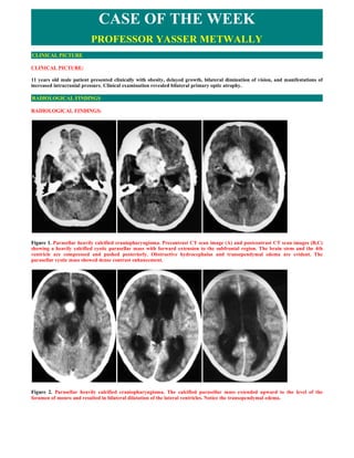
  • 1. CASE OF THE WEEK PROFESSOR YASSER METWALLY CLINICAL PICTURE CLINICAL PICTURE: 11 years old male patient presented clinically with obesity, delayed growth, bilateral diminution of vision, and manifestations of increased intracranial pressure. Clinical examination revealed bilateral primary optic atrophy. RADIOLOGICAL FINDINGS RADIOLOGICAL FINDINGS: Figure 1. Parasellar heavily calcified craniopharyngioma. Precontrast CT scan image (A) and postcontrast CT scan images (B,C) showing a heavily calcified cystic parasellar mass with forward extension to the subfrontal region. The brain stem and the 4th ventricle are compressed and pushed posteriorly. Obstructive hydrocephalus and transependymal edema are evident. The parasellar cystic mass showed dense contrast enhancement. Figure 2. Parasellar heavily calcified craniopharyngioma. The calcified parasellar mass extended upward to the level of the foramen of monro and resulted in bilateral dilatation of the lateral ventricles. Notice the transependymal edema.
  • 2. Figure 3. CT scan precontrast A, and postcontrast B,C,D. A heavily calcified suprasellar cystic craniopharyngioma, with subfrontal extension, the lesion also showed suprasellar extension to the foramen of monro causing obstructive hydrocephalus. Dense contrast enhancement occurred in some parts of the tumour. DIAGNOSIS: DIAGNOSIS: PARASELLAR CRANIOPHARYNGIOMA DISCUSSION DISCUSSION:  Introduction Craniopharyngiomas represent 3% of all intracranial neoplasms. 17 They are common suprasellar and intrasellar tumors in childhood. They are most frequently seen in children between the ages of 5 and 10 years. A second age peak is encountered between the fifth and seventh decades. The tumors are mostly suprasellar in location, but the tumor also can originate within the sella and extend through the sella to the suprasellar region. These tumors originate from the squamous epithelial remnant of Rathke's pouch at the junction of the infundibulum and the pituitary gland. Figure 4. Suprasellar Adamantinomatous craniopharyngioma compressing the hypothalamus, and the brain stem Microscopically the tumour is composed of a solid and a cystic component. The solid parts of the tumour are composed of sheets of epithelial cells arranged in several layers; the periphery is formed by a basal layer of palisading cells beneath which several layers of stratified epithelial cells are found resulting in the formation of horny pearls composed of keratinized cells,among these epithelial areas small cysts lined by palisading cells may be seen as well as deposits of lamellar bone. These epithelial areas are separated by connective tissues rich in blood vessels ,microcysts, macrophage, cholesterol crystals and lymphocytic infiltrates. The cystic portions of the tumour are lined by stratified squamous epithelium. The cystic fluid is rich in cholesterol crystals and protein substance.
  • 3. Figure 5. histopathological picture of a craniopharyngioma,notice the squamous epithelium,microcysts and cholesterol crystals Figure 6. A, Craniopharyngioma, compressing the optic chiasma, hypothalamus and extending upward into the lateral ventricle. The tumour is partially cystic with calcified material. B, A sagittal section of the brain shows a large craniopharyngioma below the cerebral ventricle. Note the stippled pattern of the tumor. Craniopharyngiomas are divided into two types based on clinical and microscopic features-ordinary or adamantinomatous and papillary. Both are midline tumors that arise in the region of the sella. Adamantinomatous craniopharyngiomas occur more caudally, often with intrasellar as well as suprasellar involvement, whereas papillary tumors are suprasellar and often extend to involve the third ventricle. 5,14 About half occur in children and adolescents, and half occur in adults. 4,7 The cells of origin of craniopharyngiomas are controversial. As the alternative name, adamantinoma, implies, some bear strong resemblance to the adamantinoma of the jaw and to the calcifying odontogenic cyst. This resemblance has prompted the consideration that craniopharyngiomas arise from rests of enamel organ epithelium. 2,10,12 Squamous epithelial rests of Rathke's pouch found in the pars tuberalis of the pituitary stalk are a more likely candidate. 1,9,10,13,14,15,17,18 The significant differences and the different location and clinical behavior raise the possibility of a different origin for the adamantinomatous and papillary variants-1. Adamantinomatous craniopharyngiomas are more common and predominately Adamantinomatous craniopharyngiomas have a cystic with frequent calcification. characteristic microscopic appearance. 6,13,17 A palisading layer of basaloid epithelium surrounds irregularly arranged cells that resemble the stellate reticulum of the epidermis. These nests may be solid but often form a complex trabecular network of microcysts. Bands of fibrous tissue weave between nests of epithelial cells and around cysts. The squamous epithelium of craniopharyngiomas demonstrates typical immunoreactivity for keratin and has no special immunohistochemical features. It usually does not undergo surface maturation with keratinization and desquamation of flattened epithelial cells, but this pattern may be present in the flattened epithelium of larger cysts, where it may mimic an epidermoid cyst. Instead, microscopic nodules of keratin debris form from the degeneration of plump squamous cells, the outlines of which impart a distinctive appearance: Although the cysts appear empty after tissue processing and sectioning, in vivo they contain cholesterol-rich oil that is highly irritating and produces striking fibroglial and foreign body reactions. These findings alone are sufficient for a presumptive diagnosis of craniopharyngioma in a limited biopsy specimen that does not demonstrate epithelial elements. Microcalcifications and necrotic foci often are present. Papillary craniopharyngiomas are something of a misnomer. Squamous epithelial nests that surround loose fibrovascular tissue rather than microcysts create a solid tumor with a pseudopapillary pattern. The peripheral epithelium lacks a basaloid appearance. Keratin nodules are not seen.
  • 4. Figure 7.Classic adamantinoma-like appearance of a craniopharyngioma. The pink region corresponds to brain tissue (BT) at the interface with the tumor and is mainly composed of reactive astrocytes. The tumor itself is composed of epithelial cells arranged around cystic spaces. The cells proximal to the spaces are cuboidal in shape and arranged in palisades, above this cell layer the epithelium became squamous. Some cysts are empty but others contain keratin debris (K). B, Gross pathological specimen of a craniopharyngioma Figure 8. Epithelial lesion with peripheral palisading of basal squamous epithelium surrounding loosely arranged epithelial cells, the so-called quot;stellate reticulumquot; and nodules of keratin and variable calcification are typical histologic features of a craniopharyngioma. Craniopharyngiomas are slow-growing, indolent tumors and are classified as WHO grade I. Similar to chordomas, their classification by some as low-grade malignant tumors is more clinical than biologic because of the difficulty of surgical cure. The tumors spread locally along intracranial surfaces and do not metastasize. As they spread, they form strongly adherent fibroglial attachments to any structures they encounter, including brain, cranial nerves, and critical vascular structures. Complete surgical removal is the optimal treatment for a craniopharyngioma. 4,7 Their pattern of spread among the complex surfaces of the skull base, however, often makes complete removal impossible or, with involvement of the hypothalamus and third ventricle, clinically too costly. Consequently, postoperative radiation therapy is employed for local control of the tumor. 4,7,14 About 50% of patients remain free of disease, with therapeutic failure resulting in a slowly progressive course. 4,7 Papillary craniopharyngiomas have a slightly worse prognosis, probably more as a result of their location than biologic aggressiveness. Rarely, carcinomatous transformation occurs in recurrent tumors. 9 Figure 9. Papillary craniopharyngioma: In contrast to the adamantinomatous variant, papillary craniopharyngiomas do not show the complex heterogeneous architecture seen in the former but rather are composed of simple squamous epithelium and fibrovascular islands of connective tissue (hematoxylin-eosin, x40) (A). Papillary craniopharyngiomas: Under high power, only simple squamous epithelium is seen in papillary craniopharyngioma. The distinctive peripheral nuclear palisading, internal stellate reticulum, and nodules of quot;wetquot; keratin, that typify the adamantinomatous variant, are not seen in the papillary variant (hematoxylin-eosin, x100). (B)
  • 5. Figure 10. Adamantinomatous craniopharyngiomas: Another distinctive feature of the adamantinomatous variant is scattered nodules of keratin. These nodules are referred to as quot;wetquot; keratin because of the plump appearance of the keratinocytes; this is in contrast to the flat, flaky keratin seen in epidermoid and dermoid cysts (hematoxylin-eosin, x100) (A). Adamantinomatous craniopharyngiomas: Nodules of quot;wetquot; keratin frequently calcify; in aggregate, this calcification can often be detected on CT scans, which is a recognized radiologic feature of craniopharyngiomas (hematoxylin-eosin, x100).(B) Figure 11. The adamantinomatous craniopharyngioma is a histologically complex epithelial lesion with several, very distinctive morphologic features. (hematoxylin-eosin, x40) (A). Adamantinomatous craniopharyngiomas: Peripheral palisading of the epithelium is a pronounced feature (hematoxylin-eosin, x100) (B). Adamantinomatous craniopharyngiomas: Frequently, the inner epithelium beneath the superficial palisade undergoes hydropic vacuolization and is referred to as the stellate reticulum (hematoxylin-eosin, x100) (C). The gross features and growth patterns of both types of craniopharyngioma are similar, even if their locations are not. Craniopharyngiomas attach to and spread along the surfaces of structures of the suprasellar region. Bony infiltration is less common than in chordomas. Their external surfaces are lobular and appear smooth. They have interdigitating interfaces with surrounding soft tissue, including brain, where a striking glial response is provoked. 8 The cut surfaces demonstrate a complex network of fibrous septa and nodules. Cysts are prominent in adamantinomatous tumors, where they are filled with cholesterol- rich fluid or soft necrotic debris. In some cases, a single large cyst predominates. 6,13 Papillary tumors are predominantly solid. Calcification is variably prominent, again more so in adamantinomatous tumors. Table 1. Differences between adamantinomatous and papillary craniopharyngiomas Adamantinomatous craniopharyngiomas Papillary craniopharyngiomas More common Less common Occur at a younger age Occur at an older age Commonly calcified Calcification is less common Commonly cystic and filled with cholesterol-rich fluid or soft Commonly solid necrotic debris A palisading layer of basaloid epithelium surrounds irregularly  Squamous epithelial nests that surround loose fibrovascular  arranged cells that resemble the stellate reticulum of the tissue rather than microcysts create a solid tumor with a epidermis. These nests may be solid but often form a complex pseudopapillary pattern. trabecular network of microcysts. Bands of fibrous tissue weave between nests of epithelial cells and around cysts. Keratin nodules are commonly seen. Keratin nodules are not seen. The differential diagnosis of craniopharyngioma is limited. The gross appearance is generally distinctive. Microscopically the principal considerations are epidermoid cyst, discussed earlier, and metastatic squamous carcinoma. Some authors believe that the distinction between craniopharyngioma and epidermoid cysts is artificial. 13,17 Infiltration of brain parenchyma induces an exuberant gliosis, often with Rosenthal fibers, that can resemble a hypothalamic pilocytic astrocytoma. Figure 12a. Rosenthal fibers in neuropil surrounding the craniopharyngioma. The brain parenchyma that surrounds both variants of craniopharyngioma is typically gliotic and often shows profuse numbers of eosinophilic Rosenthal fibers. The latter structures are composed of densely compacted bundles of glial filaments and are typically seen in astrocytic cell processes of neuropil that has been subjected to chronic compression from slowly expanding mass lesions. Rosenthal fibers are a characteristic feature of juvenile pilocytic astrocytomas (JPAs), which may also arise in the suprasellar/third ventricular region. Hence, a biopsy that samples only the surrounding neuropil of a craniopharyngioma may yield an erroneous diagnosis of JPA, if the pathologist is unaware of the close association of craniopharyngioma with Rosenthal fiber formation (hematoxylin-eosin, x100).
  • 6. Figure 12b. A, Craniopharyngiomas account for about 3% of all intraranial tumors. They are most frequently seen in children and adolescents. These tumors are suprasellar in location and it is thought that they arise from epithelial remnants of Rathke's pouch that are trapped in the stalk of the pituitary gland. Grossly, the tumor appears partially solid and partially cystic and compresses the optic chiasm in front, the pituitary below, and the third ventricle above. B, Craniopharyngiomas are composed of sheets of squamous epithelial cells and keratin set in a loose connective tissue stroma. Note in this image that sheets of epithelial cells (E) surround cysts, some of which contain keratin (K).  CT scan imaging of craniopharyngiomas Craniopharyngiomas occur almost exclusively in the region of the sella turcica and third ventricle and account for 1.2% to 3% of intracranial tumors. 24 They are tumors of epithelial origin and can be divided into adamantinomatous craniopharyngiomas and squamous-papillary craniopharyngiomas based on histopathology. 24 Mixed tumors, having histologic features of both types, occur 15% of the time. The clinical and imaging features of mixed tumors are the same as those of the adamantinomatous type. 24 As a group, craniopharyngiomas have a bimodal age distribution. Most are encountered in children and adolescents, with a peak incidence between 6 and 10 years of age. 20 A second, smaller peak occurs in the 50s. Half of all suprasellar tumors in children are craniopharyngiomas. 25 Figure 13. A, This photograph of a lateral X-ray of the skull shows the expansion of the sella turcica and calcification in mass (look between arrows). B, Suprasellar cystic craniopharyngioma with peripheral enhancement Figure 14. Suprasellar heavily calcified solid craniopharyngioma with intrasellar extension,notice ballooning of the sella
  • 7. Craniopharyngioma, which develops from remnants of Rathke's pouch, may arise within the sella but is most characteristically centered above the dorsum sellae. Suprasellar calcification on plain x-rays is evident in over 90 per cent of childhood tumours but in under 50 per cent of adult craniopharyngiomas. The tip of the dorsum sellae may be truncated by the longstanding suprasellar mass; occasionally the outlet of the sella may be widened with the calcification extending down into it, this is called an open sella. Figure 15. A solid heavily calcified craniopharyngioma,with the characteristic open sella type The pathologic features of craniopharyngiomas may be quite varied. This is reflected in the diversity of CT findings. The characteristic type of craniopharyngiomas consists of a cystic lesion. In this case, CT shows a hypodense round lesion with a solid calcification or a peripheral rim of punctate or continuous rim of calcification. The hypodense lesion is frequently demonstrated to be lower attenuation than CSF, reflecting the presence of thick proteinaceous material. In other cases, the CT evidence of calcification may be absent. There may be thin peripheral or variable thickness rim of enhancement in craniopharyngioma. The finding of a hypodense lesion of a craniopharyngioma may be similar to that seen with subarachnoid cysts, epidermoid cysts, or cystic pituitary adenomas. Figure 16. Parasellar craniopharyngioma: A, Precontrast CT scan (left) and postcontrast CT scan (right), B, postcontrast MRI T1 image (left) and precontrast MRI T1 image (right). Notice the calcification, the mild postcontrast enhancement, the MRI signal heterogeneity and the associated hydrocephalus. Craniopharyngiomas that are predominantly solid appear as irregularly marginated heterogeneous hyperdense lesions with irregular and complex enhancement; however, in other cases, there may be homogeneous dense sharply marginated enhancement. In rare cases, craniopharyngiomas may be isodense without evidence of calcification. However complete absence of calcified elements is very rare in children, but over 20 per cent of tumours presenting in adults are uncalcified. If this lesion does not enhance, it may only be delineated by the finding of anterior third ventricular distortion and effacement of the suprasellar cistern. In these isodense craniopharyngiomas, metrizamide CT cisternography or MRI may be necessary to demonstrate the presence of the tumor.
  • 8. Figure 17. Craniopharyngioma intraoperatively The tumour may be large, displacing the adjacent brain substance and extending through the region of the third ventricle to the foramina of Monro causing hydrocephalus. The relationship of the tumour to the optic chiasm and third ventricle is well shown on sagittal reformatted sections. Angiography is the only additional study which is likely to be required before radical surgery, mainly to demonstrate the relationship of the Posterior communicating artery to the mass and to estimate the relative importance of the contributions of this vessel and the basilar artery to the blood supply to the tumour.  MR imaging of craniopharyngiomas On MR imaging, craniopharyngiomas exhibit variable appearance, depending on the solid and cystic components of the mass. The solid lesions appear hypointense on Tl- weighted images and hyperintense on T2- weighted images, with areas of dark signal intensity contributed by calcifications. A unique MR imaging appearance of craniopharyngioma is produced by machine oil content of the cyst, in which the cyst appears hyperintense both in precontrast Tl-weighted and in T2- weighted images. The hyperintensity of the cyst within craniopharyngioma in Tl- weighted images has been attributed to a high concentration of protein and cholesterol. Figure 18. Suprasellar craniopharyngioma, precontrast MRI T1 images (A,C) and postcontrast MRI T1 image (B). Notice the partial tumour enhancement, the T1 hyperintensity in the precontrast images is either due to increased lipid content of the sac or previous haemorrhage, also notice the associated hydrocephalus
  • 9. Figure 19. Craniopharyngioma. A 6-year-old boy presenting with nausea, vomiting, and conjugate gaze disturbance. A, CT image reveals a mass, isodense to the brain, compressing the cerebral peduncles and third ventricle. There is resulting hydrocephalus. B, The mass has high signal intensity on a Tl-weighted sagittal image. It is entirely suprasellar in location and effaces the third ventricle. C, The lesion has homogeneously high signal intensity on the T2-weighted image. Again seen is mass effect on the pons and evidence of hydrocephalus. Adamantinomatous craniopharyngiomas are the more common variety. These tumors frequently present as suprasellar masses causing symptoms of increased intracranial pressure: headache, nausea, and vomiting. The lesions are lobulated masses and almost always have a cystic component. The cystic component is generally hyperintense on T2-weighted images but can also be hypointense depending on the cyst contents. 24 It contains proteinaceous fluid that typically causes Tl shortening and hyperintense signal on Tl-weighted images. The solid component of the lesion demonstrates intense enhancement with contrast administration. Calcification is common and is better seen on CT. Encasement of adjacent vessels is a feature of adamantinomatous craniopharyngiomas that is not seen in the squamous papillary type. Adamantinomatous craniopharyngiomas must be distinguished from other parasellar lesions demonstrating increased signal on T1-weighted images. These include Rathke's cleft cysts, lipomas, dermoids, aneurysms, and cases of an ectopic posterior pituitary bright spot. Squamous-papillary craniopharyngiomas are less likely to have calcifications than adamantinomatous craniopharyngiomas and are primarily solid rather than cystic lesions. They often are rounded in shape as opposed to lobulated. 24 On MR imaging, their appearance is nonspecific. The solid portion of these tumors is isointense to gray matter on Tl-weighted images and hyperintense on T2-weighted images. These tumors demonstrate marked contrast enhancement. They are more common in adult patients and often originate in the third ventricle. In contrast to adamantinomatous craniopharyngiomas, these lesions rarely recur after excision . 24 Figure 20. Suprasellar cystic craniopharyngioma with intrasellar extension, notice the cystic hypointensity with compression of the optic chiasma superiorly, MRI T1 postcontrast Figure 21. Suprasellar cystic craniopharyngioma with intrasellar extension, notice the cystic hyperintensity which is attributable to increased lipid or protein content of the cyst, MRI T1 precontrast An ectopic posterior pituitary bright spot can be seen in patients with pituitary gland hypoplasia as well as after trauma and surgery. 22,23 The ectopic posterior pituitary bright spot is a relatively small rounded lesion. Although this abnormality has
  • 10. increased signal on T1-weighted images, signal on T2- weighted images is not as intense as that seen in a craniopharyngioma. Partially thrombosed aneurysms may have regions of increased Tl and T2 signal. These lesions, however, typically demonstrate signal misregistration artifact in the phase encoding direction because of pulsatile flow as well as a characteristic flow void. 19,21 SUMMARY SUMMARY Craniopharyngioma is a slow-growing, extra-axial, epithelial-squamous, calcified, cystic tumor arising from remnants of the craniopharyngeal duct and/or Rathke cleft and occupying the (supra)sellar region. Two main hypotheses explain the origin of craniopharyngioma—embryogenetic and metaplastic; they complement each other and explain the craniopharyngioma spectrum.26  The embryogenetic theory26 The embryogenetic theory relates to the development of the adenohypophysis and transformation of the remnant ectoblastic cells of the craniopharyngeal duct and the involuted Rathke pouch. Both the Rathke pouch and the infundibulum develop during the fourth week of gestation and together form the hypophysis. Both elongate and come in contact during the second month. The infundibulum is a downward invagination of diencephalons; Rathke pouch is an upward invagination of the primitive oral cavity (stomodeum). The craniopharyngeal duct is the neck of the pouch, connecting to the stomodeum, which narrows, closes, and separates the pouch from the primitive oral cavity by the end of the second month. Thus, the pouch becomes a vesicle, which flattens and surrounds the anterior and lateral surfaces of the infundibulum. Walls of this vesicle form different structures of the hypophysis. Finally, this vesicle involutes into a mere cleft and may disappear completely. Rathke cleft, together with remnants of the craniopharyngeal duct, can be the site of origin of craniopharyngiomas.  The metaplastic theory 26 The metaplastic theory relates to the residual squamous epithelium (derived from stomodeum and normally part of the adenohypophysis), which may undergo metaplasia. The dual theory explains the craniopharyngioma spectrum attributing the adamantinous type (most prevalent in childhood) to embryonic remnants and the adult type (squamous papillary) to metaplastic foci derived from mature cells of the anterior hypophysis (prevalence of the adult type increases with each decade of life and is almost never found in children). Other cystic lesions may originate from remnants of the stomodeum and pharyngohypophyseal duct as well, such as Rathke cleft cysts, epithelial cysts, epidermoid cysts, and dermoid cysts. 26  Addendum  A new version of this PDF file (with a new case) is uploaded in my web site every week (every Saturday and remains available till Friday.)  To download the current version follow the link quot;http://pdf.yassermetwally.com/case.pdfquot;.  You can also download the current version from my web site at quot;http://yassermetwally.comquot;.  To download the software version of the publication (crow.exe) follow the link: http://neurology.yassermetwally.com/crow.zip  The case is also presented as a short case in PDF format, to download the short case follow the link: http://pdf.yassermetwally.com/short.pdf  At the end of each year, all the publications are compiled on a single CD-ROM, please contact the author to know more details.  Screen resolution is better set at 1024*768 pixel screen area for optimum display.  For an archive of the previously reported cases go to www.yassermetwally.net, then under pages in the right panel, scroll down and click on the text entry quot;downloadable case records in PDF formatquot; REFERENCES
  • 11. References 1. Asa SL, Kovacs K, Bilbao JM, et al: Immunohistochemical localization of keratin craniopharyngiomas and squamous cell nests of the human pituitary. Acta Neuropathol 54:257-260, 1981 2. Badger KV, Gardner DG: The relationship of adamantinomatous craniopharyngioma to ghost cell ameloblastoma of the jaws: A histopathologic and immunohistochemical study. J Oral Pathol Med 26:349-355,1997 3. Banjeree AK, Sharma BS, Kak VK, et al: Gliosarcoma with cartilage formation. Cancer 63:518-523, 1989 9. Banna M: Craniopharyngioma in adults. Surg Neurol 1:202-204,1973 4. Baskin DS, Wilson CB: Surgical management of craniopharyngiomas. J Neurosurg 65:22-27, 1986 5. Crotty TB, Scheithauer BW, Young WF Jr, et al: Papillary craniopharyngioma: A clinicopathological study of 48 cases. J Neurosurg 83:206-214, 1995 6. Goldberg GM, Eshbaugh DE: Squamous cell nests of the pituitary gland as related to the origin of craniopharyngiomas. Arch Pathol 70:293-299,1960 7. Hoff JT, Patterson RH Jr: Craniopharyngioma in children and adults. J Neurosurg 36:299-302,1972 8. Kasai H, Hirano A, Liena JF, et al: A histopathological study of craniopharyngioma with special reference to its stroma and surrounding tissue. Brain Tumor Pathol 14:41-45, 1997 9. Nelson GA, Bastian FO, Schfitt M, et al: Malignant transformation in craniopharyngioma. Neurosurgery 22:427-429,1988 10. Oka H, Kawano N, Yagishita S, et al: Ciliated craniopharyngioma indicates histogenetic relationship to Rathke cleft epithelium. Clin Neuropathol 16:103- 106,1997 11. Paulus W, Stockel C, Drauss J, et al: Odontogenic classification of craniopharyngiomas: A clinicopathological study of 54 cases. Histopathology 30:172- 176,1997 12. Petito CK: Craniopharyngioma: Prognostic importance of histologic features [comments]. AJNR Am j Neuroradiol 17:1427- 1439, 1996 13. Petito CK, DeGirolami U, Earle KM: Craniopharyngiomas: A clinical and pathological review. Cancer 37:1944-1952,1976 14. Pollack IF, Lunsford MD, Slamovits TL, et al: Stereotaxic intracavitary irradiation for cystic craniopharyngiomas. J Neurosurg 68:227-233,1988 14. Pusey E, Kortman KE, Flannigan BD, et al: MR of craniopharyngioma. Neurosurgery 422:427-429, 1988 16. Sartoretti-Schefer S, Wichmann W, Aguzzi A, et al: MR differentiation of adamantinomatous and squamous- papillary craniopharyngiomas. AJNR Am j Neuroradiol 18:77-87, 1977 17. Russell DS , Rn Lj: Pathology of Tumors of the Nervous System, ed 5. Baltimore, Williams & Wilkins, 1989 18. Boggan JE, Davis RL, Zorman G, et al: Intrasellar epidermoid cyst: Case report. I Neurosurg 58:411-415, 1983 19. Biondi A, Scialfa G, Scotti G: Intracranial aneurysms: MR imaging. Neuroradiology 30:214-218,1988 20. Carmel PW, Antunes JL, Chang CH: Craniopharyngiomas in children. Neurosurgery 11:382-389,1982 21. Donovan JL, Nesbit GM: Distinction of masses involving the sella and suprasellar space: Specificity of imaging features. AJR Am J Roentgenol 167:597-603, 1996 22. Fujisawa 1, Kikuchi K, Nishimura K, et al: Transection of the pituitary stalk: Development of an ectopic posterior lobe assessed with MR imaging. Radiology 165:487-489, 1987 23. Kelly WM, Kucharczyk W, Kucharczyk J, et al: Posterior pituitary ectopia: An MR feature of pituitary dwarfism. AJNR Am j Neuroradiol 9:453-460,1988 24. Sartoretti-Schefer S, Wichmann W, Aguzzi A: MR differentiation of adamantinomatous and squamous-papillary craniopharyngiomas. AJNR Am j Neuroradiol 18:77-87,1997 25. Osborn AG: Brain tumors and tumorlike masses: Classification and differential diagnosis. In Diagnostic Neuroradiology. St Louis, Mosby, 1994 26. Metwally, MYM: Textbook of neuroimaging, A CD-ROM publication, (Metwally, MYM editor) WEB-CD agency for electronic publication, version 9.2a April 2008