What Are The Drone Anti-jamming Systems Technology?
Lanl science 1995 history of radiation standards
1. Radiation and Risk–A Hard Look at the Data
A Brief History of Radiation
H
ealth physics is concerned with protecting people from the harmful effects
of ionizing radiation while allowing its beneficial use in medicine, science,
and industry. Since the discovery of radiation and radioactivity 100 years
ago, radiation protection standards and the philosophy governing those standards
have evolved in somewhat discrete intervals. The changes have been driven by
two factors—new information on the effects of radiation on biological systems
and changing attitudes toward acceptable
risk. The earliest limits were based on
preventing the onset of such obvious effects as skin ulcerations that appeared
after intense exposure to radiation fields.
Later limits were based on preventing delayed effects such as cancer that had been
observed in populations of people receiving high doses, particularly from medical
exposures and from the atomic-bomb exposures in Hiroshima and Nagasaki.
During the evolution of standards, the
general approach has been to rely on risk estimates that have
little chance of underestimating the consequences of radiation exposure. It is important to realize that most of the effects observed in human populations have occurred at high
doses and high dose rates. The information gathered from
those populations must be scaled down to low doses and
low dose rates to estimate the risks that occur in occupational settings.
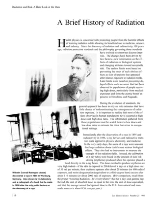
Wilhelm Conrad Roentgen (above)
discovered x rays in 1895 in Wurzburg,
Germany. Also shown is his laboratory
and a radiograph of a hand that he made
in 1896 after his only public lecture on
the discovery of x rays.
116
Immediately after the discoveries of x rays in 1895 and
radioactivity in 1896, x-ray devices and radioactive materials were applied in physics, chemistry, and medicine.
In the very early days, the users of x rays were unaware
that large radiation doses could cause serious biological
effects. They also had no instruments to measure the
strength of the radiation fields. Instead, the calibration
of x-ray tubes were based on the amount of skin reddening (erythema) produced when the operator placed a
hand directly in the x-ray beam. The doses needed to produce erythema are
very high indeed—if the skin is exposed to 200-kilovolt x rays at a high dose rate
of 30 rad per minute, then erythema appears after about 20 minutes (or 600 rad) of
exposure, and moist desquamation (equivalent to a third-degree burn) occurs after
about 110 minutes (or about 2000 rad) of exposure. (For comparison, recall from
the primer “Ionizing Radiation—It’s Everywhere!” that for x rays and gamma rays
the rad, the unit of absorbed dose, is equal to the rem, the unit of dose-equivalent,
and that the average annual background dose in the U.S. from natural and manmade sources is about 0.36 rem per year.)
Los Alamos Science Number 23 1995
2. Radiation and Risk–A Hard Look at the Data
Protection Standards
William C. Inkret, Charles B. Meinhold, and John C. Taschner
Early ignorance of the hazards of radiation resulted in
numerous unexpected injuries to patients, physicians,
and scientists, and as a result, some researchers took
steps to publicize the hazards and set limits on exposure. In July 1896, only one month after the discovery of x rays, a severe case of x-ray-induced dermatitis was published, and in 1902, the first dose limit of
about 10 rad per day (or 3000 rad per year), was recommended. The 10 rad-per-day limit was based not
on biological data but rather on the lowest amount
that could be easily detected, namely, the amount required to produce an observable exposure, or fogging,
on a photographic plate. By 1903, animal studies had
shown that x rays could produce cancer and kill living tissue and that the organs most vulnerable to radiation damage were the skin, the blood-forming organs, and the reproductive organs. Table 1 contains
estimates of dose rates encountered by radiation
workers in the early part of the 20th century.
In September 1924 at a meeting of the American
Roentgen Ray Society, Arthur Mutscheller was the
first person to recommend a “tolerance” dose rate for radiation workers, a dose
rate that in his judgement could be tolerated indefinitely. He based his recommendation on observations of physicians and technicians who worked in shielded work
areas. He estimated that the workers had received about one-tenth of an erythema
dose per month (or about 60 rem per month) as measured by the x-ray-tube current and voltage, the filtration of the beam, the distance of the workers from the
Antoine Henri Becquerel discovered
radioactivity in 1896 in Paris. He is
shown here in his laboratory.
Table 1. Dose Rates for Radiation Workers in the Early Part of the 20th Century
Occupation
Approximate Dose Rate
(rad min-1)
fluoroscopist
0.6 - 6 (hands)
0.006 - 0.06 (body)
x-ray therapy technician
0.006 (body)
radium therapist or technician
0.006 - 0.06 (body)
x-ray tube, and the exposure time. He also observed that none of the individuals
had shown any signs of radiation injury. He concluded that the dose-rate levels in
the shielded rooms were acceptable, but in proposing a tolerance dose, he applied
a safety factor of ten and recommended that the tolerance limit be set at one-hunNumber 23 1995 Los Alamos Science
117
3. Radiation and Risk–A Hard Look at the Data
dredth of an erythema dose per month (equivalent to about 70 rem per year). A
tolerance dose was "assumed to be a radiation dose to which the body can be subjected without production of harmful effects.” Mutscheller presented his recommendation in a paper entitled, “Physical Standards of Protection Against Roentgen
Ray Dangers,” which was published in 1925. Quite fortuitously, F. M. Sievert arrived at about the same limits using a similar approach.
In 1934, the U.S. Advisory Committee on X-ray and Radium Protection proposed
the first formal standard for protecting people from radiation sources. By then the
quantitative measurement of ionizing radiation had become standardized in units
of roentgens,* and therefore, the recommended limit on dose rate was expressed as
0.1 roentgen per day. That value was in line with Mutscheller’s recommendation
of one-hundredth of an erythema dose per month, and in fact, the two tolerance
limits differed only by a factor of two. Whether that difference was due to a
rounding factor or a technical difference in the way the roentgen was measured in
the U.S. versus Europe is open to interpretation.
It was common for the hands of the
early radiologists to receive exceptionally high radiation doses. The loss of
It is worth emphasizing that those early limits on exposure to x rays were not arrived at through quantitative observation of biological changes but rather through a
judgement call based on the absence of observed biological harm.
fingers, as shown in the photograph
above, was sometimes the result. Such
conditions are ultimately caused by
outright killing of many cells. In the
case above, dermal basal cells and
blood vessels were critically injured in
the fingers, scar tissue probably
plugged the blood vessels and stopped
the flow of blood. The loss of blood
The dose limits for radiation sources outside of the body (external sources) were
augmented in 1941 by a limit on the amount of radium a person could tolerate inside the body (radium tends to be retained by the body, and because of its long radioactive half-life, it thereby becomes a relatively constant internal source of radiation). The devastating experiences of the radium-dial painters and the origin of
the radium standard are described in “Radium—The Benchmark for Internal Alpha
Emitters” (see page 224). Decade-long clinical observations of twenty-seven persons who were exposed internally to radium, in combination with quantitative
supply ultimately led to the death of tissue in the fingers and the loss of those
extremities.
118
*The roentgen, the first formal radiation unit, was adopted in 1928 and specifies the quantity of ionizing radiation in terms of the amount of electrostatic charge it produces passing through a volume of
air. In particular, the Roentgen is defined as that amount of ionizing radiation that produces 1 electrostatic unit of negative charge in 0.00129 gram of air (1 cubic centimeter of air at standard temperature
and pressure). For x rays, 1 rad = 1 rem = 0.96 roentgen.
Los Alamos Science Number 23 1995
4. Radiation and Risk–A Hard Look at the Data
measurements of their radium body burdens, were the basis for the radium standard. In particular, it appeared that the retention of 1.0 microgram or more was
required to produce deleterious effects. Applying a safety factor of ten to that result, the committee members responsible for recommending a standard (many of
whom had performed the clinical research on the radium patients) suggested that
0.1 microgram (or 0.1 microcurie) of radium would be an appropriate tolerance
limit. Again, the ultimate criteria used was a judgement call: They all agreed that
they would feel comfortable even if their own children had that amount in their
bodies. That initial standard has essentially remained in effect up to the present.
In 1944, the radium standard was used as a basis for setting the first tolerance
limit for internal retention of plutonium. A working-lifetime limit of 5 micrograms (0.3 microcuries) was proposed on the basis that plutonium was long-lived
and would be a boneseeker like radium and that the alpha-particle emissions from
5 micrograms of plutonium would deposit ionizing energy at the same rate as the
alpha emissions from the allowed 0.1 microgram of radium. In 1945, as a result
of animals studies on the relative toxicity of plutonium and radium and on their
relative distribution in the body, the Manhattan Engineer District reduced the plutonium limit a factor of 5 to 0.06 microcuries. The Hanford Site, where plutonium
was being produced in reactors, reduced the limit even further to 0.03 microcuries.
Although today’s standards are expressed in terms of an annual inhalation limit
rather than a maximum permissible body burden, the current limit recommended
by the International Commission on Radiation Protection (ICRP) translates to a
body burden that is about the same as the working-lifetime limit set at Hanford
during World War II. The concern for limiting and monitoring intakes of radium
and plutonium were the beginnings of the field of internal radiation dosimetry.
In the 1930s, Robley D. Evans developed the first quantitative technique
for making in vivo measurements of
radium body burdens. Those measurements were the basis for the
A great deal of research, particularly animal studies, on the biological effects of
radiation were carried out during and immediately after World War II. In 1949
the United States, Canada, and Great Britain held a conference at Chalk River,
Ontario, on permissible doses and then published the Tripartite report in which all
radiation protection information that had been gathered was discussed and collated.
A number of new concepts concerning the measurement of dose had been developed through animal studies. These included absorbed dose (measured in rad),
dose-equivalent (measured in rem), relative biological effectiveness (RBE), which
relates the rad to the rem for different types of radiations, the absorbed dose as a
function of photon energy and depth in tissue (depth dose), the radiotoxicity of
plutonium, and the concept of a reference anatomical human. The Tripartite report
also recommended standards for internal and external radiation protection, including a plutonium body-burden limit of 0.03 microcuries, a limit on the bone-marrow dose of 300 millirem per week (about 15 rem per year), and a limit on the
skin dose of 600 millirem per week (a factor of 2 lower than the value initially
recommended by Mutscheller in his 1925 publication). With the exception of the
plutonium limit, those values were adopted by the ICRP and the National Council
on Radiation Protection and Measurements (NCRP, the new name for the old U.S.
Advisory Committee) in 1953 and 1954, respectively. (The plutonium limit recommended by the ICRP was somewhat higher at 0.04 microcuries for the maximum permissible amount of plutonium-239 fixed in the body.)
radium standard set in 1941.
During the 1950s, further reductions in the standards for external radiation were
made as a result of studies on the survivors of the two nuclear weapons dropped
on Japan and studies of survivors of high-dose medical procedures. In particular,
an early analysis of data from the Japanese atomic-bomb survivors indicated an
apparent change in the ratio of the number of males to females among infants born
Number 23 1995 Los Alamos Science
119
5. Radiation and Risk–A Hard Look at the Data
Copyrighted photo could not be reproduced here
Copyrighted photo could not be reproduced here
to survivors. At the same time, data from experiments on
mammals and fruit flies demonstrated that genetic changes
could be induced from very high radiation exposures. Thus,
radiation-induced genetic effects became a dominant concern in the early 1950s and led to the first recommended
standards for annual dose limits to the public. Later analyses indicated that the early assessment of the atomic-bomb
survivors was incorrect, and to this day, radiation-induced
genetic changes in humans have never been observed.
Nevertheless, the fear of future genetic effects lingered on
and probably inspired the creation of such science fiction
characters as Godzilla, the Incredible Shrinking Man,
Spiderman, the Incredible Hulk, and many others. The
concern also led to a reduction in radiation protection
standards.
In 1957, the ICRP recommended an annual occupational dose limit of 5 rem per year, and in 1958 the
NCRP recommended a life-time occupational dose
limit of [(age in years 2 18) 3 5] rem, or a limit of
235 rem for someone who works from ages 18 to 65.
The NCRP also recommended an annual limit to the
public of 500 millirem per year. In 1960, the Federal
Radiation Council recommended an annual limit of 500
millirem per year for an individual in the general public
and a limit of 170 millirem per year as the average annual dose to a population group.
By 1961, it was generally understood that the risk of genetic effects had been overestimated in studies of the
atomic-bomb survivors, but another risk was becoming apparent—studies of cancer incidence and mortality among the survivors were beginning to show elevated rates for leukemia. As time passed, elevated rates for solid-tumor cancers
were also observed. Those findings as well as other studies led to the understanding that different cancers have different latency periods, or elapsed times,
between irradiation of the individual and clinical observation of a malignancy.
Solid tumors have latency periods of 25 to 40 years, and leukemia has a latency period of 2 to 25 years. The latency periods generally hold true irrespective of the particular agent that serves as the carcinogen.
Radiation had a big impact on the
popular imagination in the 1950s.
The unmistakable appearance of an increased rate of cancer among the atomic-bomb survivors had a profound impact on the radiation protection community—it brought into focus the possibility that even low levels of exposure
might induce cancers. Of course, the data regarding malignancies were obtained from populations receiving high doses at high dose rates. Risks estimates for low doses could only be made by extrapolating the high-dose
data, and that procedure suggested that the cancer risks from low doses
were small. Nevertheless, there were no data to suggest the existence of a
threshold dose for radiogenic cancers, so the small risk per person at low doses
had to be considered in relation to the large number of workers who were receiving those doses.
Those considerations resulted in a philosophical shift from mere compliance with
dose limits and the avoidance of deterministic effects (such as cataracts and per-
120
Los Alamos Science Number 23 1995
6. Radiation and Risk–A Hard Look at the Data
manent damage to organs) to an emphasis on reducing overall cancer risks to
working populations. The ICRP defined a system of dose control consisting of
three parts: justification, optimization, and limitation. Justification requires that
no new practice involving radiation shall be allowed unless its introduction produces a positive net benefit. Optimization requires that all doses shall be kept as
Annual occupational limit
6
10
Dose-equivalent (millirem)
5
10
70 rem (1924)
30 rem (1934)
15 rem (bone marrow 1949)
4
10
5 rem (1958)
2 rem (1990)
3
10
1 rem (1993)
Annual public limit (infrequent exposure)
500 millirem (1960)
Annual public limit (continuous exposure)
150 millirem (1960)
2
10
1
10
1890
100 millirem (continuous exxposure 1989) (infrequent exposure 1993)
1900
1910
1920
1930
1940
1950
1960
1970
1980
1990
2000
Year
low as reasonably achievable (ALARA) taking into account the relevant economic
and social factors. Limitation requires that any individual dose not exceed limits
set for appropriate circumstances. In today’s applications of the dose-control concept, justification and optimization dominate. (More to the point, subjective judgements of regulators rather than the mathematics of optimization often drive the
dose limits to lower and lower levels; economic factors are often ignored; and the
net result is to make operations involving radiation and radioactive materials extremely expensive.)
Figure 1. Radiation Dose Limits
over the Past Century
This logarithmic plot of the recommended limits on annual exposures to
radiation shows a continual decrease
from the beginning of the century to
the present. The 1993 NCRP recommendation for occupational dose limits
allows for an average of about 1.5 rem
In 1977, the ICRP adopted a more formal risk-based approach to setting standards.
That approach required that the average incremental risk of death from radiation
exposure to workers in radiation industries be no larger than the average incremental risk of death from traumatic injuries to workers in “safe” industries. The incremental risk of death in safe industries is one in ten-thousand, or 10-4, per year.
Studies of the atomic-bomb survivors had shown that the risk coefficient for radiation-induced cancer mortality was about 10-4 per rem. Based on that risk coefficient, the ICRP recommended a maximum annual dose limit to a radiation worker
of 5 rem per year. The 5-rem annual limit was set under the assumption that the
Number 23 1995 Los Alamos Science
per year over a working life from age
18 to age 65 (that is, a lifetime limit for
an individual 65 years old is 65 rem;
this dose distributed over a 47 year period yields about 1.5 rem per year).
The ICRP does not recommend a lifetime dose limit; rather, an annual limit
of 2 rem per year averaged over any 5year period is recommended.
121
7. Radiation and Risk–A Hard Look at the Data
average dose would be less than 1 rem per year, and, thus, the average risk of
death would be the same as for safe industries. Thus, the new 1977 limit was unchanged from the 1957 limit, but it was now justified in terms of a risk-based
philosophy.
The 1993 NCRP limits on annual radiation doses relate both to stochastic effects, such as cancer and genetic effects, and to deterministic effects, such
as cataracts or permanent damage to
an organ. Stochastic effects, by definition, arise from random processes. The
During the 1980s, estimates of the doses received by the atomic-bomb survivors
were adjusted downward based on new estimates of the ratio of neutrons to
gamma rays in the radiation produced by the bomb. Also, new data on cancer incidence and mortality among the survivors indicated higher rates for some cancers
than previously thought. That meant the risk per unit dose, or the risk coefficient,
was higher, and in fact, it was calculated to be 4 3 10-4 per rem. Based on that
increase, the ICRP released a new set of international recommendations in 1990.
They recommended limiting radiation exposure to 10 rem over any 5-year period
and 5 rem in any one year. The public limit was set at a 100 millirem per year
averaged over any 5-year period.
probability of their occurrence increases with increasing dose, but their severity does not. Moreover, there is no
threshold dose below which the risk is
zero. In contrast, there is a threshold
dose for deterministic effects. That is,
doses below the threshold will not kill
enough cells to cause dysfunction in a
tissue or organ.
The NCRP released its own new set of national recommendations in 1993. Those
limits and the associated risks are listed in Table 2. They relate both to stochastic effects, such as cancer and genetic effects, and to deterministic effects. The
present limits for deterministic effects are not much different than the first recommendations: 50 rem per year to any tissue or organ and 15 rem to the lens of the
eye to avoid cataract formation. The recommended limits on whole-body doses
for stochastic effects, first set at 5 rem per year in 1958, are now set at no more
than 5 rem in any one year and a lifetime average of no more than 1.5 rem per
year.
Table 2. Current Standards and Associated Estimates of Risk (NCRP Report Number 116, 1993)
Category
5 rem (stochastic)
Recommended
Risk Coefficient
Estimated Risk
at the Annual Limit
4 3 10-4 rem-1
(for fatal cancer)
2 in 1,000 per year
8 3 10-5 rem-1
(for severe genetic
effects)
Occupational annual whole-body
limit for stochastic effects
Annual Limit
4 in 10,000 per year
Occupational lifetime limit
1 rem 3 age (years)
—
3 in 100 at age 70
Occupational annual limit for
deterministic effects
15 rem to lens of eye
50 rem to any other
organ or tissue system
—
no risk if limits
not exceeded
Public annual whole body
limit for continuous exposure
100 mrem
500 mrem
Negligible individual dose
(annual whole-body dose per
source or practice)
1 mrem
122
1 in 10,000 per year
1 3 10-4 rem-1
(for severe genetic
effects)
Public annual whole-body
limit for infrequent exposure
5 3 10-4 rem-1
(for fatal cancer)
1 in 100,000 per year
1 3 10-4 rem-1
1 in 10,000 per year
—
no discernable effects
(5 in 10,000,000)
Los Alamos Science Number 23 1995
8. Radiation and Risk–A Hard Look at the Data
The current limits represent a culmination of intensive epidemiology and radiobiological research. However, there are still many open questions regarding the detailed mechanisms that cause biological effects. What are the relative risks of different types of radiations, acute versus chronic exposures, age of exposure, and
chronic exposure to low doses? Those concerns dominate discussions on the future evolution of radiation protection standards. s
Charles B. Meinhold has been the President of the National Council on Radiation
Protection (NCRP) since 1991. He is also a
Senior Scientist and Deputy Division Head
of the Radiological Sciences Division at
Brookhaven National Laboratory. Charle’s
field of expertise is the application of radiological physics and radiobiological data to
radiation protection. He served as Chairman of NCRP Scientific Committee I on
Basic Radiation Protection Criteria from
1988 to 1992 and was a co-author of the
basic recommendations of the NCRP and
ICRP. Charles has been a member of the
International Commission of Radiological
Protection (ICRP) Main Commission since
1978 and is presently its Vice Chairman.
He was Chairman of Committee 2 on Basic
Standards of the NCRP from 1985 to 1992.
Charles is President of the International Radiation Protection Association (IRPA) and
has been a member of the IRPA Executive
Council since 1984. He has served on the
oversight committees for Rocky Flats and
for the Indian Point, Shorham, and Pilgrim
nuclear power stations, and was appointed
by the NRC to serve on the Blue Ribbon
panel for Three Mile Island Unit 2. Charles
has a B.S. in physics from Providence College and studied radiological physics at the
University of Rochester under an AEC Fellowship. He is certified by the American
Board of Health Physic, and is an Honorary
Professor of the China Institute of Atomic
Energy.
Number 23 1995 Los Alamos Science
John C. Taschner joined the Laboratory in
1992 as a technical staff member in the Environment, Safety and Health Division
(ESH-10) and is involved in radiological
transportation accident exercise planning.
In 1994, he joined the Laboratory’s Human
Studies Project Tea, and was the Project
Leader for the RaLa/Bayo Canyon Project.
Prior to coming to Los Alamos, John was
Deputy Director of the Navy’s Radiological
Controls Program Office in Washington,
D.C., and has held numerous key health
physics management positions with the U.S.
Navy and the U. S. Air Force. Over the
past thirty years, John has served on several
Radiation Protection Standards Committees.
Since 1992, John has been the Vice Chairman of the American National Standards
Institute’s N43 Committee, which writes radiation safety standards for non-medical radiation producing equipment. He has been
a member of the Health Physics Society
since 1958 and is a member of the American Academy of Health Physics. John
earned his M.S. in radiation biophysics
from the University of Kansas in 1966 and,
in 1973, received his certification in Health
Physics by the American Board of Health
Physics.
William C. Inkret See biography at the
end of “On the Front Lines.”
123