
Recommended
More Related Content
What's hot
What's hot (20)
Viewers also liked
Viewers also liked (20)
Similar to Early management of ACS
Similar to Early management of ACS (20)
More from Vitrag Shah
More from Vitrag Shah (16)
Recently uploaded
Recently uploaded (20)
Early management of ACS
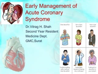
- 1. Early Management of Acute Coronary Syndrome Dr.Vitrag H. Shah Second Year Resident Medicine Dept. GMC,Surat
- 2. Introduction • The management of patients with acute myocardial ischemia and infarction is one of the few practices in critical care medicine that can save lives on a continued basis, but only when appropriate interventions are used early (often within hours after the initial contact with the patient). • Those interventions are described in this slide using information from practice guidelines published by the American College of Cardiology and American Heart Association (ACC/AHA).
- 3. Spectrum of ACS ST-segment elevation myocardial infarction (STEMI) Non-ST-segment elevation myocardial infarction (Non- STEMI) Unstable Angina (UA) Slient MI : More common in elderly and diabetic patients. • The first condition (STEMI) is the result of complete and sustained thrombotic coronary occlusion, while the last two conditions (non-STEMI and UA) are the result of either partial thrombotic coronary occlusion or transient complete occlusion with spontaneous revascularization.
- 4. Terminology • Electrocardiographic-pathologic correlations are far from perfect and terms such as Q-wave MI, non-Q-wave MI, transmural MI, and nontransmural MI, have been replaced by STEMI and NSTEMI. • Contemporary studies using MRI suggest that the development of a Q wave on the ECG is more dependent on the volume of infarcted tissue rather than the transmurality of infarction.
- 6. Epidemiology • The relative incidence of UA/NSTEMI compared to STEMI appears to be increasing. • More than one-third of patients with UA/NSTEMI are women, while less than one-fourth of patients with STEMI are women. • Approximately six million persons per year in the United States present to hospital emergency departments (EDs) with a complaint of chest pain or other symptoms suggestive of ACS. A diagnosis of an ACS is established in 20 to 25% of such patients.
- 7. Defination Stable Angina Pectoris : Chest or arm discomfort that may not be described as pain but is reproducibly associated with physical exertion or stress and is relieved within 5–10 minutes by rest and/or sublingual nitroglycerin. UA : Angina pectoris or equivalent ischemic discomfort with at least one of three features: • (1) it occurs at rest (or with minimal exertion), usually lasting >10 minutes; • (2) it is severe and of new onset (i.e., within the prior 4–6 weeks); and/or • (3) it occurs with a crescendo pattern (i.e., distinctly more severe, prolonged, or frequent than previously). NSTEMI : UA + elevated cardiac biomarkers
- 8. CHARACTERISTICS OF TYPICAL ANGINAL CHEST PAIN (ADAPTED FROM ROSEN’S EMERGENCY MEDICINE) CHARACTERISTIC SUGGESTIVE OF ANGINA LESS SUGGESTIVE OF ANGINA TYPE OF PAIN DULL SHARP/STABBING PRESSURE/CRUSHING PAIN DURATION 2-5 MIN, <20 MIN SECONDSTO HOURS/CONTINUOUS ONSET GRADUAL RAPID LOCATION/CHEST WALL SUBSTERNAL, NOT LATERAL CHEST TENDERNESS TENDER TO PALP. WALL/TENDER TO PALP. REPRODUCIBALITY WITH WITH EXERTION/ACTIVITY BREATHING/MOVING AUTONOMIC SYMPTOMS PRESENT USUALLY ABSENT
- 10. • The joint task force further refined the definition of MI by developing a clinical classification according to the assumed proximate cause of the myocardial ischemia: • Type 1: MI consequent to a pathologic process in the wall of the coronary artery (e.g. plaque erosion/rupture, fissuring, or dissection) • Type 2: MI consequent to increased oxygen demand or decreased supply (e.g. coronary artery spasm, coronary artery embolus, anemia, arrhythmias, hypertension or hypotension) • Type 3: Sudden unexpected cardiac death before blood samples for biomarkers could be drawn or before their appearance in the blood • Type 4a: MI associated with PCI • Type 4b: MI associated with stent thrombosis • Type 5: MI associated with CABG
- 11. Cardiac Biomarkers • Direct relationship between the degree of troponin elevation and mortality. • Both Trop-T & I are equivalent except in renal failure, Trop-T is less sensitive. Trop-I is cheaper than Trop-T. • However, in patients without a clear clinical history of myocardial ischemia, minor troponin elevations have been reported and can be caused by congestive heart failure (CHF), myocarditis, or pulmonary embolism, or they may be false-positive readings. • Thus, in patients with an unclear history, small troponin elevations may not be diagnostic of an ACS. • A ratio (relative index) of CKMB mass: CK activity ≥ 2.5 suggests but is not diagnostic of a myocardial rather than a skeletal muscle source for the CKMB elevation.
- 13. Role of ECG in ACS other than diagnosis Leads Location of MI Infarct Related Artery II, III, aVF Inferior wall of LV RCA/Dominant LCx V1-V4* Anteroseptal wall of LV LAD I, aVL, V5-V6/V4-V6 Lateral wall of LV Circumflex artery(LCx) V4R (V3R-V6R) Right ventricle Proximal RCA V7-V9** Posterior wall LCx/RCA (Posterolateral/ Inferoposterior MI) *V1-V2: Ventricular septum. V2-V4: Anterior wall of the LV. V2 overlaps the septum and anterior wall. V1-V3: Anteroseptal wall of the LV. **V7is located at the left posterior axillary line V8 at the tip of the left scapula V9 at the left of the spinal column in the same horizontal plane as V4-V6.
- 14. Inferior Wall MI due to RCA vs LCx ECG RCA LCx ST Elevation III > II II > III T wave III > II II > III Reciprocal ST depression aVL > I May be present in V2- V3. I & aVL either isoelectric or may be ST elevation V4R T wave upright T wave inversion RVMI May occur in Proximal - RCA obstruction Posterior MI - May occur
- 15. Posterior Wall MI • Posterior MI can be confirmed by placing extra electrodes in V7, V8, and V9 • Posterolateral MI: Occlusion of the LCx artery can cause posterior, straight posterior, or posterolateral MI. It is suspected in the 12-lead ECG when there is ST depression in V1 to V3. • Inferoposterior MI: ST elevation in leads II, III, and aVF with ST depression in V1 to V3. Reciprocal ST depression in V1-V3 indicates the presence of a posterolateral MI. Tall R waves may develop in V1 or V2, although this usually occurs much later several hours after the acute episode.
- 16. Right Ventricular Myocardial Infarction • RVMI is a common complication of acute inferior MI. If the initial ECG confirms the diagnosis of acute inferior MI, right-sided precordial leads should be recorded immediately . • Right sided precordial leads are recorded by repositioning the precordial leads V3, V4, V5, and V6 to the right side of the chest in the same standard location as that on the left. • Right-sided precordial leads are not routinely recorded if there is no evidence of acute inferior MI.
- 17. Thrombolysis in Myocardial Infarction (TIMI) Score Other risk factors include diabetes mellitus, left ventricular dysfunction, renal dysfunction and elevated levels of brain natriuretic peptides and C-reactive protein.
- 19. Early Management of ACS (CCU) • Coronary thrombosis is responsible for the tissue injury in acute myocardial infarction led to the adoption of several therapeutic measures designed to limit thrombus formation and alleviate thrombotic obstruction. • These measures include antiplatelet therapy (aspirin, platelet glycoprotein inhibitors), anticoagulant therapy (heparin), chemical dissolution of clots (fibrinolytic agents), and mechanical disruption of clots (coronary angioplasty).
- 21. Routine Measures : Relieving Chest Pain Nitroglycerin • Decreases myocardial oxygen demand (by lowering preload) and increasing myocardial oxygen supply (by dilating infarct-related coronary vessels or collateral vessels) • Nitroglycerin (0.3-0.6 mg sublingual tablets or aerosol/buccal spray) is given for up to three doses (each 5 minutes apart) to relieve chest pain. If the pain subsides, intravenous nitroglycerin can be started for continued pain relief. • If the chest pain persists after 3 doses of nitroglycerin, immediate administration of morphine is indicated.
- 22. Nitroglycerin • Intravenous nitroglycerin is also indicated for persistent or recurrent chest pain due to unstable angina and for acute coronary syndromes associated with hypertension or pulmonary congestion. Start with 5-10 µg/min & may be increased by 10 µg/min every 3-5min max upto 100- 200µg/min until symptoms are relieved or fall in SBP<100 mmhg. • Caution/Contraindication: • Right ventricular infarction (because of the risk of hypotension - aggressive volume loading is needed in this situation to counteract the venodilating effects of NTG) • In patients who have taken a phosphodiesterase(PDE-5) inhibitor for erectile dysfunction within the past 24 hours (Because of risk of hypotension) • IV Atrpine if idiosyncratic sudden marked hypotension
- 23. Morphine • Drug of choice for chest pain that is refractory to nitroglycerin • The initial dose is usually 4 mg, given by slow intravenous push (e.g., 1 mg/minute), and this can be repeated every 5 to 10 minutes if necessary. • It may lead to fall in blood pressure due to decrease in sympathetic nervous system activity and is not a pathologic process. A drop in blood pressure to hypotensive levels usually indicates hypovolemia and can be corrected by volume infusion & leg elevation. Pressor agents should NEVER be used to correct morphine-induced decrease in blood pressure.
- 25. Antiplatelet Therapy : Aspirin • Chewable aspirin in a dose of 162 to 325 mg should be given to all patients with ACS who have not taken aspirin prior to presentation. • Non-enteric-coated aspirin is preferred because of enhanced buccal absorption. The initial aspirin dose (162 to 325 mg) should be followed by a daily dose of 75 to 162 mg(Enteric/Non-Enteric coated) , which is continued indefinitely. • Aspirin causes irreversible inhibition of platelet aggregation by inhibiting thromboxane production, and aspirin therapy (either alone or in combination with thrombolytic therapy) has been shown to reduce mortality and decrease the rate of re-infarction. • All NSAIDS should be discontinued except ASA.
- 26. Antiplatelet Therapy : Thienopyridines • Irreversibly block surface receptors involved in ADP- induced platelet aggregation. This mechanism of action differs from that of aspirin, which means that the antiplatelet effects of aspirin and the thienopyridines are additive. • The anti-platelet activity of the thienopyridines requires drug activation in the liver, so these drugs are not recommended in patients with liver failure. • Available Drugs in this class: Ticlodipine, Clopidogrel, Prasugrel
- 27. Clopidogrel • Inactive prodrug that is converted into an active metabolite • The recommended dose of clopidogrel in ACS is 300- 600 mg initially, followed by 75 mg daily • Clopidogrel seems to be preferred because of fewer side effects • Should certainly be continued along with aspirin for atleast one year in patients with a drug-eluting stent.
- 28. Prasugrel • More rapid onset, and higher level of platelet inhibition than clopidogrel. • Increased risk of major bleeding • It has been used in ACS patients following angiography in whom PCI is planned. • 60 mg loading dose followed by 10 mg/d for up to 15 months • Contraindicated in patients with prior stroke or transient ischemic attack
- 29. Antiplatelet Therapy : Ticagrelor • Reversible ADP inhibitor • Approved by US FDA on July 20, 2011 • Initial 180mg loading dose (with 325 mg loading dose of aspirin) f/b 90 mg bid, initiated 12 hours after loading dose with low dose aspirin (75-100 mg/day) • Continued for atleast 1 year in combination with aspirin • Reduced mortality rate compared with clopidogrel
- 30. Antiplatelet Therapy : Aspirin & Clopidogrel resistance • "Aspirin resistance" has been noted in 5–10% of patients and more frequently in patients treated with lower doses of aspirin, but frequently has been related to noncompliance. • Up to one-third of patients have low response to clopidogrel, and a substantial proportion of these are related to a genetic variant of the cytochrome P450 system. • Alternative agent should be used in such patients.
- 31. Beta-Receptor Blockade • Reduce cardiac work and decrease myocardial energy requirements. Targeted to HR of 50-60 bpm. • Limit infarct size, reduce risk of reinfarction & VF • Early institution of beta-blocker therapy is recommended for all patients with ACS who do not have a contraindication. Usual contraindications • Severe sinus bradycardia with heart rate < 40 bpm • Second- or third-degree heart block • Decompensated systolic heart failure • Hypotension • Reactive airways disease ( Relative contraindication) • Also not advised for cocaine-induced myocardial infarction because of the potential for aggravated coronary vasospasm from unopposed a-receptor activity.
- 32. Beta-Receptor Blockade • Oral beta blocker therapy is suitable for most cases of ACS.Intravenous therapy is more appropriate for patients with hypertension or troublesome tachyarrhythmias. • The agents used most often in clinical trials of ACS are atenolol and metoprolol.(Both are selective Beta-1 Receptor antagonist) Oral regimen • Start with intravenous dose of 2.5 to 5 mg and repeat every 5 minutes if needed to a total dose of 10 mg. Fifteen minutes after the last IV dose, start oral therapy with 50 mg every 6 hours for 48 hours, then 100 mg BID. IV regimen • Add 5 mg metoprolol to 50 mL D5W and infuse over 15 to 30 minutes every 6 hours
- 33. Angiotensin-Converting-Enzyme Inhibition • Vasodilators that reduce cardiac work and decrease myocardial energy requirements & have an inhibitory effect on the cardiac remodeling that occurs after coronary artery reperfusion and contributes to post-MI heart failure. • Oral therapy started in the first 24 hours after onset of ACS provides a significant survival benefit in patients with anterior MI and acute MI associated with symptomatic heart failure, left ventricular dysfunction (LVEF <0.40) and tachycardia. • Contraindications to ACE inhibitor therapy include hypotension, renal failure (creatinine >2.5 mg/dL), K+≥5.0 and bilateral renal artery stenosis. • To minimize the risk of hypotension, only oral therapy is recommended, and the starting dose is usually reduced and then increased over the next 48 hours.
- 34. Angiotensin-Receptor Blockers • Produce a survival benefit equivalent to ACE inhibitors • ARBs are considered as a suitable alternative for patients who do not tolerate ACE inhibitors. • One example of a successful ARB regimen is oral valsartan, 20 mg initially, then gradually increase to a final dose of 160 mg twice daily by the end of the hospitalization. • The contraindications for ARBs are the same as those mentioned previously for ACE inhibitors.
- 35. Statin • Early administration of intensive statin therapy (e.g., atorvastatin 80 mg) prior to percutaneous coronary intervention (PCI) has been shown to reduce complications, suggesting that high-dose statin therapy should be started at the time of admission.
- 36. Reperfusion Therapy • In the early 1980s, two distinct modes of therapy were introduced to alleviate thrombotic obstruction and restore patency in occluded coronary arteries. • Pharmacologic dissolution of blood clots using drugs that stimulate fibrinolysis (thrombolytic therapy), and the other involves the mechanical disruption of clots using specialized balloon-tipped catheters (coronary angioplasty).
- 37. Thrombolytic Therapy • The first fibrinolytic agent studied was streptokinase. • Survival benefit depends of Initial ECG, Timing , Selection of candidates, Fibrinolytic agent. Available Drugs: • Streptokinase (SK), Alteplase (tPA), Reteplase (rPA), Tenectaplase (TNK)
- 38. Initial ECG • The survival benefit of thrombolytic therapy is greatest in patients who present with new-onset left bundle branch block and ST-segment elevation in the anterior precordial leads, while there is no survival benefit in patients with ST-segment depression on the initial ECG. • So not useful in NSTEMI & UA.
- 40. Timing (Time lost is lives lost) • The survival benefit of thrombolytic therapy is greatest when therapy is initiated in the first few hours after the onset of chest pain. Thereafter, the survival benefit declines steadily with time and is negligible or lost when the delay to initiation of therapy exceeds 12 hours. • When a patient with sudden onset of chest pain enters the emergency room, an electrocardiogram should be performed and interpreted within the next 10 minutes (door-to-ECG time <10 minutes). • Thrombolytic therapy, if indicated, should be started within 30 minutes after the patient enters the emergency room (door-to-needle time <30 minutes).
- 42. Selection of Candidates • Patients are candidates for thrombolytic therapy if coronary angioplasty is not immediately available and all of the following conditions are present: (1) chest pain for at least 30 minutes but less than 12 hours (2) a 12-lead ECG that shows ST elevation of 0.1 mV (1 mm) or more in two contiguous leads, or a new left bundle branch block; (3) the absence of hypotension or heart failure; and (4) the absence of a contraindication to thrombolytic therapy that would create an unacceptable risk of bleeding
- 43. Thrombolytic therapy in Posterior wall MI • The most recent ACC/AHA practice guidelines include true posterior MI as a condition that might benefit from thrombolytic therapy if treated within 12 hours of symptom onset. • This condition should be suspected when the ECG shows ST-segment depression with upright T waves in precordial leads V1 through V4 . The discovery of ST- segment elevation in additional precordial leads V7 - V9 will help confirm the diagnosis of posterior wall MI • .
- 45. Fibrinolytic Agents • Act by converting plasminogen to plasmin, which then breaks fibrin strands into smaller subunits. Some (streptokinase) act on circulating plasminogen and produce a systemic lytic state, while others (alteplase, reteplase and tenecteplase) act only on plasminogen that is bound to fibrin and produce clot-specific lysis. • The site of action (clot-specific versus systemic) has little clinical relevance.
- 46. Streptokinase (SK) Bacterial protein First thrombolytic agent evaluated in clinical trials Least expensive Least favored because it acts as an antigen and produces fever (in 20 to 40% of cases), allergic reactions (in 5% of cases), and accumulation of neutralizing antibodies with repeated use Because of the risk of an allergic reaction, patients should not receive streptokinase if that agent had been received within the preceding five days to two years.
- 47. Alteplase (tPA) • Tissue plasminogen activator or tPA • Molecular clone of an endogenous plasminogen activator • Does not produce allergic reactions • Improved survival with alteplase compared to streptokinase
- 48. Bolus Fibrinolytics : Reteplase & Tenecteplase Reteplase (rTA): • Molecular variant of tPA that is given in 2 bolus doses 30 minutes apart. • It is easier to give than tPA and produces more rapid clot lysis. Tenecteplase (TNK-tPA) • Another variant of tPA that is given as a single bolus. It is the most clot-specific fibrinolytic agent and produces the most rapid clot lysis. Neither of these (tPA, rTA, TNK) attributes offers a clinical advantage because no difference in the incidence of life-threatening bleeding and no difference in mortality rate but all of these are superior than SK.
- 51. Complications Intracerebral hemorrhage (0.5-1.0%) • May be more common with alteplase when compared to streptokinase, but there is no difference between alteplase, reteplase, and tenecteplase. Extracranial bleeding that requires blood transfusions occurs in 5 to 15% of patients, regardless of the lytic agent used. • There is no correlation between the risk of hemorrhage and the degree of clot-specificity of the fibrinolytic agents.
- 52. Hemorrhagic complications : Management • Hemorrhagic complications of thrombolytic therapy are the result of systemic fibrinolysis with depletion of circulating fibrinogen levels. • If necessary, cryoprecipitate (10 to 15 bags) can be used to achieve a serum fibrinogen level of 1 g/L. • If bleeding persists, fresh frozen plasma (up to 6 units) can be administered, followed by platelet infusions (10 bags) if needed. • The use of antifibrinolytic agents such as epsilon- aminocaproic acid (5 grams given over 15 to 30 minutes) is discouraged because these agents can produce extensive thrombosis.
- 53. Reocclusion • The benefit of thrombolytic therapy is limited by the risk of reocclusion following clot lysis, which is reported in up to 25% of cases. • This may be a natural consequence of clot dissolution because the exposed thrombin (which had been enmeshed in the thrombus) has prothrombotic effects via platelet activation and an increased rate of thrombin formation . • To counteract this process, antithrombotic therapy with heparin and antiplatelet agents is given in combination with thrombolytic therapy
- 54. Assessment by Angiography TIMI grading system: • Grade 0: Complete occlusion of the infarct-related artery • Grade 1: Some penetration of the contrast material beyond the point of obstruction but without perfusion of the distal coronary bed • Grade 2: Perfusion of the entire infarct vessel into the distal bed, but with flow is delayed compared with normal • Grade 3: Full perfusion of infarct vessel with normal flow TIMI frame count : Number of frames on the cine film required for dye to flow from the origin of the infarct- related artery to a landmark in the distal vascular bed TIMI myocardial perfusion grade : Determining the rate of entry and exit of contrast dye from the microvasculature in the myocardial infarct zone
- 55. Coronary Angioplasty • The use of balloon-tipped catheters to open occluded arteries (balloon angioplasty) was adapted for use in the coronary arteries in 1977 by a Swiss physician named Andreas Gruntzig. • Percutaneous coronary angioplasty is now the preferred method of reperfusion therapy for patients with occlusive coronary thrombosis.
- 56. Primary PCI • PCI, usually angioplasty and/or stenting without preceding fibrinolysis, referred to as Primary PCI. • It appears to be more effective than fibrinolysis in opening occluded coronary arteries and, when performed by experienced operators [75 PCI cases (not necessarily primary) per year] in dedicated medical centers (36 primary PCI cases per year), is associated with better short-term and long-term clinical outcomes. • Compared with fibrinolysis, Primary PCI is generally preferred when the diagnosis is in doubt, cardiogenic shock is present, bleeding risk is increased, or symptoms have been present for at least 2–3 h when the clot is more mature and less easily lysed by fibrinolytic drugs.
- 57. Angioplasty vs. Lytic Therapy
- 58. Timing • The beneficial effects of coronary angioplasty, like those of thrombolytic therapy, are time-dependent. • Angioplasty should be performed within 90 minutes after the patient arrives in the emergency department . • This, of course, only applies to the use of angioplasty for patients with ST-elevation MI who present within 12 hours of symptom onset.
- 59. Interhospital Transfer • The major limitation of coronary angioplasty is availability. Less than 25% of hospitals in the United States have facilities for coronary angioplasty, and in Europe, fewer than 10% of hospitals have this capability. The current recommendations for interhospital transfer • If the symptom duration is less than 3 hours, thrombolytic therapy is recommended unless interhospital transfer will not add more than a one-hour delay to treatment. • If the symptom duration is longer than 3 hours, interhospital transfer for angioplasty is recommended. The total door-to-balloon time, including the transfer time, should be close to 90 minutes to achieve the optimal benefit of angioplasty.
- 61. Summary : Timing • If patient presents within 12 hours: • Door to ECG : 10min • Door to Needle : 30min • Door to Balloon : 90min • Golder Hour : First 60 min • Total Ischemic Time : Within 120 min
- 62. Class-I indications of CABG in NSTEMI • Significant left main coronary artery stenosis. • Left main equivalent disease, defined as ≥70 percent stenosis of the proximal left anterior descending (LAD) and proximal left circumflex arteries. • Ongoing ischemia that is not responsive to maximal nonsurgical therapy in patients in whom revascularization with percutaneous coronary intervention is suboptimal or not possible.
- 63. Class-I indications of CABG in STEMI The following are indications for emergent or urgent CABG: 1. Failed percutaneous coronary intervention (PCI) with persistent pain or hemodynamic instability if coronary anatomy is suitable for surgery. 2. Persistent or recurrent ischemia refractory to medical therapy if coronary anatomy is suitable for surgery, a significant area of myocardium is at risk, and the patient is not a candidate for PCI. 3. At the time of surgical repair of postinfarction ventricular septal rupture or mitral regurgitation. 4. Cardiogenic shock in patients less than 75 years of age who develop shock within 36 hours of MI and are suitable and appropriate candidates for revascularization that can be performed within 18 hours of shock. 5. Life-threatening ventricular arrhythmias in the presence of at least 50 percent left main stenosis and/or triple-vessel disease. • If possible, an internal mammary artery graft should be used to bypass a significantly stenosed left anterior descending artery.
- 64. Adjuncts to Reperfusion Therapy • Antithrombotic therapy with antiplatelet agents and heparin has a proven benefit when used with or without reperfusion therapy. • When added to reperfusion therapy (particularly thrombolytic therapy), antithrombotic therapy can help to prevent reocclusion and recurrent infarction. LMWH/UFH , Fodaparinux Bivalirudin Aspirin Platelet Glycoprotein Inhibitors
- 65. Heparin : UFH vs LMWH • Particularly advantageous in patients who receive fibrinolytic agents to reduce the risk of reocclusion from the prothrombotic effects of thrombin exposed during clot dissolution. LMWH is preferred to UFH for patients with unstable angina (UA) and non-ST-segment elevation myocardial infarction (non-STEMI). UFH and LMWH are considered equivalent in patients with ST-segment elevation myocardial infarction (STEMI) who do not undergo reperfusion therapy. Despite promising results with LMWH, UFH is recommended for patients with STEMI who undergo reperfusion therapy with fibrinolytic agents or angioplasty.
- 66. Recommended Dose Regimens : LMWH Enoxaparin • 0.6 ml = 60 mg • Start with intravenous bolus of 30-40 mg, and follow with subcutaneous injection of 1 mg/kg twice daily for 2-8 days (Usually 5 days). Max 100 mg for 1st two doses. • Reduced dosing is necessary in renal insufficiency. • Give Enoxaparin 15 min before or 30 min after Fibrinolytic therapy. • First SubQ dose should be administered with IV bous. • Don’t give IV Bolus in patients of NSTEMI/UA & age >75 yrs. • In patients >75 yrs, 0.75 mg/kg every 12 hours and Max 75 mg for 1st two doses.
- 68. Recommended Dose Regimens : UFH UFH: Start with intravenous bolus of 60-70 Units/kg, and follow with infusion of 12-15 Units/kg/hr. UFH with fibrinolytic agents: Start with intravenous bolus of 60 Units/kg (Max 4000U), and follow with infusion of 12 Units/kg/hr (Max 1000U/hr). UFH with angioplasty: • If not planning to use GPIIb/IIIa inhibitor, intravenous bolus of 70-100 Units/kg & 50-70 Units/kg if planning to use GPIIb/IIIa inhibitor and follow with infusion of 12-15 Units/kg/hr. • Adjust infusion to maintain activated partial thromboplastin time (aPTT) at 1.5 to 2 times control.The aPTT should be checked 3 hours after starting the infusion and 6 hours after each dose adjustment. In addition, platelet levels should be checked daily in all patients receiving heparin (because of the risk of heparin-induced thrombocytopenia)
- 69. Fondaparinux • The indirect Factor Xa inhibitor • Equivalent for early efficacy compared with enoxaparin • Lower risk of major bleeding.
- 70. Bivalirudin • Direct thrombin inhibitor • Similar in efficacy to either UFH or LMWH among patients treated with a GP IIb/IIIa inhibitor • Use of bivalirudin alone causes less bleeding than the combination of heparin and a GP IIb/IIIa inhibitor in patients with UA/NSTEMI undergoing catheterization and/or PCI
- 71. Role of Warfarin • Patients with an anterior location of the infarction, severe LV dysfunction, heart failure, a history of embolism, two- dimensional echocardiographic evidence of mural thrombus, or atrial fibrillation are at increased risk of systemic or pulmonary thromboembolism. • Such individuals should receive full therapeutic levels of anticoagulant therapy (LMWH or UFH) while hospitalized, followed by at least three months of warfarin therapy.
- 73. Platelet Glycoprotein Inhibitors • Block platelet receptors involved in platelet aggregation. • When platelets are activated, specialized glycoproteins on the platelet surface (called IIb/IIIa receptors) change configuration and begin to bind fibrinogen. When fibrinogen molecules bind to adjacent platelets, platelet aggregation occurs. The platelet glycoprotein (IIb/IIIa) inhibitors bind to the surface receptors on platelets and prevent the binding of fibrinogen. The result is inhibition of platelet aggregation. • The IIb/IIIa receptors are the final common pathway for platelet aggregation, so the IIb/IIIa inhibitors are the most powerful antiplatelet agents available (and are sometimes called superaspirins• ).
- 74. Drug Administration • Abciximab, eptifibatide, and tirofiban –Given by IV infusion. • Abciximab is a monoclonal antibody that is the most potent, most expensive, and longest-acting drug in the group. After discontinuing abciximab, bleeding times can take 12 hours to normalize , and this prolonged action can be a disadvantage when emergency bypass surgery is contemplated. • Eptifibatide (a synthetic peptide) and tirofiban (a tyrosine derivative) are short-acting agents that are cleared by the kidneys. After discontinuing these drugs, bleeding times return to normal in 15 minutes for eptifibatide and 4 hours for tirofiban. Dose adjustments in renal insufficiency are recommended for both. • Dose adjustments in renal failure are not necessary for abciximab because it is an antibody and is presumably cleared by the reticuloendothelial system.
- 76. Indications • Primarily used in patients with UA & Non-STEMI when the following conditions are present: When coronary angioplasty is planned in the next 24 to 48 hours. When there is evidence of continuing myocardial ischemia (e.g., recurrent angina or angina at rest with transient ST- segment changes). When there are risk factors for recurrent ischemic events, such as age >75 years, heart failure, new or worsening mitral regurgitation, markedly elevated cardiac troponin levels, and cardiogenic shock.
- 77. Indications • Greatest benefits occur when these agents are used in conjunction with angioplasty. • Abciximab is recommended only when angioplasty is planned. In the cath lab, the initial bolus of abciximab is given after the arterial sheath is placed, and the abciximab infusion is continued for 12 hours after the procedure. • Gaining popularity in patients with STEMI and are usually given in combination with angioplasty or thrombolytic therapy. • In the future, expect platelet glycoprotein inhibitors to be combined with low-dose fibrinolytic agents as a prelude to coronary angioplasty (so-called Facilitated Angioplasty• ).
- 78. Adverse Effects & Contraindications • Bleeding : The incidence of bleeding from these agents is difficult to assess because they are often used in combination with aspirin and heparin. • Most of the bleeding is mucocutaneous, and intracranial hemorrhage is not a risk with these agents. • Thrombocytopenia : up to 2% of patients • Active bleeding is an absolute contraindication while Relative contraindications include major surgery within the past 3 months, stroke in the past 6 months, systolic blood pressure >180 mm Hg or diastolic pressure >110 mm Hg, and severe thrombocytopenia.
- 79. Invasive versus Conservative Strategy
- 80. Invasive versus Conservative Strategy • In high risk patients, following treatment with anti- ischemic and antithrombotic agents, coronary arteriography is carried out within 48 h of admission, followed by coronary revascularization (PCI or coronary artery bypass grafting), depending on the coronary anatomy. • In low-risk patients, anti-ischemic and antithrombotic therapy followed by "watchful waiting," and in which coronary arteriography is carried out only if rest pain or ST-segment changes recur or there is evidence of ischemia on a stress test.
- 85. Hospital Phase Management : Activity • Bed rest for the first 12 h • In the absence of complications, resume an upright posture by dangling their feet over the side of the bed and sitting in a chair within the first 24 h. This practice is psychologically beneficial and usually results in a reduction in the pulmonary capillary wedge pressure. • In the absence of hypotension & other complications, by the 2nd or 3rd day, patients typically are ambulating in their room with increasing duration and frequency, and they may shower or stand at the sink to bathe. • By day 3 after infarction, patients should be increasing their ambulation progressively to a goal of 185 m (600 ft) at least 3 times a day.
- 86. Hospital Phase Management : Diet • Because of the risk of emesis and aspiration soon after STEMI, patients should receive either nothing or only clear liquids by mouth for the first 4–12 h. • The typical coronary care unit diet should provide ≤ 30% of total calories as fat and have a cholesterol content of ≤300 mg/d. • Complex carbohydrates should make up 50–55% of total calories. • Portions should not be unusually large, and the menu should be enriched with foods that are high in potassium, magnesium, and fiber, but low in sodium.
- 87. Hospital Phase Management : Bowel Management • Bed rest and the effect of the narcotics used for the relief of pain often lead to constipation. • A bedside commode rather than a bedpan, a diet rich in bulk, and the routine use of a stool softener such as dioctyl sodium sulfosuccinate (200 mg/d) are recommended. • If the patient remains constipated despite these measures, a laxative can be prescribed.
- 88. Hospital Phase Management : Sedation • Diazepam (5 mg), oxazepam (15–30 mg), or lorazepam (0.5–2 mg), given 3–4 times daily, is usually effective. • An additional dose may be given at night to ensure adequate sleep. • Specially important during the first few days in the CCU, where the atmosphere of 24-h vigilance may interfere with the patient's sleep. • However, sedation is no substitute for reassuring, quiet surroundings.
- 89. Prescription on discharge Drug Duration/Indication Aspirin + Clopidogrel/Other Atleast 1 year Aspirin Lifelong Betablockers Atleast 2 years ACE inhibitors Lifelong if CHF, EF ≤ 40%, Large wall motion abnormality, recurrent ischemic events & Diabetes Nitroglycerin In CHF* after MI, SOS if chest pain Statins Highdose atleast for initial 30days and then according to lipid profile Sedatives SOS if sleep disturbance/anxiety Warfarin As described earlier * Preffered over diuretics (as Nitrates decrease preload without decreasing plasma volume, so no risk of decreasing coronary perfusion)
- 90. Solving Myth : MI due to Hypoxia or Clot?? • The discovery that acute MI is the result of blood clots that obstruct coronary arteries disputes the traditional teaching that myocardial infarction is the result of a generalized imbalance between myocardial O2 delivery and O2 consumption. This distinction is important because the O2-imbalance paradigm is the basis for the overzealous use of oxygen breathing and blood transfusions in patients with coronary artery disease. • Blood clots (from ruptured atherosclerotic plaques) cause heart attacks, not hypoxia or anemia. • If you have ever wondered why heart attacks are uncommon in patients with progressive shock and multiorgan failure, you now have the answer.
- 91. Refereces: American Heart Association & American College of Cardiology Guidelines (http://www.cardiosource.org) THE ICU Book, 3rd Edition - Paul L. Marino Harrison’s PRINCIPLES OF INTERNAL MEDICINE : Eighteenth Edition Basic and Bedside Electrocardiography, 1st Edition - Baltazar, Romulo F. Braunwald's Heart Disease : A Textbook of Cardiovascular Medicine : Ninth Edition Washington Manual of Critical Care : 2nd Edition UpToDate (http://www.uptodate.com) eMedicine (http://www.emedicine.com)
- 92. Thank You