SlideShare ist ein Scribd-Unternehmen logo
1 von 9
Downloaden Sie, um offline zu lesen
Acta Otolaryngol 2004; Suppl 552: 16Á/24


Age-related Hearing Loss and its Association with Reactive Oxygen Species and
Mitochondrial DNA damage*
MICHAEL D. SEIDMAN1, NADIR AHMAD1, DIPA JOSHI2, JAKE SEIDMAN3,
SUJATHA THAWANI4 and WAYNE S. QUIRK5
From the 1Henry Ford Health System, West Bloomfield, Michigan, 2Yale University, New Haven, Connecticut, 3Birmingham Groves, 4University
of Michigan, Ann Arbor, Michigan and 5Central Washington University, Ellensburg, Washington, USA


Seidman MD, Ahmad N, Joshi D, Seidman J, Thawani S, Quirk WS. Age-related hearing loss and its association with reactive
oxygen species and mitochondrial DNA damage. Acta Otolaryngol 2004; Suppl 552: 16 Á/24.
Age-related hearing loss, known as presbyacusis, is characterized by the progressive deterioration of auditory sensitivity
associated with the aging process and is the leading cause of adult auditory deficiency in the USA. Presbyacusis is described
as a progressive, bilateral, high-frequency hearing loss that is manifested on audiometric assessment by a moderately sloping
pure tone audiogram. Approximately 23% of the population between 65 and 75 years of age, and 40% of the population
older than 75 years of age are affected by this condition. It was estimated in 1980 that 11% of the population was 76 years or
older and this number is expected to almost double by the year 2030. When one considers that the population over 65 years
of age is experiencing the most accelerated development of hearing loss, the potential socioeconomic ramifications are
staggering. Curiously, the frequency of presbyacusis varies across different societies. This discrepancy has been attributed to
many factors including genetics, diet, socioeconomic factors, and environmental variables. The purpose of this article is to
review the various molecular mechanisms underlying presbyacusis and to offer insights into potential methods of mitigating
the effects of aging on hearing impairment Key words: aging, mitochondrial DNA mutations, presbyacusis, reactive oxygen
species.


INTRODUCTION                                                               deletions reach a certain level, the mitochondria
Aging is associated with a wide array of physiological,                    become bioenergetically inefficient. This inefficiency
biochemical, and molecular changes including pro-                          can be measured through reductions in specific
gressive DNA damage, deterioration of mitochondrial                        enzymes (cytochrome oxidase (COX), succinate dehy-
function, reduction of cellular water concentrations,                      drogenase (SDH), nicotinamide adenine dinucleotide
ionic changes, vascular insufficiency, and decreased                       (NADH)), as well as by using flow cytometry to
elasticity of cellular membranes. Studies have pro-                        measure reductions in mitochondrial membrane
posed that many factors contribute to this process,                        potentials (MMP) (Fig. 1), which serves as a marker
such as: altered vascular characteristics; reduced red                     of mitochondrial function, and by the measurement of
blood cell velocity and vascular plasticity; increased                     specific deletions or point mutations in the mitochon-
vascular permeability (1, 2); reductions in oxygen and                     drial DNA genome. Fig. 1A and 1B are typical
nutrient delivery and waste elimination (3 Á/6); genetic                   histograms of cell fluorescence in young and old rat
mutations; and a significant presence of reactive                          lymphocytes (106) incubated with 0.5 nM DiOC6.
oxygen species (ROS), otherwise known as free                              Preliminary results revealed a two- to threefold greater
radicals. Concurrent with the increase in ROS is a                         intensity of DiOC6 staining of peripheral blood
decreased production or function of the endogenous                         lymphocytes (PBL) of young compared with old rats
enzymes that protect the cell against ROS damage                           (Fig. 1C). Reductions in mitochondrial function and
including superoxide dismutase, catalase and glu-                          increases in mtDNA deletions have been identified
tathione peroxidase. These ROS have been shown to                          within heart, brain, liver and skeletal muscle samples
contribute in part to producing mitochondrial DNA                          in aged rats, mice, monkeys and humans
(mtDNA) damage by causing deletions in the mito-                           (7 Á/11).
chondrial genome. These deletions are an important                            Aging thus involves the growing accumulation of
component of the age-associated diminution in audi-                        metabolic and physiologic changes associated with an
tory sensitivity. Specific deletions are known to be                       increasing susceptibility to disease. There are four
directly proportional to aging such as the common                          main sources for failure of biological systems that may
aging deletion in humans, which is 4977 base pairs in                      in part contribute to the aging processes.
length (4834 base pairs in rats). When mtDNA
                                                                           1. Gene repression: essentially, this is a systematic,
                                                                              and probably programmed, age-dependent gene
* Presented in part to: the Institute of Experimental Medicine
  Academy of Sciences, Czech Republic, 30 April 2003; and the
                                                                              deterioration. Semsei et al. have demonstrated
  Conference on Sensorineural Hearing Loss: Incites of Clinical               that increased intracellular potassium results in
  Advances, Prague, 1 May 2003.                                               chromatin condensation with a consequent decline

# Taylor & Francis 2004. ISSN 0365-5237                                                                         DOI 10.1080/03655230410017823
Age-related hearing loss     17

                                                                  i.e. intima thickening, lipofuscin accumulation, and
                                                                  damaged collagen.
                                                               4. Age-related depletional defects: over time there is
                                                                  progressive breakdown of cellular function. When
                                                                  this accumulates with age, entire organ systems lose
                                                                  their normal function, often leading to disease
                                                                  states as well as normal physiologic aging (14).

                                                               There are many hypotheses in the current literature
                                                               providing explanations for senescence. Three of the
                                                               most convincing theories are the telomerase theory of
                                                               aging, the dysdifferentiation hypothesis of aging and
                                                               the membrane hypothesis of aging (MHA; also
                                                               referred to as the ‘mitochondrial clock theory’ of
                                                               aging).
                                                                  The telomerase theory of aging suggests that there is
                                                               a reduction in telomere length over time. The end of a
Fig. 1. Mitochondrial membrane potential (MMP) was
studied by flow cytometry. 3,3?-Dihexyloxacarbocyanine          chromosome is made up of a structure called the
(DiOC6), which is used for the quantitative measurement        telosome. The tip of the telosome is a region of DNA
of MMP, is a lipophilic, cationic, fluorescent probe. Periph-   repeat sequences and associated proteins collectively
eral blood lymphocytes (PBL) of young (6 months) and old       known as the telomere. It is hypothesized that DNA
(24 months) (106) rats were equilibrated with very low
concentrations (0.5 nM) of DiOC6. (A) and (B) Typical
                                                               transcription and replication are affected by position
histograms of cell fluorescence in young and old rat            effects mediated by the telomere. Reduction in the
lymphocytes (106) incubated with 0.5 nM DiOC6. There is        length of the telomere and alterations in its chromatin
a two- to threefold greater intensity of DiOC6 staining of     assembly may explain the instability that occurs
PBL of young compared with old rats (C). The mean              during senescence as well as the immortalization
fluorescence of cells treated with and without carbonyl
cyanide m-chlorophenylhydrazone (CCCP) (20 mM) is              process in vitro (15). In tumor-derived cell lines,
shown in (C). Cells treated with CCCP consisted of low         telomeres are maintained by the ribonucleoprotein
fluorescence, about 30% of control.                             enzyme, telomerase. However, telomerase activity is
                                                               repressed in practically all normal human somatic
   in RNA synthesis (12). This general decline in              cells. Thus, there is an inherent problem of end-
   protein synthesis may in part account for the age-          replication creating a tendency toward progressive
   dependent decrease in gene expression, leading to           telomere shortening. Cumulatively, this predilection
   the turning off of specific biological systems.             leads to limited replicative capacity, chromatin in-
   Hence, a threshold exists beyond which home-                stability and eventually, cellular senescence. Viral
                                                               oncogenes or certain somatic mutations can block
   ostasis begins to fail.
                                                               cellular aging probably by activating telomerase.
2. Intracellular communication: this is based upon the
                                                               Telomere shortening is therefore effectively prevented
   fact that cells require specific spatial arrangement
                                                               and this appears to be an important mechanism for
   which provides for the rapid transfer of molecules
                                                               sustaining the cellular growth of tumors (16). Low-
   to neighboring cells. Any change in this spatial
                                                               level telomerase activity has also been demonstrated in
   arrangement may lead to breakdown in cellular
                                                               normal human T and B cells, which was also shown to
   communication with resultant tissue aging, disease          decrease with aging (17). Although many aspects of
   and death. It is known that mutations in connexin-          telomerase activity remain undefined, it has been
   26, a gap junction protein, are responsible for a           hypothesized that the balance between telomere short-
   major form of non-syndromic recessive deafness              ening and telomerase activity may underlie cellular
   and a more limited form of non-syndromic domi-              aging processes. Therefore, although unproven,
   nant hearing loss (13). It is possible that a similar       changes in telomeres may also predispose to the
   mechanism may be involved in age-related hearing            development of presbyacusis.
   loss.                                                          The dysdifferentiation theory suggests that aging is
3. Waste accumulation: over time there are significant         a continuum of programmed differentiation leading to
   increases in waste products. The ability of cells to        either a cessation of normal gene activity or a
   handle and process these products progressively             systematic activation of genes whose effects are
   decreases, hence, an increase in accretional defects,       deleterious to cellular function. Support for this
18     M. D. Seidman et al.

theory is provided by apoptosis (programmed cell             of ROS damages cellular integrity, which may lead to
death) studies in Caenorhabditis elegans (C. elegans )       alterations in gene expression, including telomere
(earthworm). These experiments elegantly elaborated          shortening and activation of Bax genes, resulting in
the genetic mechanisms responsible for controlling cell      aging, presbyacusis and ultimately death.
death. The maintenance of homeostasis for cellular              As presbyacusis represents one aspect of aging, it is
metabolism and function consumes a large fraction of         imperative thus to understand these molecular me-
total body energy expenditure. This is engineered by         chanisms of aging. In our laboratory, studies have
the delicate balance between cellular proliferation and      focused on the mitochondrial theory of aging and its
death. The Bcl-2 gene, for example, exists in progeni-       applicability to presbyacusis. There are, however,
tor and long-lived cells and therefore appears to have a     several animal models that have been used to study
key function in the cells of the developing embryo. The      presbyacusis. These models have served to illuminate
gene appears to prevent oxidative damage to cellular         the various molecular mechanisms underlying age-
organelles and lipid membranes. Studies have demon-          related hearing loss. One such model is the C57BL/6J
strated that Bcl-2 protects cells from the toxicity of       strain of mice, which demonstrates an age-dependent,
H2O2 or t-butyl hydroperoxide in a dose-dependent            high-frequency, sloping loss (27). The adult hearing
manner (18, 19). Bcl-2 -deficient mice demonstrate           loss (Ahl) gene, characterized as recessive and mapped
changes expected of more rapid cell death, including         to chromosome 10, has been identified in the C57
fulminant lymphoid apoptosis of systemic organs (20).        mouse strain and is the presumed cause of progressive
Bcl-2 also inhibits other types of apoptotic cell death,     hearing loss in this species. A proposed mechanism for
implying a common mechanism of lethality. Studies            this progressive hearing loss is that the Ahl gene
have demonstrated, for example, that Bcl-2 protected         mediates, through mRNA expression, a decrease in
cells from H2O2 and menadione-induced oxidative              protective enzymes and therefore an increase in levels
deaths (18, 21). Another protein that appears to             of oxidative stress within the period of time that the
operate as an accessory to Bcl-2 , is a 21 kd protein        C57 mouse strain undergoes increased exposure to free
referred to as Bax . In experiments by Hockenberry           radicals and its most significant diminution in hearing
and colleagues, the suggested model of the ratio             (28). More recently, the Ahl gene has also been
between Bcl-2 and Bax appears to determine survival          implicated in noise-induced hearing loss (NIHL) (29).
or death following an apoptotic stimulus. Specifically,         In other studies, cochlear microphonic (CM) sensi-
elevated expression of Bcl-2 appears to be preventa-         tivity has been shown to vary with age, with a gradual
tive, while that of Bax favors the apoptotic process.        decrease in sensitivity from 12 to 24 months of age
   Lastly, the membrane hypothesis of aging states that      (30). Rat cochlear action potential (CAP) sensitivity
aging is related to decreasing effectiveness of cellular     has also been shown to decrease with age, and changes
protective and reparative mechanisms. This yields            in CAP waveform with age have been shown (31). In a
biochemical and metabolic errors, which progressively        study of rat auditory brainstem response (ABR), delay
accumulate resulting in cell death (22). The membrane        in conduction times and waveform amplitude de-
hypothesis of aging (MHA) further postulates that            creases were noted to occur in older animals (32).
cellular senescence is attributable to the cross-linking     Our laboratory has studied groups of rats varying in
action of free oxygen radicals within the cellular           age and has documented evidence of presbyacusis
membrane. Additionally, ROS lead to lipid peroxida-          using changes in cochlear microcirculation, electro-
tion, polysaccharide depolymerization, nucleic acid          physiology, molecular biology and histology (33, 34).
disruption, and oxidation of sulfhydryl groups leading       Furthermore, histologic analysis of presbyacusis sub-
to enzyme inactivation (23). Therefore, the MHA              jects demonstrate age-related hair cell degeneration
suggests that ROS-induced cell membrane structural           and loss (35). Human studies reveal a preferential loss
damage is the primary mediator in cellular aging (24,        of outer hair cells (OHCs) (36, 37). Loss of OHCs is
25).                                                         greatest in the first half of the basal turn (36, 38). This
   Careful analysis of the above mechanisms suggests         correlates tonotopically with the high-frequency loss
that certain aspects of the three leading theories of        seen in human presbyacusis. In the rat, age-related
aging may be interrelated. That is, free radical species     hair cell loss is also predominantly OHC in nature. In
lead to genetic and cellular alterations resulting in        a study of elderly rats, inner hair cell (IHC) losses
cellular dysfunction, and consequently senescence, and       ranged from 3.1% to 9.2%, while OHC losses ranged
perhaps presbyacusis. It is even more intriguing to          from 7.4% to 46.8%. The IHC loss was greatest in the
realize that a trigger for the Bax gene are the ROS          upper apex while OHC losses were greatest at the basal
(26). Thus, a critical analysis of the prominent             turn (39). While there are species-specific variations in
hypotheses of aging suggests that aspects of all three       cochleotopic patterns of hair cell loss, the rat is an
theories are likely to apply. Specifically, the generation   appropriate aging model (30, 32, 39) (Fig. 2). Several
Age-related hearing loss        19

                                                                              Fig. 2. Photomicrograph (40 )/) of the
                                                                              cochlea from a representative animal. It
                                                                              shows a surface mount preparation of
                                                                              the basillar membrane of the right organ
                                                                              of Corti with osmium tetroxide staining
                                                                              from a 4-month old (A) and an 18-
                                                                              month old (B) rat. Outer hair cell rows
                                                                              are shown by 1, 2, and 3. ‘I’ denotes the
                                                                              single inner hair cell row. Loss of hair
                                                                              cells is denoted by arrows. No obvious
                                                                              hair cell pathology is noted in (A). Hair
                                                                              cell loss is shown in (B).


human studies have also demonstrated accelerated            atherosclerosis and delay death from myocardial
hearing loss in patients with Alzheimer’s disease and       infarction, presumably by inhibiting lipid peroxidation
other cognitive and neurodegenerative disorders             (45). Carotenoids, such as b-carotene and other plant
(40 Á/42). There is a genetic predilection for Alzhei-      pigments, may also have preventive effects against
mer’s disease and perhaps these findings can be             cancer and cardiovascular disease (45 Á/47).
extrapolated to auditory dysfunction as well. As the           Available data also suggest that many of the
formation of reactive oxygen species (ROS) and their        pathological correlates of Alzheimer’s disease are
subsequent effects on mitochondrial integrity are an        precipitated by oxidative stress-induced mechanisms
integral mechanism in presbyacusis, it is important to      (48 Á/50). Similarly, Parkinson’s disease has also been
consider these processes in greater detail.                 associated with oxidative stress, increased lipid perox-
                                                            idation, reduced levels of glutathione (GSH), high
                                                            concentrations of iron, and free radical generation via
REACTIVE OXYGEN SPECIES (ROS)                               auto-catalytic mechanisms within neuromelanin con-
Reactive oxygen species (ROS) contain an unpaired           taining catecholaminergic neurons (51, 52). Recent
number of electrons, making them chemically reactive        studies have further demonstrated that aging in the
and extremely toxic to cellular and subcellular struc-      central nervous system is directly correlated with
tures. ROS have been implicated in more than 100            oxidative stress. In the harlequin mouse model, muta-
clinical conditions (43). They are produced in vivo         tions in the apoptosis-inducing factor (AIF) gene
during mitochondrial respiration, as well as via auto-      resulted in greater susceptibility to ROS-induced
oxidation of chemical and biological molecules. ROS         damage (53). A lack or down-regulation of AIF in
are also environmental contaminants and can be              neuronal cells renders them more sensitive to per-
formed from ionizing and ultraviolet radiation. The         oxides and to a much larger extent vulnerable to
                                                     (
most common ROS are superoxide anion (O2 ),                 progressive increases in ROS-induced damage. This
                        (                        (
hydroxyl radical (OH ), hypochloride (OCL ) and             observation provides a molecular mechanism by which
nitric oxide (NO (). Not only is the pathogenic role of     free radical damage can lead to neuronal death.
ROS becoming acceptable in the realm of human                  Many intrinsic enzyme systems protect cells from
disease, but their contribution as active participants in   oxidative damage. These include superoxide dismutase
determining disease activity is also becoming increas-      (SOD), glutathione peroxidase, and the glutathione
ingly recognized (44). Despite this primacy, ROS are        transferases, and catalase. Additionally, a variety of
notoriously difficult to quantify in biological systems.    small molecules in the human diet are required for
However, the use of modern assay techniques has             antioxidant defenses. Experimentally and clinically, it
made this less of a limitation (43).                        is well known that a primary source of ROS generation
   There is extensive support in the literature for the     is through oxidative phosphorylation, ischemia/reper-
protective effects of antioxidants and ROS scavengers.      fusion or prolonged hypoperfusion, such as that seen
For example, tocopherols decrease the process of            in myocardial infarction, cerebrovascular accidents,
20    M. D. Seidman et al.

aging, and possibly sudden sensorineural hearing loss      energy state explains their important role in many
and presbyacusis. It is clear that in the aging cochlea    human diseases (57). MtDNA deletions accumulate
there is a significant reduction of blood supply and the   preferentially in post-mitotic tissues such as the inner
ongoing need for energy generation through oxidative       ear, retina, skeletal muscle and brain. This appears to
phosphorylation (2). Thus, these two processes, as well    account for the increased susceptibility of these tissues
as others, allow for the generation of ROS within the      to hypoxia, ischemia and aging. Richter has shown
cochlea.                                                   that extensive oxidative damage occurs to mitochon-
                                                           drial and nuclear DNA, and has identified the
                                                           presence of 8-hydroxydeoxyguanosine (OH8dG) in
MITOCHONDRIAL DNA DELETIONS (MTDNA                         mtDNA and nuclear DNA (nDNA). This compound
DEL) AND HEARING LOSS                                      is formed by excited oxygen species and is considered a
Mitochondria are unique organelles possessing their        marker for DNA damage. Mitochondria treated with
own DNA as well as their own enzymatic constituents        prooxidants have an increased level of OH8dG. This
to allow for transcription and translation of genetic      increased level may be caused by several factors,
information into proteins. Each mtDNA codes for a          including extensive oxygen metabolism, inefficient
complete set of ribosomal (rRNA) and transfer RNA          DNA repair and lack of mitochondrial histones.
(tRNA). Additionally, mtDNA codes for 13 of the            Studies have shown that increasing amounts of ROS
approximately 60 polypeptides necessary for oxidative      occur as a function of age, which lead to an increase in
phosphorylation. These include seven of the 25 sub-        membrane peroxide content and, thus, rapidly exceeds
units of respiratory complex I (ND1-4, 4L, 5, and          the capacity of homeostatic protection (66). Thus,
6)(NADH-ubiquinol-oxidoreductase), one of the ap-          there is extensive support that mtDNA deletions
proximately nine subunits (cyt b) of respiratory           accumulate with age and disease (67 Á/69).
complex III (ubiquinol-cytochrome c oxidoreductase),          Deafness is clearly shown to have an association
three of the 13 subunits of respiratory complex IV         with mtDNA mutations. It has been suggested that
(COI, COII, and COIII) (cytochrome c oxidase), and         mitochondrial diseases should be considered in cases
two of the 12 subunits of respiratory complex V            of progressive sensorineural hearing loss, especially
(ATP6 and 8) (ATP synthase). The remaining subunits        those associated with multisystem involvement (70,
of these complexes are encoded by nuclear DNA              71). A 10.4 kb mtDNA deletion has been identified in
(54 Á/56).                                                 association with maternally transmitted diabetes and
   It has been proposed that mitochondrial genomic         deafness, without ophthalmoplegia or mitochondrial
mutations may be a major cause of human diseases. In       myopathies, which were the hallmark of mtDNA
1959, the first patient with a mtDNA deletion as a         deletion syndromes (72). Other studies have identified
cause of their illness was reported by Ernster. Luft is    mutations in the tRNA Leu(UUR) gene in a large
often credited with recognizing the importance of          pedigree with maternally inherited diabetes mellitus
mitochondrial medicine in 1962. Mitochondrial muta-        type II and deafness (73). A 3243 point mutation
tions and subsequent cytoplasmic segregation contri-       (A 0/G) has been demonstrated in a patient with
bute to neuromuscular disorders such as Kearns Á/          sensorineural deafness without diabetes (74). Several
Sayre/chronic external ophthalmoplegia plus syn-           human studies have demonstrated an association of
drome (57 Á/60), mitochondrial encephalomyopathy,          mitochondrial DNA mutations and presbyacusis,
lactic acidosis, and stroke-like episodes (MELAS)          including a study which demonstrated that older
(61), subacute necrotizing encephalomyopathies             patients with presbyacusis had a higher frequency of
(SNE, Leigh syndrome), progressive neuronal degen-         the common aging deletion (4977 bp) compared with
eration of childhood (Alpers syndrome) (59), and           similar aged patients without presbyacusis (75). It has
myoclonic epilepsy associated with ragged-red muscle       been demonstrated, using human archival temporal
fibers (MERRF) (54, 62). Interestingly, up to 67% of       bones, that 14 of 17 aged patients with presbyacusis
patients with mtDNA disorders also manifest sensor-        had the 4977 bp deletion compared with 8 of 17
ineural hearing loss (63).                                 control patients with normal hearing. This difference
   Mitochondrial DNA deletions with subsequent             was statistically significant (p B/0.05) (76).
segregation and enrichment of the deletions through-          Through experiments conducted in our laboratory,
out life are contributing factors in the aging process     various nutrient compounds have been identified that
(9, 64, 65). MtDNA deletions in the human heart are        enhance mitochondrial function by their ability to
age-dependent (9), and these mitochondrial mutations       protect and repair age-induced damage to cochlear
accumulate progressively until death. Cells that accu-     mtDNA (Fig. 3). One such compound is acetyl L-
mulate large numbers of mitochondrial mutations            carnitine. In one study involving Fischer rats, it was
become bioenergetically deficient. This compromised        noted that auditory thresholds actually improved over
Age-related hearing loss         21

                                                                   using PCR. Harlan-Fischer rats, aged 18Á/20 months
                                                                   (n 0/14), were divided into two groups. The experi-
                                                                   mental group was supplemented orally for 6 months
                                                                   with lecithin, a purified extract of soybean phospho-
                                                                   lipid (Nutritional Therapeutics, Allendale, NJ, USA).
                                                                   The data obtained were compared with the control
                                                                   group. ABR were recorded at 2-month intervals and
                                                                   showed significant preservation of hearing sensitivities
Fig. 3. (A) Data for the identification of ND1-16S rRNA             in the treated subjects. Flow cytometry revealed
from stria vascularis. This represents a 601 bp product and        mitochondrial membrane potentials to be significantly
verifies the integrity of the PCR and the presence of               higher in the treated subjects, suggesting preserved
mitochondrial DNA. Lane 1: negative control; lane 2:               mitochondrial function. Finally, the common aging
placebo; lane 3: diet restriction; lane 4: vitamin C; lane 5:      mitochondrial DNA deletion (mtDNA4834) were am-
vitamin E; lane 6: melatonin; lane 7: lazaroid; lane 8:
molecular weight standards. (B) Data identifying the 4834          plified from brain and cochlear tissue, including stria
bp deletion (common aging deletion) from stria vascularis.         vascularis and auditory nerve. This specific deletion
This represents a 597 bp product. Lane 1: negative control;        was quantitatively found to be fewer in all tissues in
lane 2: placebo; lane 3: diet restriction; lane 4: vitamin C;      the treated group compared with the controls. Based
lane 5: vitamin E; lane 6: melatonin; lane 7: lazaroid; lane 8:    on these findings, these experiments support our
molecular weight standards. The DNA concentration was
normalized to 150 ng for all samples.                              hypothesis and provide evidence that lecithin may
                                                                   preserve cochlear mitochondrial function and protect
                                                                   from hearing loss associated with aging.
the course of 6 weeks, suggesting a strong positive role              Caloric restriction has also been shown to retard the
for these compounds (77) (Fig. 4). In another study,               aging process. In a study conducted by Lee et al. (78),
our laboratory investigated the effects of lecithin on             a control and calorie-restricted group of mice were
                                                                   compared over a 30-month period, and the results
aging and age-associated hearing loss in rats by
                                                                   revealed that age-related changes in gene expression
measuring hearing sensitivities using auditory brain-
                                                                   profiles were remarkably attenuated by caloric restric-
stem responses (ABR). In addition, mitochondrial
                                                                   tion. During aging there is a lower expression of
function, as a measure of aging, was assessed by
                                                                   metabolic and biosynthetic genes, and caloric restric-
determining mitochondrial membrane potentials. This                tion was noted to decrease the normal age-induced
was achieved using flow cytometry and by amplifying                genetic alterations by causing a metabolic shift toward
mitochondrial DNA deletions associated with aging                  increased protein turnover and decreased macromole-


Fig. 4. Auditory threshold measurements in (A) placebo and (B) diet-restricted groups. Over the lifespan of 24 Á/27 months, the
thresholds shifted the least in diet-restricted groups. The difference between diet-restricted groups and placebo was statistically
significant (p B/0.01).
22    M. D. Seidman et al.

cular damage, hence creating a more bioenergetically       aging and presbyacusis, with the ultimate goal of
efficient cell. Another study by Lee et al. (79)           enhancing our understanding of the mechanisms that
demonstrated that caloric-restricted mice produced         surround senescence, both as a phenomenon and as an
fewer proteins associated with the oxidative stress        intrinsic biological property.
response and heat shock factors.


DISCUSSION
                                                           REFERENCES
Presbyacusis, or the progressive deterioration of hear-
ing associated with aging, is the most common cause         1. Prazma J, Carrasco VN, Butler B, Waters G, Anderson
of adult auditory deficiency in the USA. This condi-           T, Pillsbury HC. Cochlear microcirculation in young and
tion creates a significant burden not only for the             old gerbils. Arch Otolaryngol Head Neck Surg 1990;
sufferers, but also for those who communicate with             116: 932 Á/6.
                                                            2. Seidman MD, Khan MJ, Dolan DF, Quirk WS. Age-
them. The medical and socioeconomic costs are
                                                               related differences in cochlear microcirculation and
staggering, and with the expansion of the world                auditory brain stem response. Arch Otolaryngol Head
population and the numbers of elderly individuals              Neck Surg 1996; 122: 1221 Á/6.
expecting to more than double by the year 2030, this        3. Gacek MR, Schuknecht HF. Pathology of presbyacusis.
problem is escalating. However, significant progress           Int Audiol 1969; 8: 199 Á/209.
has been made to identify the molecular mechanisms          4. Harkins SW. Effects of age and interstimulus interval on
                                                               brainstem auditory evoked potential. Int J Neurosci
of age-related hearing loss, and this in turn has led to       1981; 15: 107 Á/18.
research on methods of mitigating the adverse effects       5. Rosenhall U, Pederson K, Dotevall M. Effects of
on the auditory system. Among the various mechan-              presbyacusis and other types of hearing loss on auditory
isms that are postulated to result in age-related              brain stem responses. Scand Audiol 1986; 15: 179 Á/85.
hearing loss in the current literature, the one that is     6. Hoeffding V, Feldman ML. Changes with age in the
                                                               morphology of the cochlear nerve in rats: light micro-
arguably the most intriguing, is the membrane hypoth-
                                                               scopy. J Comp Neurol 1988; 276: 537 Á/46.
esis of aging (MHA). Also known as the mitochon-            7. Cortopassi GA, Arnheim N. Detection of a specific
drial clock theory of aging, the premise of this theory        mitochondrial DNA deletion in tissues of older humans.
is that with aging, hypoperfusion of the cochlear tissue       Nucleic Acids Res 1990; 18: 6927 Á/33.
occurs, which leads to ischemia and the formation of        8. Corral-Debrinski M, Stepien G, Shoffner JM, et al.
ROS. These species are highly toxic substances that            Hypoxemia is associated with mitochondrial DNA
                                                               damage and gene induction: implications for cardiac
adversely affect the auditory neuroepithelia. More             disease. JAMA 1991; 266: 1812 Á/6.
specifically, these ROS damage mitochondrial DNA            9. Hattori K, Tanaka M, Sugiyama S, et al. Age-dependent
(mtDNA), resulting in both the production of specific          increase in deleted mitochondrial DNA in the human
mtDNA deletions, as well as reducing the mitochon-             heart: possible contributory factor to presbyacusis. Am
drial membrane potential (MMP). The resultant effect           Heart J 1991; 121(6, Pt 1): 1735 Á/42.
                                                           10. Seidman MD, Quirk WS, Nuttall AL, Schweitzer VG.
is to render the mitochondria bioenergetically ineffi-         The protective effects of allopurinol and superoxide
cient. These specific mtDNA deletions are also known           dismutase-polyethylene glycol on ischemic and reperfu-
as the common aging deletion. Studies are increasingly         sion-induced cochlear damage. Otolaryngol Head Neck
supportive of this relationship between presbyacusis           Surg 1991; 105: 457 Á/63.
and the common aging deletion.                             11. Wallace DC. Diseases of the mitochondrial DNA. Annu
   The results of these studies have further led to            Rev Biochem 1992; 61: 1175 Á/212.
                                                           12. Semsei I, Szeszek F, Nagy I. In vivo studies of age
investigations concerning the role of antioxidants,            dependent decrease of the rates of total and mRNA
nutritional supplementation, and dietary restriction           synthesis in the brain cortex of rats. Arch Gerontol
on hearing loss and the aging process in general               Geriatr 1982; 1: 29 Á/42.
There is compelling evidence to suggest that the           13. Kudo T, Ikeda K, Oshima T, et al. GJB2 (connexin 26)
beneficial effect of these compounds is to slow down           mutations and childhood deafness in Thailand. Otol
                                                               Neurotol 2001; 22: 858 Á/61.
the ‘mitochondrial clock’, by decreasing the amount        14. Strehler BL. Genetic instability as the primary cause of
of mtDNA deletions and causing less of a reduction in          human aging. Exp Gerontol 1986; 21: 283 Á/319.
mitochondrial membrane potential. Currently, our           15. Pommier JP, Lebeau J, Ducray C, et al. Chromosomal
laboratory is investigating the role of resveratrol, a         instability and alteration of telomere repeat sequences.
constituent of the red wine grape, in attenuating the          Biochimie 1995; 77: 817 Á/25.
                                                           16. Shay JW, Wright WE. Telomerase activity in human
age-induced reductions in auditory sensitivity; specifi-
                                                               cancer. Curr Opin Oncol 1996; 8: 66 Á/71.
cally, its antioxidant effect on the mitochondrial         17. Hiyama K, Hirai Y, Kyoizumi S, et al. Activation of
apparatus. This study will hopefully provide strong            telomerase in human lymphocytes and hematopoietic
empirical evidence of an integrated hypothesis of              progenitor cells. J Immunol 1995; 155: 3711 Á/5.
Age-related hearing loss        23

18. Hockenberry DM, Otlvai ZN, Yin XM, Milliman CL,            36. Johnsson LG, Hawkins JE Jr. Sensory and neural
    Korsmeyer SJ. Bcl-2 functions in an antioxidant path-          degeneration with aging, as seen in microdissections of
    way to prevent apoptosis. Cell 1993; 75: 241 Á/52.             the human inner ear. Ann Otol Rhinol Laryngol 1972;
19. Kane DJ, Sarafian TA, Anton R, et al. Bcl-2 inhibition          81: 179 Á/93.
    of neural cell death: decreased generation of reactive     37. Nadol JB Jr. Application of electron microscopy to
    oxygen species. Science 1993; 262: 1274 Á/7.                   human otopathology Ultrastructural findings in neural
20. Korsmeyer SJ, Yin XM, Oltvai ZN, Veis-Novack DJ,               presbyacusis, Meniere’s disease and Usher’s syndrome.
    Linette GP. Reactive oxygen species and regulation of          Acta Otolaryngol 1988; 105: 411 Á/9.
    cell death by the Bcl-2 gene family. Biochim Biophys       38. Willott JF, Milbrandt JC, Bross LS, Caspary DM.
    Acta 1995; 1271: 63 Á/6.                                       Glycine immunoreactivity and receptor binding in the
21. Oltvai ZN, Milliman CL, Korsmeyer SJ. Bcl-2 hetero-            cochlear nucleus of C57BL/6J and CBA/CaJ mice:
    dimerizes in vivo with a conserved homolog, Bax, that          effects of cochlear impairment and aging. J Comp
    accelerates programmed cell death. Cell 1993; 74: 609 Á/       Neurol 1997; 385: 405 Á/14.
    19.                                                        39. Keithley EM, Feldman ML. Hair cell counts in an age-
22. Sohal RS, Allen RG. Relationship between metabolic             graded series of rat cochleas. Hear Res 1982; 8: 249 Á/62.
    rate, free radicals, differentiation and aging: a unified   40. Kurylo DD, Corkin S, Allard T, Zatorre RJ, Growdon
    theory. Basic Life Sci 1985; 35: 75 Á/104.                     JH. Auditory function in Alzheimer’s disease. Neurology
23. Southorn PA, Powis G. Free radicals in medicine, I:            1993; 43: 1893 Á/9.
    chemical nature and biologic reactions. Mayo Clin Proc     41. Sinha UK, Hollen KM, Rodrigues R, Miller CA.
    1988; 63: 381 Á/9.                                             Auditory system degeneration in Alzheimer’s disease.
24. Zy-Nagy I, Semsei I. Centrophenoxine increases the             Neurology 1993; 43: 779 Á/85.
    rates of total and mRNA synthesis in the brain cortex of   42. Gates GA, Karzon RK, Garcia P, et al. Auditory
    old rats: an explanation of its action in terms of the         dysfunction in aging and senile dementia of the Alzhei-
    membrane hypothesis of aging. Exp Gerontol 1984; 19:           mer’s type. Arch Neurol 1995; 52: 626 Á/34.
    171 Á/8.                                                   43. Halliwell B, Gutteridge JM, Cross CE. Free radicals,
25. Zs-Nagy I, Cutler RG, Semsei I. Dysdifferentiation             antioxidants, and human disease: where are we now? J
    hypothesis of aging and cancer: a comparison with the          Lab Clin Med 1992; 119: 589 Á/620.
    membrane hypothesis of aging. Ann N Y Acad Sci 1988;       44. Halliwell B. Free radicals and antioxidants: a personal
    521: 215 Á/25.                                                 view. Nutr Rev 1994; 52(8 Pt 1): 253 Á/65.
26. Moon H, Baek D, Lee B, et al. Soybean ascorbate            45. Byers T, Bowman B. Vitamin E supplements and
                                                                   coronary heart disease. Nutr Rev 1993; 51: 33 Á/6.
    peroxidase suppresses Bax-induced apoptosis in yeast by
                                                               46. Comstock GW, Helziouser KJ, Bush TL. Prediagnostic
    inhibiting oxygen radical generation. Biochem Biophys
                                                                   serum levels of carotenoids and vitamin E as related to
    Res Commun 2002; 290: 457 Á/62.
                                                                   subsequent cancer in Washington County, Maryland.
27. Johnson KR, Erway LC, Cook SA, Willott JF, Zheng
                                                                   Am J Clin Nutr 1991; 53(1 Suppl): 260S Á/4.
    QY. A major gene affecting age-related hearing loss in
                                                               47. Ziegler RG. Vegetables, fruits and carotenoids and the
    C57BL/6J mice. Hear Res 1997; 114: 83 Á/92.                    risk of cancer. Am J Clin Nutr 1991; 53(1 Suppl):
28. Staeker H, Zheng QY, Van de Water TR. Oxidative                251S Á/9.
    stress in aging in the C57B16/J mouse cochlea. Acta        48. Smith MA, Perry G. Free radical damage, iron and
    Otolaryngol 2001; 121: 666 Á/72.                               Alzheimer’s disease. J Neurol Sci 1995; 134(Suppl):
29. Davis RR, Newlander JK, Ling X, Cortopassi GA,                 92 Á/4.
    Krieg EF, Erway LC. Genetic basis for susceptibility to    49. Lyras L, Cairns NJ, Jenner A, Jenner P, Halliwell B. An
    noise-induced hearing loss in mice. Hear Res 2001; 155:        assessment of oxidative damage to proteins, lipids and
    82 Á/90.                                                       DNA in brain from patients with Alzheimer’s disease. J
30. Crowley DE, Swain RE, Schramm VL, Swanson SN.                  Neurochem 1997; 68: 2091 Á/9.
    Analysis of age-related changes in electric responses      50. Sheehan JP, Swerdlow RH, Miller SW, et al. Calcium
    from the inner ear of rats. Ann Oto Rhinol Laryngol            homeostasis and reactive oxygen species production in
    1972; 81: 739 Á/46.                                            cells transformed by mitochondria from individuals with
31. Crowley DE, Schramm VL, Swain RE, Swanson SN.                  sporadic Alzheimer’s disease. J Neurosci 1997; 17:
    Age-related waveform changes of 8th nerve action               4612 Á/22.
    potentials in rats. Laryngoscope 1973; 83: 264 Á/75.       51. Choi BH. Oxygen, antioxidants and brain dysfunction.
32. Simpson GV, Knight RT, Brailowsky S, Prospero-Garcia           Yonsei Med J 1993; 34: 1 Á/10.
    O, Scabini D. Altered peripheral and brainstem auditory    52. Jenner P, Olanow CW. Oxidative stress and the patho-
    function in aged rats. Brain Res 1985; 348: 28 Á/35.           genesis of Parkinson’s disease. Neurology 1996; 47
33. Seidman MD, Shivapuja BG, Quirk WS. The protective             (6 Suppl 3): S161 Á/70.
    effects of allopurinol and superoxide dismutase on         53. Klein JA, Longo-Guess CM, Rossmann MP, et al. The
    noise-induced cochlear damage. Otolaryngol Head                harlequin mouse mutation downregulates apoptosis-
    Neck Surg 1993; 109: 1052 Á/6.                                 inducing factor. Nature 2002; 419: 367 Á/74.
34. Seidman MD, Bai U, Khan MJ, Quirk WS. Mitochon-            54. Wallace DC, Zheng X, Lott MT, et al. Familial
    drial DNA deletions associated with aging and presby-          mitochondrial encephalomyopathy (MERRF): genetic,
    acusis. Arch Otolaryngol Head Neck Surg 1997; 123:             pathophysiological, and biochemical characterization of
    1039 Á/45.                                                     a mitochondrial DNA disease. Cell 1988; 55: 601 Á/10.
35. Soucek S, Michaels L, Frohlich A. Pathological changes     55. Wallace DC, Lott MT, Lezza AM, et al. Mitochondrial
    in the organ of Corti in presbyacusis as revealed by           DNA mutations associated with neuromuscular dis-
    microslicing and staining. Acta Otolaryngol Suppl 1987;        eases: analysis and diagnosis using polymerase chain
    436: 93 Á/102.                                                 reaction. Pediatric Res 1990; 28: 525 Á/8.
24     M. D. Seidman et al.

56. Yen TC, Chen YS, King KL, et al. Liver mitochondrial         71. Miyabayashi S, Hanamizu H, Endo H, et al. A new type
    respiratory functions decline with age. Biochem Biophys          of mitochondrial DNA deletion in patients with en-
    Res Commun 1989; 165: 944 Á/1003.                                cephalomyopathy. J Inherit Metab Disease 1991; 14:
57. Lindane AW, Marzuki S, Ozawa T, et al. Mitochondrial             805 Á/12.
    DNA mutations as an important contributor to aging           72. Ballinger SW, Shoffner JM, Hedaya EV, et al. Mater-
    and degenerative diseases. Lancet 1985; i: 642 Á/5.              nally transmitted diabetes and deafness associated with a
58. Shoffner JM, Lott MT, Voljavec AS, et al. Spontaneous            10.4 Kb mitochondrial DNA deletion. Nat Genet 1992;
    Kearn Á/Sayre/Chronic External Ophthalmoplegia Plus              1: 11 Á/5.
    Syndrome associated with a mitochondrial DNA dele-           73. Van den Ouweland JM, Lemkes HH, Ruitenbeek W,
    tion: a slip replication model and metabolic therapy.            Sankuijl LA. Mutation in mitochondrial tRNA(Leu)
    Proc Natl Acad Sci U S A 1989; 86(20): 7952 Á/6.                 (UUR) gene in large pedigree with maternally trans-
59. DiMauro S, Bonilla E, Lombes A, et al. Mitochondrial             mitted type II diabetes mellitus and deafness. Nat Genet
    encephalomyopathies. Neurol Clin 1990; 8: 483 Á/506.             1992; 1: 368 Á/71.
60. Grossman LI. Mitochondrial DNA in sickness and               74. Sawada S, Takeda T, Kakigi A, et al. Audiological
    health. Am J Hum Genet 1990; 46: 415 Á/7.                        findings of sensorineural deafness associated with a
61. Pavlakis SG, Phillips PC, DiMauro S, et al. Mitochon-            mutation in the mitochondrial DNA. Am J Otol 1997;
    drial myopathy, encephalopathy, lactic acidosis, and             18: 332 Á/5.
    stroke-like episodes: a distinctive clinical syndrome.       75. Ueda N, Oshima T, Ikeda K, et al. Mitochondrial DNA
    Ann Neurol 1984; 16: 481 Á/8.                                    deletion is a predisposing cause for sensorineural hear-
62. Seibel P, Degoul F, Romero N, et al. Identification of            ing loss. Laryngoscope 1998; 108(4 Pt 1): 580 Á/4.
    point mutations by mispairing PCR as exemplified by           76. Bai U, Seidman MD, Hinojosa R, Quirk WS. Mito-
    MERRF disease. Biochem Biophys Res Commun 1990;                  chondrial DNA deletions associated with aging and
    173: 561 Á/5.                                                    possibly presbyacusis: a human archival temporal bone
63. Gold M, Rapin I. Non-Mendelian mitochondrial in-                 study. Am J Otol 1997; 18: 449 Á/53.
    heritance as a cause of progressive geneticsensori-neural    77. Seidman MD, Khan MJ, Bai U, Shirwany N, Quirk WS.
    hearing loss. Int J Pediatr Otorhinolaryngol 1994; 30:           Biologic activity of mitochondrial metabolites on aging
    91 Á/104.                                                        and age-related hearing loss. Am J Otol 2000; 21: 161 Á/7.
64. Ames BN. Dietary carcinogens and anticarcinogens.            78. Lee CK, Klopp RG, Weindruch R, Prolla TA. Gene
    Oxygen radicals and degenerative diseases. Science               expression profile of aging and its retardation by caloric
    1983; 221: 1256 Á/64.                                            restriction. Science 1999; 285: 1390 Á/3.
65. Wallace DC. Mitochondrial DNA in aging and disease.          79. Lee CK, Weindruch R, Prolla TA. Gene-expression
    Sci Am 1997; 277: 40 Á/7.                                        profile of the ageing brain in mice. Nat Genet 2000; 25:
66. Nohl H, Hegner D. Do mitochondria produce oxygen                 294 Á/7.
    radicals in vivo? Eur J Biochem 1978; 82: 563 Á/7.
67. Piko L, Hougham AJ, Bulpitt AJ. Studies of sequence
    heterogeneity of mitochondrial DNA from rat and
    mouse tissues: evidence for an increased frequency of        Address for correspondence:
    deletions/additions with aging. Mech Ageing Dev 1988;        Michael D. Seidman, MD, FACS
    43: 279 Á/93.                                                Department of Otolaryngology Á/Head & Neck Surgery
68. Muller Á/Hocker J. Cytochromec-oxidase deficient cardi-       Director Á/Division tologic/Neurotologic Surgery
    omyocytes in the human heart Á/ an age-related phe-          Henry Ford Health System
    nomenon. A histochemical, ultracytochemical study.           Director Á/Complementary/Integrative Medicine
    Am J Pathol 1989; 134: 1167 Á/73.                            6777 W. Maple Road
69. Bandy B, Davison AJ. Mitochondrial mutations may             W. Bloomfield
    increase oxidative stress: implications for carcinogenesis   MI 48323
    and aging? Free Radic Biol Med 1990; 8: 523 Á/39.            USA
70. Gold M, Rapin I, Shankse S. Mitochondrial inheritance        Tel: '/1 248 661 7211
    of acquired deafness. Ann N Y Acad Sci 1991; 630:            Fax: '/1 248 661 6456
    301 Á/2.                                                     E-mail: mseidma1@hfhs.org

Weitere ähnliche Inhalte

Was ist angesagt?

Telomere telomerase and aging
Telomere telomerase and agingTelomere telomerase and aging
Telomere telomerase and agingRanjithaKM1
 
Clinical use of telomere measurement in preventive and personalized medicine ...
Clinical use of telomere measurement in preventive and personalized medicine ...Clinical use of telomere measurement in preventive and personalized medicine ...
Clinical use of telomere measurement in preventive and personalized medicine ...Life Length
 
Telomere length a 21st century biomarker
Telomere length  a 21st century biomarkerTelomere length  a 21st century biomarker
Telomere length a 21st century biomarkerChris O'Reilly
 
Hallmarks of Aging summary
Hallmarks of Aging summaryHallmarks of Aging summary
Hallmarks of Aging summarySteve Cepa
 
TA 65 - Telomeres and Telomerase - Los Angeles 2015
TA 65 - Telomeres and Telomerase - Los Angeles 2015TA 65 - Telomeres and Telomerase - Los Angeles 2015
TA 65 - Telomeres and Telomerase - Los Angeles 2015telomerescience
 
Cellular Senescence Presentation
Cellular Senescence PresentationCellular Senescence Presentation
Cellular Senescence PresentationStephen Liu
 
Telomerase its role in aging and cancer
Telomerase its role in aging and cancerTelomerase its role in aging and cancer
Telomerase its role in aging and cancerHimadri Nath
 
Telomerase Inhibition as Novel Cancer Therapeutic Method
Telomerase Inhibition as Novel Cancer Therapeutic MethodTelomerase Inhibition as Novel Cancer Therapeutic Method
Telomerase Inhibition as Novel Cancer Therapeutic MethodVincensanicko
 
Ageing and its relationship with longevity genes
Ageing and its relationship with longevity genesAgeing and its relationship with longevity genes
Ageing and its relationship with longevity genesNazish_Nehal
 

Was ist angesagt? (20)

Ta Sciences - How TA-65 can Help You Today!
Ta Sciences - How TA-65 can Help You Today! Ta Sciences - How TA-65 can Help You Today!
Ta Sciences - How TA-65 can Help You Today!
 
Lifecycle
LifecycleLifecycle
Lifecycle
 
Telomere telomerase and aging
Telomere telomerase and agingTelomere telomerase and aging
Telomere telomerase and aging
 
Clinical use of telomere measurement in preventive and personalized medicine ...
Clinical use of telomere measurement in preventive and personalized medicine ...Clinical use of telomere measurement in preventive and personalized medicine ...
Clinical use of telomere measurement in preventive and personalized medicine ...
 
Telomere length a 21st century biomarker
Telomere length  a 21st century biomarkerTelomere length  a 21st century biomarker
Telomere length a 21st century biomarker
 
About Tolemerase
About TolemeraseAbout Tolemerase
About Tolemerase
 
Telomeres
TelomeresTelomeres
Telomeres
 
Jerry Shay, Telomere Testing
Jerry Shay, Telomere TestingJerry Shay, Telomere Testing
Jerry Shay, Telomere Testing
 
Telomere ppt
Telomere pptTelomere ppt
Telomere ppt
 
Hallmarks of Aging summary
Hallmarks of Aging summaryHallmarks of Aging summary
Hallmarks of Aging summary
 
TA 65 - Telomeres and Telomerase - Los Angeles 2015
TA 65 - Telomeres and Telomerase - Los Angeles 2015TA 65 - Telomeres and Telomerase - Los Angeles 2015
TA 65 - Telomeres and Telomerase - Los Angeles 2015
 
Cellular Senescence Presentation
Cellular Senescence PresentationCellular Senescence Presentation
Cellular Senescence Presentation
 
Cellular ageing
Cellular ageingCellular ageing
Cellular ageing
 
Cell aging
Cell agingCell aging
Cell aging
 
Aging- the biology of senescence
Aging- the biology of senescenceAging- the biology of senescence
Aging- the biology of senescence
 
Biochemistry of Aging
Biochemistry of AgingBiochemistry of Aging
Biochemistry of Aging
 
Telomeres kiran br
Telomeres kiran brTelomeres kiran br
Telomeres kiran br
 
Telomerase its role in aging and cancer
Telomerase its role in aging and cancerTelomerase its role in aging and cancer
Telomerase its role in aging and cancer
 
Telomerase Inhibition as Novel Cancer Therapeutic Method
Telomerase Inhibition as Novel Cancer Therapeutic MethodTelomerase Inhibition as Novel Cancer Therapeutic Method
Telomerase Inhibition as Novel Cancer Therapeutic Method
 
Ageing and its relationship with longevity genes
Ageing and its relationship with longevity genesAgeing and its relationship with longevity genes
Ageing and its relationship with longevity genes
 

Andere mochten auch (6)

Wp seidman 08
Wp seidman 08Wp seidman 08
Wp seidman 08
 
Wp seidman 10
Wp seidman 10Wp seidman 10
Wp seidman 10
 
Wp seidman 06
Wp seidman 06Wp seidman 06
Wp seidman 06
 
Wp seidman 11
Wp seidman 11Wp seidman 11
Wp seidman 11
 
Wp seidman 09
Wp seidman 09Wp seidman 09
Wp seidman 09
 
Wp seidman 12
Wp seidman 12Wp seidman 12
Wp seidman 12
 

Ähnlich wie Wp seidman 07

Biological Basis of Aging
Biological Basis of AgingBiological Basis of Aging
Biological Basis of AgingFarhad Zargari
 
Aging and the telomere connection dr. Jerry w. Shay - april 2012
Aging and the telomere connection   dr. Jerry w. Shay - april 2012Aging and the telomere connection   dr. Jerry w. Shay - april 2012
Aging and the telomere connection dr. Jerry w. Shay - april 2012Life Length
 
Cellular aging
Cellular agingCellular aging
Cellular agingabdul aziz
 
ROLE OF TELOMERE AND IN CELL AGEING
ROLE OF TELOMERE  AND IN CELL AGEING ROLE OF TELOMERE  AND IN CELL AGEING
ROLE OF TELOMERE AND IN CELL AGEING abhishek tiwatane
 
Biology of healthy aging and longivity-2018
Biology of healthy aging and longivity-2018Biology of healthy aging and longivity-2018
Biology of healthy aging and longivity-2018dr_ekbalabohashem
 
Development and implementation of a novel interactome platform in studies on ...
Development and implementation of a novel interactome platform in studies on ...Development and implementation of a novel interactome platform in studies on ...
Development and implementation of a novel interactome platform in studies on ...Ewelina Maliszewska-Cyna, PhD
 
Cell injury & Cell death
Cell injury & Cell deathCell injury & Cell death
Cell injury & Cell deathSuraj Dhara
 
The moringa antioxidant assortment
The moringa antioxidant assortmentThe moringa antioxidant assortment
The moringa antioxidant assortmentfb.com/malaysiamlm
 
Age changes in dental tissues bhavan
Age changes in dental tissues   bhavanAge changes in dental tissues   bhavan
Age changes in dental tissues bhavanIndian dental academy
 
Biotechnology and ageing. pptx.
Biotechnology and ageing.           pptx.Biotechnology and ageing.           pptx.
Biotechnology and ageing. pptx.AbhishekUppal18
 
ageing final.pptx
ageing final.pptxageing final.pptx
ageing final.pptxanintamelie
 

Ähnlich wie Wp seidman 07 (20)

Biological Basis of Aging
Biological Basis of AgingBiological Basis of Aging
Biological Basis of Aging
 
Wp seidman 02
Wp seidman 02Wp seidman 02
Wp seidman 02
 
Aging
AgingAging
Aging
 
Biology of ageing
Biology of ageingBiology of ageing
Biology of ageing
 
Aging and the telomere connection dr. Jerry w. Shay - april 2012
Aging and the telomere connection   dr. Jerry w. Shay - april 2012Aging and the telomere connection   dr. Jerry w. Shay - april 2012
Aging and the telomere connection dr. Jerry w. Shay - april 2012
 
Wp seidman 05
Wp seidman 05Wp seidman 05
Wp seidman 05
 
Cellular aging
Cellular agingCellular aging
Cellular aging
 
Age changes in periodontium
Age changes in periodontium Age changes in periodontium
Age changes in periodontium
 
Cell senescence
Cell senescenceCell senescence
Cell senescence
 
ROLE OF TELOMERE AND IN CELL AGEING
ROLE OF TELOMERE  AND IN CELL AGEING ROLE OF TELOMERE  AND IN CELL AGEING
ROLE OF TELOMERE AND IN CELL AGEING
 
Biology of healthy aging and longivity-2018
Biology of healthy aging and longivity-2018Biology of healthy aging and longivity-2018
Biology of healthy aging and longivity-2018
 
Development and implementation of a novel interactome platform in studies on ...
Development and implementation of a novel interactome platform in studies on ...Development and implementation of a novel interactome platform in studies on ...
Development and implementation of a novel interactome platform in studies on ...
 
Cell injury & Cell death
Cell injury & Cell deathCell injury & Cell death
Cell injury & Cell death
 
The moringa antioxidant assortment
The moringa antioxidant assortmentThe moringa antioxidant assortment
The moringa antioxidant assortment
 
MSc dissertation
MSc dissertationMSc dissertation
MSc dissertation
 
Theory of ageing
Theory of ageingTheory of ageing
Theory of ageing
 
Bhawan age changes
Bhawan age changesBhawan age changes
Bhawan age changes
 
Age changes in dental tissues bhavan
Age changes in dental tissues   bhavanAge changes in dental tissues   bhavan
Age changes in dental tissues bhavan
 
Biotechnology and ageing. pptx.
Biotechnology and ageing.           pptx.Biotechnology and ageing.           pptx.
Biotechnology and ageing. pptx.
 
ageing final.pptx
ageing final.pptxageing final.pptx
ageing final.pptx
 

Mehr von Visalus Bay Area (14)

Wp seidman 03
Wp seidman 03Wp seidman 03
Wp seidman 03
 
Wp seidman 01
Wp seidman 01Wp seidman 01
Wp seidman 01
 
Bimmer club
Bimmer clubBimmer club
Bimmer club
 
Bbvi community challenge
Bbvi community challengeBbvi community challenge
Bbvi community challenge
 
201007 ndexperience
201007 ndexperience201007 ndexperience
201007 ndexperience
 
Kosher vishape
Kosher vishapeKosher vishape
Kosher vishape
 
Kosher neuro
Kosher neuroKosher neuro
Kosher neuro
 
Bbvi sell sheet
Bbvi sell sheetBbvi sell sheet
Bbvi sell sheet
 
Bbvi faq
Bbvi faqBbvi faq
Bbvi faq
 
Vitality
VitalityVitality
Vitality
 
Regional success tour_2010
Regional success tour_2010Regional success tour_2010
Regional success tour_2010
 
201003 concert
201003 concert201003 concert
201003 concert
 
D1047 us compplanbook
D1047 us compplanbookD1047 us compplanbook
D1047 us compplanbook
 
Art 200807 chiropractic_products
Art 200807 chiropractic_productsArt 200807 chiropractic_products
Art 200807 chiropractic_products
 

Wp seidman 07

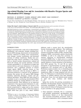
  • 1. Acta Otolaryngol 2004; Suppl 552: 16Á/24 Age-related Hearing Loss and its Association with Reactive Oxygen Species and Mitochondrial DNA damage* MICHAEL D. SEIDMAN1, NADIR AHMAD1, DIPA JOSHI2, JAKE SEIDMAN3, SUJATHA THAWANI4 and WAYNE S. QUIRK5 From the 1Henry Ford Health System, West Bloomfield, Michigan, 2Yale University, New Haven, Connecticut, 3Birmingham Groves, 4University of Michigan, Ann Arbor, Michigan and 5Central Washington University, Ellensburg, Washington, USA Seidman MD, Ahmad N, Joshi D, Seidman J, Thawani S, Quirk WS. Age-related hearing loss and its association with reactive oxygen species and mitochondrial DNA damage. Acta Otolaryngol 2004; Suppl 552: 16 Á/24. Age-related hearing loss, known as presbyacusis, is characterized by the progressive deterioration of auditory sensitivity associated with the aging process and is the leading cause of adult auditory deficiency in the USA. Presbyacusis is described as a progressive, bilateral, high-frequency hearing loss that is manifested on audiometric assessment by a moderately sloping pure tone audiogram. Approximately 23% of the population between 65 and 75 years of age, and 40% of the population older than 75 years of age are affected by this condition. It was estimated in 1980 that 11% of the population was 76 years or older and this number is expected to almost double by the year 2030. When one considers that the population over 65 years of age is experiencing the most accelerated development of hearing loss, the potential socioeconomic ramifications are staggering. Curiously, the frequency of presbyacusis varies across different societies. This discrepancy has been attributed to many factors including genetics, diet, socioeconomic factors, and environmental variables. The purpose of this article is to review the various molecular mechanisms underlying presbyacusis and to offer insights into potential methods of mitigating the effects of aging on hearing impairment Key words: aging, mitochondrial DNA mutations, presbyacusis, reactive oxygen species. INTRODUCTION deletions reach a certain level, the mitochondria Aging is associated with a wide array of physiological, become bioenergetically inefficient. This inefficiency biochemical, and molecular changes including pro- can be measured through reductions in specific gressive DNA damage, deterioration of mitochondrial enzymes (cytochrome oxidase (COX), succinate dehy- function, reduction of cellular water concentrations, drogenase (SDH), nicotinamide adenine dinucleotide ionic changes, vascular insufficiency, and decreased (NADH)), as well as by using flow cytometry to elasticity of cellular membranes. Studies have pro- measure reductions in mitochondrial membrane posed that many factors contribute to this process, potentials (MMP) (Fig. 1), which serves as a marker such as: altered vascular characteristics; reduced red of mitochondrial function, and by the measurement of blood cell velocity and vascular plasticity; increased specific deletions or point mutations in the mitochon- vascular permeability (1, 2); reductions in oxygen and drial DNA genome. Fig. 1A and 1B are typical nutrient delivery and waste elimination (3 Á/6); genetic histograms of cell fluorescence in young and old rat mutations; and a significant presence of reactive lymphocytes (106) incubated with 0.5 nM DiOC6. oxygen species (ROS), otherwise known as free Preliminary results revealed a two- to threefold greater radicals. Concurrent with the increase in ROS is a intensity of DiOC6 staining of peripheral blood decreased production or function of the endogenous lymphocytes (PBL) of young compared with old rats enzymes that protect the cell against ROS damage (Fig. 1C). Reductions in mitochondrial function and including superoxide dismutase, catalase and glu- increases in mtDNA deletions have been identified tathione peroxidase. These ROS have been shown to within heart, brain, liver and skeletal muscle samples contribute in part to producing mitochondrial DNA in aged rats, mice, monkeys and humans (mtDNA) damage by causing deletions in the mito- (7 Á/11). chondrial genome. These deletions are an important Aging thus involves the growing accumulation of component of the age-associated diminution in audi- metabolic and physiologic changes associated with an tory sensitivity. Specific deletions are known to be increasing susceptibility to disease. There are four directly proportional to aging such as the common main sources for failure of biological systems that may aging deletion in humans, which is 4977 base pairs in in part contribute to the aging processes. length (4834 base pairs in rats). When mtDNA 1. Gene repression: essentially, this is a systematic, and probably programmed, age-dependent gene * Presented in part to: the Institute of Experimental Medicine Academy of Sciences, Czech Republic, 30 April 2003; and the deterioration. Semsei et al. have demonstrated Conference on Sensorineural Hearing Loss: Incites of Clinical that increased intracellular potassium results in Advances, Prague, 1 May 2003. chromatin condensation with a consequent decline # Taylor & Francis 2004. ISSN 0365-5237 DOI 10.1080/03655230410017823
  • 2. Age-related hearing loss 17 i.e. intima thickening, lipofuscin accumulation, and damaged collagen. 4. Age-related depletional defects: over time there is progressive breakdown of cellular function. When this accumulates with age, entire organ systems lose their normal function, often leading to disease states as well as normal physiologic aging (14). There are many hypotheses in the current literature providing explanations for senescence. Three of the most convincing theories are the telomerase theory of aging, the dysdifferentiation hypothesis of aging and the membrane hypothesis of aging (MHA; also referred to as the ‘mitochondrial clock theory’ of aging). The telomerase theory of aging suggests that there is a reduction in telomere length over time. The end of a Fig. 1. Mitochondrial membrane potential (MMP) was studied by flow cytometry. 3,3?-Dihexyloxacarbocyanine chromosome is made up of a structure called the (DiOC6), which is used for the quantitative measurement telosome. The tip of the telosome is a region of DNA of MMP, is a lipophilic, cationic, fluorescent probe. Periph- repeat sequences and associated proteins collectively eral blood lymphocytes (PBL) of young (6 months) and old known as the telomere. It is hypothesized that DNA (24 months) (106) rats were equilibrated with very low concentrations (0.5 nM) of DiOC6. (A) and (B) Typical transcription and replication are affected by position histograms of cell fluorescence in young and old rat effects mediated by the telomere. Reduction in the lymphocytes (106) incubated with 0.5 nM DiOC6. There is length of the telomere and alterations in its chromatin a two- to threefold greater intensity of DiOC6 staining of assembly may explain the instability that occurs PBL of young compared with old rats (C). The mean during senescence as well as the immortalization fluorescence of cells treated with and without carbonyl cyanide m-chlorophenylhydrazone (CCCP) (20 mM) is process in vitro (15). In tumor-derived cell lines, shown in (C). Cells treated with CCCP consisted of low telomeres are maintained by the ribonucleoprotein fluorescence, about 30% of control. enzyme, telomerase. However, telomerase activity is repressed in practically all normal human somatic in RNA synthesis (12). This general decline in cells. Thus, there is an inherent problem of end- protein synthesis may in part account for the age- replication creating a tendency toward progressive dependent decrease in gene expression, leading to telomere shortening. Cumulatively, this predilection the turning off of specific biological systems. leads to limited replicative capacity, chromatin in- Hence, a threshold exists beyond which home- stability and eventually, cellular senescence. Viral oncogenes or certain somatic mutations can block ostasis begins to fail. cellular aging probably by activating telomerase. 2. Intracellular communication: this is based upon the Telomere shortening is therefore effectively prevented fact that cells require specific spatial arrangement and this appears to be an important mechanism for which provides for the rapid transfer of molecules sustaining the cellular growth of tumors (16). Low- to neighboring cells. Any change in this spatial level telomerase activity has also been demonstrated in arrangement may lead to breakdown in cellular normal human T and B cells, which was also shown to communication with resultant tissue aging, disease decrease with aging (17). Although many aspects of and death. It is known that mutations in connexin- telomerase activity remain undefined, it has been 26, a gap junction protein, are responsible for a hypothesized that the balance between telomere short- major form of non-syndromic recessive deafness ening and telomerase activity may underlie cellular and a more limited form of non-syndromic domi- aging processes. Therefore, although unproven, nant hearing loss (13). It is possible that a similar changes in telomeres may also predispose to the mechanism may be involved in age-related hearing development of presbyacusis. loss. The dysdifferentiation theory suggests that aging is 3. Waste accumulation: over time there are significant a continuum of programmed differentiation leading to increases in waste products. The ability of cells to either a cessation of normal gene activity or a handle and process these products progressively systematic activation of genes whose effects are decreases, hence, an increase in accretional defects, deleterious to cellular function. Support for this
  • 3. 18 M. D. Seidman et al. theory is provided by apoptosis (programmed cell of ROS damages cellular integrity, which may lead to death) studies in Caenorhabditis elegans (C. elegans ) alterations in gene expression, including telomere (earthworm). These experiments elegantly elaborated shortening and activation of Bax genes, resulting in the genetic mechanisms responsible for controlling cell aging, presbyacusis and ultimately death. death. The maintenance of homeostasis for cellular As presbyacusis represents one aspect of aging, it is metabolism and function consumes a large fraction of imperative thus to understand these molecular me- total body energy expenditure. This is engineered by chanisms of aging. In our laboratory, studies have the delicate balance between cellular proliferation and focused on the mitochondrial theory of aging and its death. The Bcl-2 gene, for example, exists in progeni- applicability to presbyacusis. There are, however, tor and long-lived cells and therefore appears to have a several animal models that have been used to study key function in the cells of the developing embryo. The presbyacusis. These models have served to illuminate gene appears to prevent oxidative damage to cellular the various molecular mechanisms underlying age- organelles and lipid membranes. Studies have demon- related hearing loss. One such model is the C57BL/6J strated that Bcl-2 protects cells from the toxicity of strain of mice, which demonstrates an age-dependent, H2O2 or t-butyl hydroperoxide in a dose-dependent high-frequency, sloping loss (27). The adult hearing manner (18, 19). Bcl-2 -deficient mice demonstrate loss (Ahl) gene, characterized as recessive and mapped changes expected of more rapid cell death, including to chromosome 10, has been identified in the C57 fulminant lymphoid apoptosis of systemic organs (20). mouse strain and is the presumed cause of progressive Bcl-2 also inhibits other types of apoptotic cell death, hearing loss in this species. A proposed mechanism for implying a common mechanism of lethality. Studies this progressive hearing loss is that the Ahl gene have demonstrated, for example, that Bcl-2 protected mediates, through mRNA expression, a decrease in cells from H2O2 and menadione-induced oxidative protective enzymes and therefore an increase in levels deaths (18, 21). Another protein that appears to of oxidative stress within the period of time that the operate as an accessory to Bcl-2 , is a 21 kd protein C57 mouse strain undergoes increased exposure to free referred to as Bax . In experiments by Hockenberry radicals and its most significant diminution in hearing and colleagues, the suggested model of the ratio (28). More recently, the Ahl gene has also been between Bcl-2 and Bax appears to determine survival implicated in noise-induced hearing loss (NIHL) (29). or death following an apoptotic stimulus. Specifically, In other studies, cochlear microphonic (CM) sensi- elevated expression of Bcl-2 appears to be preventa- tivity has been shown to vary with age, with a gradual tive, while that of Bax favors the apoptotic process. decrease in sensitivity from 12 to 24 months of age Lastly, the membrane hypothesis of aging states that (30). Rat cochlear action potential (CAP) sensitivity aging is related to decreasing effectiveness of cellular has also been shown to decrease with age, and changes protective and reparative mechanisms. This yields in CAP waveform with age have been shown (31). In a biochemical and metabolic errors, which progressively study of rat auditory brainstem response (ABR), delay accumulate resulting in cell death (22). The membrane in conduction times and waveform amplitude de- hypothesis of aging (MHA) further postulates that creases were noted to occur in older animals (32). cellular senescence is attributable to the cross-linking Our laboratory has studied groups of rats varying in action of free oxygen radicals within the cellular age and has documented evidence of presbyacusis membrane. Additionally, ROS lead to lipid peroxida- using changes in cochlear microcirculation, electro- tion, polysaccharide depolymerization, nucleic acid physiology, molecular biology and histology (33, 34). disruption, and oxidation of sulfhydryl groups leading Furthermore, histologic analysis of presbyacusis sub- to enzyme inactivation (23). Therefore, the MHA jects demonstrate age-related hair cell degeneration suggests that ROS-induced cell membrane structural and loss (35). Human studies reveal a preferential loss damage is the primary mediator in cellular aging (24, of outer hair cells (OHCs) (36, 37). Loss of OHCs is 25). greatest in the first half of the basal turn (36, 38). This Careful analysis of the above mechanisms suggests correlates tonotopically with the high-frequency loss that certain aspects of the three leading theories of seen in human presbyacusis. In the rat, age-related aging may be interrelated. That is, free radical species hair cell loss is also predominantly OHC in nature. In lead to genetic and cellular alterations resulting in a study of elderly rats, inner hair cell (IHC) losses cellular dysfunction, and consequently senescence, and ranged from 3.1% to 9.2%, while OHC losses ranged perhaps presbyacusis. It is even more intriguing to from 7.4% to 46.8%. The IHC loss was greatest in the realize that a trigger for the Bax gene are the ROS upper apex while OHC losses were greatest at the basal (26). Thus, a critical analysis of the prominent turn (39). While there are species-specific variations in hypotheses of aging suggests that aspects of all three cochleotopic patterns of hair cell loss, the rat is an theories are likely to apply. Specifically, the generation appropriate aging model (30, 32, 39) (Fig. 2). Several
  • 4. Age-related hearing loss 19 Fig. 2. Photomicrograph (40 )/) of the cochlea from a representative animal. It shows a surface mount preparation of the basillar membrane of the right organ of Corti with osmium tetroxide staining from a 4-month old (A) and an 18- month old (B) rat. Outer hair cell rows are shown by 1, 2, and 3. ‘I’ denotes the single inner hair cell row. Loss of hair cells is denoted by arrows. No obvious hair cell pathology is noted in (A). Hair cell loss is shown in (B). human studies have also demonstrated accelerated atherosclerosis and delay death from myocardial hearing loss in patients with Alzheimer’s disease and infarction, presumably by inhibiting lipid peroxidation other cognitive and neurodegenerative disorders (45). Carotenoids, such as b-carotene and other plant (40 Á/42). There is a genetic predilection for Alzhei- pigments, may also have preventive effects against mer’s disease and perhaps these findings can be cancer and cardiovascular disease (45 Á/47). extrapolated to auditory dysfunction as well. As the Available data also suggest that many of the formation of reactive oxygen species (ROS) and their pathological correlates of Alzheimer’s disease are subsequent effects on mitochondrial integrity are an precipitated by oxidative stress-induced mechanisms integral mechanism in presbyacusis, it is important to (48 Á/50). Similarly, Parkinson’s disease has also been consider these processes in greater detail. associated with oxidative stress, increased lipid perox- idation, reduced levels of glutathione (GSH), high concentrations of iron, and free radical generation via REACTIVE OXYGEN SPECIES (ROS) auto-catalytic mechanisms within neuromelanin con- Reactive oxygen species (ROS) contain an unpaired taining catecholaminergic neurons (51, 52). Recent number of electrons, making them chemically reactive studies have further demonstrated that aging in the and extremely toxic to cellular and subcellular struc- central nervous system is directly correlated with tures. ROS have been implicated in more than 100 oxidative stress. In the harlequin mouse model, muta- clinical conditions (43). They are produced in vivo tions in the apoptosis-inducing factor (AIF) gene during mitochondrial respiration, as well as via auto- resulted in greater susceptibility to ROS-induced oxidation of chemical and biological molecules. ROS damage (53). A lack or down-regulation of AIF in are also environmental contaminants and can be neuronal cells renders them more sensitive to per- formed from ionizing and ultraviolet radiation. The oxides and to a much larger extent vulnerable to ( most common ROS are superoxide anion (O2 ), progressive increases in ROS-induced damage. This ( ( hydroxyl radical (OH ), hypochloride (OCL ) and observation provides a molecular mechanism by which nitric oxide (NO (). Not only is the pathogenic role of free radical damage can lead to neuronal death. ROS becoming acceptable in the realm of human Many intrinsic enzyme systems protect cells from disease, but their contribution as active participants in oxidative damage. These include superoxide dismutase determining disease activity is also becoming increas- (SOD), glutathione peroxidase, and the glutathione ingly recognized (44). Despite this primacy, ROS are transferases, and catalase. Additionally, a variety of notoriously difficult to quantify in biological systems. small molecules in the human diet are required for However, the use of modern assay techniques has antioxidant defenses. Experimentally and clinically, it made this less of a limitation (43). is well known that a primary source of ROS generation There is extensive support in the literature for the is through oxidative phosphorylation, ischemia/reper- protective effects of antioxidants and ROS scavengers. fusion or prolonged hypoperfusion, such as that seen For example, tocopherols decrease the process of in myocardial infarction, cerebrovascular accidents,
  • 5. 20 M. D. Seidman et al. aging, and possibly sudden sensorineural hearing loss energy state explains their important role in many and presbyacusis. It is clear that in the aging cochlea human diseases (57). MtDNA deletions accumulate there is a significant reduction of blood supply and the preferentially in post-mitotic tissues such as the inner ongoing need for energy generation through oxidative ear, retina, skeletal muscle and brain. This appears to phosphorylation (2). Thus, these two processes, as well account for the increased susceptibility of these tissues as others, allow for the generation of ROS within the to hypoxia, ischemia and aging. Richter has shown cochlea. that extensive oxidative damage occurs to mitochon- drial and nuclear DNA, and has identified the presence of 8-hydroxydeoxyguanosine (OH8dG) in MITOCHONDRIAL DNA DELETIONS (MTDNA mtDNA and nuclear DNA (nDNA). This compound DEL) AND HEARING LOSS is formed by excited oxygen species and is considered a Mitochondria are unique organelles possessing their marker for DNA damage. Mitochondria treated with own DNA as well as their own enzymatic constituents prooxidants have an increased level of OH8dG. This to allow for transcription and translation of genetic increased level may be caused by several factors, information into proteins. Each mtDNA codes for a including extensive oxygen metabolism, inefficient complete set of ribosomal (rRNA) and transfer RNA DNA repair and lack of mitochondrial histones. (tRNA). Additionally, mtDNA codes for 13 of the Studies have shown that increasing amounts of ROS approximately 60 polypeptides necessary for oxidative occur as a function of age, which lead to an increase in phosphorylation. These include seven of the 25 sub- membrane peroxide content and, thus, rapidly exceeds units of respiratory complex I (ND1-4, 4L, 5, and the capacity of homeostatic protection (66). Thus, 6)(NADH-ubiquinol-oxidoreductase), one of the ap- there is extensive support that mtDNA deletions proximately nine subunits (cyt b) of respiratory accumulate with age and disease (67 Á/69). complex III (ubiquinol-cytochrome c oxidoreductase), Deafness is clearly shown to have an association three of the 13 subunits of respiratory complex IV with mtDNA mutations. It has been suggested that (COI, COII, and COIII) (cytochrome c oxidase), and mitochondrial diseases should be considered in cases two of the 12 subunits of respiratory complex V of progressive sensorineural hearing loss, especially (ATP6 and 8) (ATP synthase). The remaining subunits those associated with multisystem involvement (70, of these complexes are encoded by nuclear DNA 71). A 10.4 kb mtDNA deletion has been identified in (54 Á/56). association with maternally transmitted diabetes and It has been proposed that mitochondrial genomic deafness, without ophthalmoplegia or mitochondrial mutations may be a major cause of human diseases. In myopathies, which were the hallmark of mtDNA 1959, the first patient with a mtDNA deletion as a deletion syndromes (72). Other studies have identified cause of their illness was reported by Ernster. Luft is mutations in the tRNA Leu(UUR) gene in a large often credited with recognizing the importance of pedigree with maternally inherited diabetes mellitus mitochondrial medicine in 1962. Mitochondrial muta- type II and deafness (73). A 3243 point mutation tions and subsequent cytoplasmic segregation contri- (A 0/G) has been demonstrated in a patient with bute to neuromuscular disorders such as Kearns Á/ sensorineural deafness without diabetes (74). Several Sayre/chronic external ophthalmoplegia plus syn- human studies have demonstrated an association of drome (57 Á/60), mitochondrial encephalomyopathy, mitochondrial DNA mutations and presbyacusis, lactic acidosis, and stroke-like episodes (MELAS) including a study which demonstrated that older (61), subacute necrotizing encephalomyopathies patients with presbyacusis had a higher frequency of (SNE, Leigh syndrome), progressive neuronal degen- the common aging deletion (4977 bp) compared with eration of childhood (Alpers syndrome) (59), and similar aged patients without presbyacusis (75). It has myoclonic epilepsy associated with ragged-red muscle been demonstrated, using human archival temporal fibers (MERRF) (54, 62). Interestingly, up to 67% of bones, that 14 of 17 aged patients with presbyacusis patients with mtDNA disorders also manifest sensor- had the 4977 bp deletion compared with 8 of 17 ineural hearing loss (63). control patients with normal hearing. This difference Mitochondrial DNA deletions with subsequent was statistically significant (p B/0.05) (76). segregation and enrichment of the deletions through- Through experiments conducted in our laboratory, out life are contributing factors in the aging process various nutrient compounds have been identified that (9, 64, 65). MtDNA deletions in the human heart are enhance mitochondrial function by their ability to age-dependent (9), and these mitochondrial mutations protect and repair age-induced damage to cochlear accumulate progressively until death. Cells that accu- mtDNA (Fig. 3). One such compound is acetyl L- mulate large numbers of mitochondrial mutations carnitine. In one study involving Fischer rats, it was become bioenergetically deficient. This compromised noted that auditory thresholds actually improved over
  • 6. Age-related hearing loss 21 using PCR. Harlan-Fischer rats, aged 18Á/20 months (n 0/14), were divided into two groups. The experi- mental group was supplemented orally for 6 months with lecithin, a purified extract of soybean phospho- lipid (Nutritional Therapeutics, Allendale, NJ, USA). The data obtained were compared with the control group. ABR were recorded at 2-month intervals and showed significant preservation of hearing sensitivities Fig. 3. (A) Data for the identification of ND1-16S rRNA in the treated subjects. Flow cytometry revealed from stria vascularis. This represents a 601 bp product and mitochondrial membrane potentials to be significantly verifies the integrity of the PCR and the presence of higher in the treated subjects, suggesting preserved mitochondrial DNA. Lane 1: negative control; lane 2: mitochondrial function. Finally, the common aging placebo; lane 3: diet restriction; lane 4: vitamin C; lane 5: mitochondrial DNA deletion (mtDNA4834) were am- vitamin E; lane 6: melatonin; lane 7: lazaroid; lane 8: molecular weight standards. (B) Data identifying the 4834 plified from brain and cochlear tissue, including stria bp deletion (common aging deletion) from stria vascularis. vascularis and auditory nerve. This specific deletion This represents a 597 bp product. Lane 1: negative control; was quantitatively found to be fewer in all tissues in lane 2: placebo; lane 3: diet restriction; lane 4: vitamin C; the treated group compared with the controls. Based lane 5: vitamin E; lane 6: melatonin; lane 7: lazaroid; lane 8: on these findings, these experiments support our molecular weight standards. The DNA concentration was normalized to 150 ng for all samples. hypothesis and provide evidence that lecithin may preserve cochlear mitochondrial function and protect from hearing loss associated with aging. the course of 6 weeks, suggesting a strong positive role Caloric restriction has also been shown to retard the for these compounds (77) (Fig. 4). In another study, aging process. In a study conducted by Lee et al. (78), our laboratory investigated the effects of lecithin on a control and calorie-restricted group of mice were compared over a 30-month period, and the results aging and age-associated hearing loss in rats by revealed that age-related changes in gene expression measuring hearing sensitivities using auditory brain- profiles were remarkably attenuated by caloric restric- stem responses (ABR). In addition, mitochondrial tion. During aging there is a lower expression of function, as a measure of aging, was assessed by metabolic and biosynthetic genes, and caloric restric- determining mitochondrial membrane potentials. This tion was noted to decrease the normal age-induced was achieved using flow cytometry and by amplifying genetic alterations by causing a metabolic shift toward mitochondrial DNA deletions associated with aging increased protein turnover and decreased macromole- Fig. 4. Auditory threshold measurements in (A) placebo and (B) diet-restricted groups. Over the lifespan of 24 Á/27 months, the thresholds shifted the least in diet-restricted groups. The difference between diet-restricted groups and placebo was statistically significant (p B/0.01).
  • 7. 22 M. D. Seidman et al. cular damage, hence creating a more bioenergetically aging and presbyacusis, with the ultimate goal of efficient cell. Another study by Lee et al. (79) enhancing our understanding of the mechanisms that demonstrated that caloric-restricted mice produced surround senescence, both as a phenomenon and as an fewer proteins associated with the oxidative stress intrinsic biological property. response and heat shock factors. DISCUSSION REFERENCES Presbyacusis, or the progressive deterioration of hear- ing associated with aging, is the most common cause 1. Prazma J, Carrasco VN, Butler B, Waters G, Anderson of adult auditory deficiency in the USA. This condi- T, Pillsbury HC. Cochlear microcirculation in young and tion creates a significant burden not only for the old gerbils. Arch Otolaryngol Head Neck Surg 1990; sufferers, but also for those who communicate with 116: 932 Á/6. 2. Seidman MD, Khan MJ, Dolan DF, Quirk WS. Age- them. The medical and socioeconomic costs are related differences in cochlear microcirculation and staggering, and with the expansion of the world auditory brain stem response. Arch Otolaryngol Head population and the numbers of elderly individuals Neck Surg 1996; 122: 1221 Á/6. expecting to more than double by the year 2030, this 3. Gacek MR, Schuknecht HF. Pathology of presbyacusis. problem is escalating. However, significant progress Int Audiol 1969; 8: 199 Á/209. has been made to identify the molecular mechanisms 4. Harkins SW. Effects of age and interstimulus interval on brainstem auditory evoked potential. Int J Neurosci of age-related hearing loss, and this in turn has led to 1981; 15: 107 Á/18. research on methods of mitigating the adverse effects 5. Rosenhall U, Pederson K, Dotevall M. Effects of on the auditory system. Among the various mechan- presbyacusis and other types of hearing loss on auditory isms that are postulated to result in age-related brain stem responses. Scand Audiol 1986; 15: 179 Á/85. hearing loss in the current literature, the one that is 6. Hoeffding V, Feldman ML. Changes with age in the morphology of the cochlear nerve in rats: light micro- arguably the most intriguing, is the membrane hypoth- scopy. J Comp Neurol 1988; 276: 537 Á/46. esis of aging (MHA). Also known as the mitochon- 7. Cortopassi GA, Arnheim N. Detection of a specific drial clock theory of aging, the premise of this theory mitochondrial DNA deletion in tissues of older humans. is that with aging, hypoperfusion of the cochlear tissue Nucleic Acids Res 1990; 18: 6927 Á/33. occurs, which leads to ischemia and the formation of 8. Corral-Debrinski M, Stepien G, Shoffner JM, et al. ROS. These species are highly toxic substances that Hypoxemia is associated with mitochondrial DNA damage and gene induction: implications for cardiac adversely affect the auditory neuroepithelia. More disease. JAMA 1991; 266: 1812 Á/6. specifically, these ROS damage mitochondrial DNA 9. Hattori K, Tanaka M, Sugiyama S, et al. Age-dependent (mtDNA), resulting in both the production of specific increase in deleted mitochondrial DNA in the human mtDNA deletions, as well as reducing the mitochon- heart: possible contributory factor to presbyacusis. Am drial membrane potential (MMP). The resultant effect Heart J 1991; 121(6, Pt 1): 1735 Á/42. 10. Seidman MD, Quirk WS, Nuttall AL, Schweitzer VG. is to render the mitochondria bioenergetically ineffi- The protective effects of allopurinol and superoxide cient. These specific mtDNA deletions are also known dismutase-polyethylene glycol on ischemic and reperfu- as the common aging deletion. Studies are increasingly sion-induced cochlear damage. Otolaryngol Head Neck supportive of this relationship between presbyacusis Surg 1991; 105: 457 Á/63. and the common aging deletion. 11. Wallace DC. Diseases of the mitochondrial DNA. Annu The results of these studies have further led to Rev Biochem 1992; 61: 1175 Á/212. 12. Semsei I, Szeszek F, Nagy I. In vivo studies of age investigations concerning the role of antioxidants, dependent decrease of the rates of total and mRNA nutritional supplementation, and dietary restriction synthesis in the brain cortex of rats. Arch Gerontol on hearing loss and the aging process in general Geriatr 1982; 1: 29 Á/42. There is compelling evidence to suggest that the 13. Kudo T, Ikeda K, Oshima T, et al. GJB2 (connexin 26) beneficial effect of these compounds is to slow down mutations and childhood deafness in Thailand. Otol Neurotol 2001; 22: 858 Á/61. the ‘mitochondrial clock’, by decreasing the amount 14. Strehler BL. Genetic instability as the primary cause of of mtDNA deletions and causing less of a reduction in human aging. Exp Gerontol 1986; 21: 283 Á/319. mitochondrial membrane potential. Currently, our 15. Pommier JP, Lebeau J, Ducray C, et al. Chromosomal laboratory is investigating the role of resveratrol, a instability and alteration of telomere repeat sequences. constituent of the red wine grape, in attenuating the Biochimie 1995; 77: 817 Á/25. 16. Shay JW, Wright WE. Telomerase activity in human age-induced reductions in auditory sensitivity; specifi- cancer. Curr Opin Oncol 1996; 8: 66 Á/71. cally, its antioxidant effect on the mitochondrial 17. Hiyama K, Hirai Y, Kyoizumi S, et al. Activation of apparatus. This study will hopefully provide strong telomerase in human lymphocytes and hematopoietic empirical evidence of an integrated hypothesis of progenitor cells. J Immunol 1995; 155: 3711 Á/5.
  • 8. Age-related hearing loss 23 18. Hockenberry DM, Otlvai ZN, Yin XM, Milliman CL, 36. Johnsson LG, Hawkins JE Jr. Sensory and neural Korsmeyer SJ. Bcl-2 functions in an antioxidant path- degeneration with aging, as seen in microdissections of way to prevent apoptosis. Cell 1993; 75: 241 Á/52. the human inner ear. Ann Otol Rhinol Laryngol 1972; 19. Kane DJ, Sarafian TA, Anton R, et al. Bcl-2 inhibition 81: 179 Á/93. of neural cell death: decreased generation of reactive 37. Nadol JB Jr. Application of electron microscopy to oxygen species. Science 1993; 262: 1274 Á/7. human otopathology Ultrastructural findings in neural 20. Korsmeyer SJ, Yin XM, Oltvai ZN, Veis-Novack DJ, presbyacusis, Meniere’s disease and Usher’s syndrome. Linette GP. Reactive oxygen species and regulation of Acta Otolaryngol 1988; 105: 411 Á/9. cell death by the Bcl-2 gene family. Biochim Biophys 38. Willott JF, Milbrandt JC, Bross LS, Caspary DM. Acta 1995; 1271: 63 Á/6. Glycine immunoreactivity and receptor binding in the 21. Oltvai ZN, Milliman CL, Korsmeyer SJ. Bcl-2 hetero- cochlear nucleus of C57BL/6J and CBA/CaJ mice: dimerizes in vivo with a conserved homolog, Bax, that effects of cochlear impairment and aging. J Comp accelerates programmed cell death. Cell 1993; 74: 609 Á/ Neurol 1997; 385: 405 Á/14. 19. 39. Keithley EM, Feldman ML. Hair cell counts in an age- 22. Sohal RS, Allen RG. Relationship between metabolic graded series of rat cochleas. Hear Res 1982; 8: 249 Á/62. rate, free radicals, differentiation and aging: a unified 40. Kurylo DD, Corkin S, Allard T, Zatorre RJ, Growdon theory. Basic Life Sci 1985; 35: 75 Á/104. JH. Auditory function in Alzheimer’s disease. Neurology 23. Southorn PA, Powis G. Free radicals in medicine, I: 1993; 43: 1893 Á/9. chemical nature and biologic reactions. Mayo Clin Proc 41. Sinha UK, Hollen KM, Rodrigues R, Miller CA. 1988; 63: 381 Á/9. Auditory system degeneration in Alzheimer’s disease. 24. Zy-Nagy I, Semsei I. Centrophenoxine increases the Neurology 1993; 43: 779 Á/85. rates of total and mRNA synthesis in the brain cortex of 42. Gates GA, Karzon RK, Garcia P, et al. Auditory old rats: an explanation of its action in terms of the dysfunction in aging and senile dementia of the Alzhei- membrane hypothesis of aging. Exp Gerontol 1984; 19: mer’s type. Arch Neurol 1995; 52: 626 Á/34. 171 Á/8. 43. Halliwell B, Gutteridge JM, Cross CE. Free radicals, 25. Zs-Nagy I, Cutler RG, Semsei I. Dysdifferentiation antioxidants, and human disease: where are we now? J hypothesis of aging and cancer: a comparison with the Lab Clin Med 1992; 119: 589 Á/620. membrane hypothesis of aging. Ann N Y Acad Sci 1988; 44. Halliwell B. Free radicals and antioxidants: a personal 521: 215 Á/25. view. Nutr Rev 1994; 52(8 Pt 1): 253 Á/65. 26. Moon H, Baek D, Lee B, et al. Soybean ascorbate 45. Byers T, Bowman B. Vitamin E supplements and coronary heart disease. Nutr Rev 1993; 51: 33 Á/6. peroxidase suppresses Bax-induced apoptosis in yeast by 46. Comstock GW, Helziouser KJ, Bush TL. Prediagnostic inhibiting oxygen radical generation. Biochem Biophys serum levels of carotenoids and vitamin E as related to Res Commun 2002; 290: 457 Á/62. subsequent cancer in Washington County, Maryland. 27. Johnson KR, Erway LC, Cook SA, Willott JF, Zheng Am J Clin Nutr 1991; 53(1 Suppl): 260S Á/4. QY. A major gene affecting age-related hearing loss in 47. Ziegler RG. Vegetables, fruits and carotenoids and the C57BL/6J mice. Hear Res 1997; 114: 83 Á/92. risk of cancer. Am J Clin Nutr 1991; 53(1 Suppl): 28. Staeker H, Zheng QY, Van de Water TR. Oxidative 251S Á/9. stress in aging in the C57B16/J mouse cochlea. Acta 48. Smith MA, Perry G. Free radical damage, iron and Otolaryngol 2001; 121: 666 Á/72. Alzheimer’s disease. J Neurol Sci 1995; 134(Suppl): 29. Davis RR, Newlander JK, Ling X, Cortopassi GA, 92 Á/4. Krieg EF, Erway LC. Genetic basis for susceptibility to 49. Lyras L, Cairns NJ, Jenner A, Jenner P, Halliwell B. An noise-induced hearing loss in mice. Hear Res 2001; 155: assessment of oxidative damage to proteins, lipids and 82 Á/90. DNA in brain from patients with Alzheimer’s disease. J 30. Crowley DE, Swain RE, Schramm VL, Swanson SN. Neurochem 1997; 68: 2091 Á/9. Analysis of age-related changes in electric responses 50. Sheehan JP, Swerdlow RH, Miller SW, et al. Calcium from the inner ear of rats. Ann Oto Rhinol Laryngol homeostasis and reactive oxygen species production in 1972; 81: 739 Á/46. cells transformed by mitochondria from individuals with 31. Crowley DE, Schramm VL, Swain RE, Swanson SN. sporadic Alzheimer’s disease. J Neurosci 1997; 17: Age-related waveform changes of 8th nerve action 4612 Á/22. potentials in rats. Laryngoscope 1973; 83: 264 Á/75. 51. Choi BH. Oxygen, antioxidants and brain dysfunction. 32. Simpson GV, Knight RT, Brailowsky S, Prospero-Garcia Yonsei Med J 1993; 34: 1 Á/10. O, Scabini D. Altered peripheral and brainstem auditory 52. Jenner P, Olanow CW. Oxidative stress and the patho- function in aged rats. Brain Res 1985; 348: 28 Á/35. genesis of Parkinson’s disease. Neurology 1996; 47 33. Seidman MD, Shivapuja BG, Quirk WS. The protective (6 Suppl 3): S161 Á/70. effects of allopurinol and superoxide dismutase on 53. Klein JA, Longo-Guess CM, Rossmann MP, et al. The noise-induced cochlear damage. Otolaryngol Head harlequin mouse mutation downregulates apoptosis- Neck Surg 1993; 109: 1052 Á/6. inducing factor. Nature 2002; 419: 367 Á/74. 34. Seidman MD, Bai U, Khan MJ, Quirk WS. Mitochon- 54. Wallace DC, Zheng X, Lott MT, et al. Familial drial DNA deletions associated with aging and presby- mitochondrial encephalomyopathy (MERRF): genetic, acusis. Arch Otolaryngol Head Neck Surg 1997; 123: pathophysiological, and biochemical characterization of 1039 Á/45. a mitochondrial DNA disease. Cell 1988; 55: 601 Á/10. 35. Soucek S, Michaels L, Frohlich A. Pathological changes 55. Wallace DC, Lott MT, Lezza AM, et al. Mitochondrial in the organ of Corti in presbyacusis as revealed by DNA mutations associated with neuromuscular dis- microslicing and staining. Acta Otolaryngol Suppl 1987; eases: analysis and diagnosis using polymerase chain 436: 93 Á/102. reaction. Pediatric Res 1990; 28: 525 Á/8.
  • 9. 24 M. D. Seidman et al. 56. Yen TC, Chen YS, King KL, et al. Liver mitochondrial 71. Miyabayashi S, Hanamizu H, Endo H, et al. A new type respiratory functions decline with age. Biochem Biophys of mitochondrial DNA deletion in patients with en- Res Commun 1989; 165: 944 Á/1003. cephalomyopathy. J Inherit Metab Disease 1991; 14: 57. Lindane AW, Marzuki S, Ozawa T, et al. Mitochondrial 805 Á/12. DNA mutations as an important contributor to aging 72. Ballinger SW, Shoffner JM, Hedaya EV, et al. Mater- and degenerative diseases. Lancet 1985; i: 642 Á/5. nally transmitted diabetes and deafness associated with a 58. Shoffner JM, Lott MT, Voljavec AS, et al. Spontaneous 10.4 Kb mitochondrial DNA deletion. Nat Genet 1992; Kearn Á/Sayre/Chronic External Ophthalmoplegia Plus 1: 11 Á/5. Syndrome associated with a mitochondrial DNA dele- 73. Van den Ouweland JM, Lemkes HH, Ruitenbeek W, tion: a slip replication model and metabolic therapy. Sankuijl LA. Mutation in mitochondrial tRNA(Leu) Proc Natl Acad Sci U S A 1989; 86(20): 7952 Á/6. (UUR) gene in large pedigree with maternally trans- 59. DiMauro S, Bonilla E, Lombes A, et al. Mitochondrial mitted type II diabetes mellitus and deafness. Nat Genet encephalomyopathies. Neurol Clin 1990; 8: 483 Á/506. 1992; 1: 368 Á/71. 60. Grossman LI. Mitochondrial DNA in sickness and 74. Sawada S, Takeda T, Kakigi A, et al. Audiological health. Am J Hum Genet 1990; 46: 415 Á/7. findings of sensorineural deafness associated with a 61. Pavlakis SG, Phillips PC, DiMauro S, et al. Mitochon- mutation in the mitochondrial DNA. Am J Otol 1997; drial myopathy, encephalopathy, lactic acidosis, and 18: 332 Á/5. stroke-like episodes: a distinctive clinical syndrome. 75. Ueda N, Oshima T, Ikeda K, et al. Mitochondrial DNA Ann Neurol 1984; 16: 481 Á/8. deletion is a predisposing cause for sensorineural hear- 62. Seibel P, Degoul F, Romero N, et al. Identification of ing loss. Laryngoscope 1998; 108(4 Pt 1): 580 Á/4. point mutations by mispairing PCR as exemplified by 76. Bai U, Seidman MD, Hinojosa R, Quirk WS. Mito- MERRF disease. Biochem Biophys Res Commun 1990; chondrial DNA deletions associated with aging and 173: 561 Á/5. possibly presbyacusis: a human archival temporal bone 63. Gold M, Rapin I. Non-Mendelian mitochondrial in- study. Am J Otol 1997; 18: 449 Á/53. heritance as a cause of progressive geneticsensori-neural 77. Seidman MD, Khan MJ, Bai U, Shirwany N, Quirk WS. hearing loss. Int J Pediatr Otorhinolaryngol 1994; 30: Biologic activity of mitochondrial metabolites on aging 91 Á/104. and age-related hearing loss. Am J Otol 2000; 21: 161 Á/7. 64. Ames BN. Dietary carcinogens and anticarcinogens. 78. Lee CK, Klopp RG, Weindruch R, Prolla TA. Gene Oxygen radicals and degenerative diseases. Science expression profile of aging and its retardation by caloric 1983; 221: 1256 Á/64. restriction. Science 1999; 285: 1390 Á/3. 65. Wallace DC. Mitochondrial DNA in aging and disease. 79. Lee CK, Weindruch R, Prolla TA. Gene-expression Sci Am 1997; 277: 40 Á/7. profile of the ageing brain in mice. Nat Genet 2000; 25: 66. Nohl H, Hegner D. Do mitochondria produce oxygen 294 Á/7. radicals in vivo? Eur J Biochem 1978; 82: 563 Á/7. 67. Piko L, Hougham AJ, Bulpitt AJ. Studies of sequence heterogeneity of mitochondrial DNA from rat and mouse tissues: evidence for an increased frequency of Address for correspondence: deletions/additions with aging. Mech Ageing Dev 1988; Michael D. Seidman, MD, FACS 43: 279 Á/93. Department of Otolaryngology Á/Head & Neck Surgery 68. Muller Á/Hocker J. Cytochromec-oxidase deficient cardi- Director Á/Division tologic/Neurotologic Surgery omyocytes in the human heart Á/ an age-related phe- Henry Ford Health System nomenon. A histochemical, ultracytochemical study. Director Á/Complementary/Integrative Medicine Am J Pathol 1989; 134: 1167 Á/73. 6777 W. Maple Road 69. Bandy B, Davison AJ. Mitochondrial mutations may W. Bloomfield increase oxidative stress: implications for carcinogenesis MI 48323 and aging? Free Radic Biol Med 1990; 8: 523 Á/39. USA 70. Gold M, Rapin I, Shankse S. Mitochondrial inheritance Tel: '/1 248 661 7211 of acquired deafness. Ann N Y Acad Sci 1991; 630: Fax: '/1 248 661 6456 301 Á/2. E-mail: mseidma1@hfhs.org