SlideShare ist ein Scribd-Unternehmen logo
1 von 5
Downloaden Sie, um offline zu lesen
ORIGINAL ARTICLE


         Proximal Interphalangeal Joint Fractures of the Hand
                  Treatment With an External Dynamic Traction Device
                    Kanthan Theivendran, BSc, MBBS, MRCS, Jonathan Pollock, BMBS, MRCS,
                            and Vaikunthan Rajaratnam, FRCS, Dip Hand Surg(Eur)


                                                                                      Intra-articular PIP joint fractures can be treated with
Abstract: The authors present a series of 11 consecutive patients
who sustained an intra-articular fracture of the proximal interpha-
                                                                               various techniques. This is dependent on the articular surface
langeal (PIP) joint and 1 patient with a fracture of the interphalan-
                                                                               involvement of the base of the middle phalanx and the degree
geal joint of the thumb treated with a Kirschner wire external
                                                                               of comminution. It is generally accepted that if there is less
dynamic traction device. Range of motion and grip strength were
                                                                               than 40% involvement of the joint surface, then the fracture
measured. We used the Michigan Hand Outcome Questionnaire
                                                                               is stable and can be treated nonoperatively.3 However, greater
(MHQ) to obtain a subjective response to the treatment. Average
                                                                               than 40% of joint surface is unstable and requires surgical
range of motion of the PIP joint was 64° and distal interphalangeal
                                                                               intervention.3
joint was 52°. The average grip strength was 86% with a mean MHQ
                                                                                      Treatment modalities include extension block splint-
score of 90. Two patients had pin site infections treated successfully         ing,4 open reduction and internal fixation,5,6 closed reduction
with antibiotics. All fractures had united at final follow up with              and percutaneous fixation, and volar plate arthroplasty. There
  1-mm articular step present. Our series highlights that these are            are 2 main problems with comminuted fractures at the PIP
difficult injuries to treat and that anatomic reduction is not always           joint: 1) the articular bony fragments are too small to openly
possible. However, this technique is simple and quick to perform               reduce and internally fix to gain anatomic reduction; and 2) if
while allowing early joint mobilization. The subjective response to            the joint is immobilized, the fibrotic changes that occur after
treatment is comparable with other studies using a similar device.             injury will cause stiffness.
                                                                                      These problems can be overcome by early mobilization
Key Words: dynamic, external fixation, fracture, proximal                       after anatomic reduction of the fracture using the principle of
interphalangeal joint                                                          ligamentotaxis.7,8 There have been various techniques de-
(Ann Plast Surg 2007;58: 625– 629)                                             scribed in the literature using these principles.4,7,9 –11 We
                                                                               report a series of 11 patients with intra-articular PIP joint
                                                                               fractures and 1 patient with an intra-articular interphalangeal
                                                                               (IP) joint fracture of the thumb treated with a Kirschner (K)
                                                                               wire dynamic external fixation device.10
P   roximal interphalangeal joint (PIP) joint fractures are
    relatively rare but can be severely disabling for the patient
and lead to stiffness and persistent pain if inadequately
treated.                                                                                                METHODS
       The stability of the PIP joint is provided by the articular                    We reviewed 11 consecutive patients who sustained an
congruency and soft tissue supports. Soft tissue stability is                  intra-articular fracture of the PIP joint and 1 patient of the IP
provided by collateral ligaments, volar plate, joint capsule,                  joint of the thumb. All were treated with a dynamic external
dorsal expansion, and extensor tendon as well as the flexor                     fixator frame by the senior author (V.R.) between 2004 and
tendons.1 These fractures commonly occur when an axial                         2005. We used the classification system by Seno et al (Fig. 1)
load is applied to the finger tip causing hyperextension                        to describe the fracture configuration at the base of the middle
leading to a dorsal fracture and/or dislocation.2                              phalanx.12 Objective measurements of the active range of
                                                                               motion (AROM) of the distal interphalangeal (DIP) and PIP
                                                                               joints of the affected finger or thumb were measured using a
                                                                               standard goniometer. The grip strength was also measured
Received June 27, 2006, and accepted for publication, after revision, August   using the Jamar dynamometer and was compared with the
   30, 2006.
From The Birmingham Hand Centre, University Hospital Birmingham Selly          uninjured side. We used the Michigan Hand Outcome Ques-
   Oak Hospital, Birmingham, U.K.                                              tionnaire13 (MHQ) to obtain a subjective response to the
Reprints: Kanthan Theivendran, BSc, MBBS, MRCS, 81 Pennine Way,                treatment. Duration of the procedure was obtained from
   Ashby-de-la-Zouch, Leicestershire LE65 1EZ, United Kingdom. E-mail:         operating room records. Bony union was confirmed by re-
   kanthan@hotmail.co.uk.
Copyright © 2007 by Lippincott Williams & Wilkins
                                                                               view of the radiographs at the latest follow up.
ISSN: 0148-7043/07/5806-0625                                                          Statistical analysis was performed using the Pearson
DOI: 10.1097/01.sap.0000245132.14908.9d                                        correlation test to identify if there was any significant rela-

Annals of Plastic Surgery • Volume 58, Number 6, June 2007                                                                                 625
Theivendran et al                                                 Annals of Plastic Surgery • Volume 58, Number 6, June 2007


FIGURE 1. Classification of base of
middle phalanx fractures by Seno
et al.12 Reproduced with permis-
sion and copyright of the British
Editorial Society of Bone and Joint
Surgery.


tionship between the time of delay to operation, the age of the   and distraction across the fracture site. Early active move-
patient, or the outcome of treatment. The SPSS 14.0 (SPSS         ment was encouraged within 24 hours and all patients re-
Inc., Chicago, IL) statistical software was used to perform the   ceived hand therapist input.
analysis with P values of less than 0.05 considered to be
significant.
                                                                                            RESULTS
Surgical Technique                                                       The mean follow up was 24 weeks (range, 12–56 weeks)
       All of the patients had local anesthetic for the proce-    and the average age of the patient was 35 years (range, 18 – 60
dure. A radiographic image intensifier (II) was used to visu-      years). There were 10 men and 2 women who sustained intra-
alize fracture manipulation and reduction. A 1.1-mm K-wire        articular digital fractures. Table 1 outlines the demographic
was inserted percutaneously and perpendicular to the long         details. The device was left in situ for an average of 4 weeks and
axis of the phalanx. This wire was directed to the center of      the mean time from injury to operation was 8 days. The mean
rotation of the head of the proximal phalanx while avoiding       AROM of the injured PIP joint was 64° and DIP joint (including
breaching the joint capsule. A second longer K-wire was           the IP joint of the thumb) was 52°. The mean grip strength
inserted distally and at right angles to the middle phalanx       of the injured side compared with the uninjured hand was
through the center of rotation of the head of the middle          86%. The average duration of the procedure was 12 minutes
phalanx while avoiding breaching the joint capsule. Both          (range, 10 –16 minutes). The mean MHQ score was 90. Eight
wires were used to manipulate and reduce the fracture under       patients were employed before their injury and all have
II guidance. The proximal wire was bent into a hook approx-       returned to their original occupations.
imately 5 mm from the skin. The distal K-wire was bent                   The time delay to operation correlated negatively with
parallel to the long axis of the phalanx pointing toward the      the grip strength (r      0.556, P 0.061); however, this was
proximal K-wire and shaped into a parabolic curve. This           not significant. There was no significant correlation between
preloaded curve was then made into a hook at its distal end       time delay to operation and the AROM of the PIP joint (r
and interlocked with the hook from the proximal wire while        0.176, P      0.583) or the delay to operation and the MHQ
applying traction to the digit (Fig. 2). This provided tension    score (r 0.084, P 0.795). No significant correlation was


FIGURE 2. Operative technique: A, Proximal K-wire through the center of the head of proximal phalanx. B, Another distal K-
wire parallel to proximal wire through center of the head of middle phalanx. C, The distal wire is bent backward and the
proximal wire is bent forward. D, A preloaded dorsal curve is created from the distal wire. E, Wire ends are shaped into hooks
that engage and maintain in-line traction. F, Fracture is reduced and maintained while allowing movement at the hooks of
the K-wire.

626                                                                                          © 2007 Lippincott Williams & Wilkins
Annals of Plastic Surgery • Volume 58, Number 6, June 2007                                                                                                                                                                                                    External Fixation of PIP Joint Fractures


                                                                                                                                                                                                                                   found between age of the patient and the outcome (r


                                                                                                                                                                                                              Pin site infection

                                                                                                                                                                                                              Pin site infection
                                                                                                                                                                                Questionnaire Complications
                                                                                                                                                                                                                                   0.04, P 0.903 for the grip strength; r 0.443, P 0.149


                                                                                                                                                                                                              Disassembly
                                                                                                                                                                                                                                   for the AROM of the PIP joint; and r 0.292, P 0.357
                                                                                                                                                                                                                                   for the MHQ).
                                                                                                                                                                                                                                          Two patients developed minor pin track infection suc-
                                                                                                                                                                                                              Nil

                                                                                                                                                                                                              Nil

                                                                                                                                                                                                              Nil
                                                                                                                                                                                                              Nil
                                                                                                                                                                                                              Nil
                                                                                                                                                                                                              Nil
                                                                                                                                                                                                              Nil

                                                                                                                                                                                                              Nil
                                                                                                                                                                                                              Nil
                                                                                                                                                                                                                                   cessfully treated with oral antibiotics. One patient knocked
                                                                                                                                                                                                                                   the device on the gearshift of a car. It became loose and
                                                                                                                                                                                 Michigan

                                                                                                                                                                                  Outcome
                                                                                                                                                                                   Hand


                                                                                                                                                                                                                                   needed reapplication at 4 weeks. In this case, the disengage-
                                                                                                                                                                                                              88
                                                                                                                                                                                                              93
                                                                                                                                                                                                              93
                                                                                                                                                                                                              96
                                                                                                                                                                                                              98
                                                                                                                                                                                                              82
                                                                                                                                                                                                              95
                                                                                                                                                                                                              85
                                                                                                                                                                                                              83
                                                                                                                                                                                                              89
                                                                                                                                                                                                              80
                                                                                                                                                                                                              94
                                                                                                                                                                                                                                   ment of the construct occurred as a result of a technical error
                                                                                                                                                                                                                                   of inadequate crimping of the ends of the wire. All patients
                                                                                                                                                                                                                                   were pain-free at the latest follow up, and there was no
                                                                                                                                                        Joint Extensor Strength
                                                                                                                                                                         Grip


                                                                                                                                                                                                                                   clinical or radiologic evidence of osteomyelitis. All fractures
                                                                                                                                                                         (%)
                                                                                                                                                                                                              100
                                                                                                                                                                                                              100
                                                                                                                                                                                                               77
                                                                                                                                                                                                               75
                                                                                                                                                                                                               80
                                                                                                                                                                                                               80
                                                                                                                                                                                                               92
                                                                                                                                                                                                               85
                                                                                                                                                                                                               86
                                                                                                                                                                                                               95
                                                                                                                                                                                                               75
                                                                                                                                                                                                               90
                                                                                                                                                                                                                                   were united at final follow up with an average extensor lag at
                                                                                                                                                                                                                                   the PIP joint of 11° with pain-free range of motion. However,
                                                                                                                                                       Interphalangeal

                                                                                                                                                        Lag (degrees)


                                                                                                                                                                                                                                   all our patients had less than a 1-mm intra-articular step present
                                                                                                                                                           Proximal


                                                                                                                                                                                                                                   on radiographs with no evidence of reduction in joint space at
                                                                                                                                                                                                               8
                                                                                                                                                                                                              12
                                                                                                                                                                                                              10
                                                                                                                                                                                                               8
                                                                                                                                                                                                               9
                                                                                                                                                                                                              12
                                                                                                                                                                                                              10
                                                                                                                                                                                                               8
                                                                                                                                                                                                              12
                                                                                                                                                                                                              15
                                                                                                                                                                                                              16
                                                                                                                                                                                                              12                   final follow up. Case no. 12 (Fig. 3) shows a Seno type 3 base
                                                                                                                                                                                                                                   of the middle phalanx fracture that shows bony union at 3
                                                                                                                                                                                                                                   months postinjury with an intra-articular step present.
                                                                                                                       Interphalangeal Interphalangeal
                                                                                                                                         Joint Active
                                                                                                                                          Range of

                                                                                                                                          (degrees)
                                                                                                                                           Motion
                                                                                                                                            Distal


                                                                                                                                                                                                                                                           DISCUSSION
                                                                                                                                                                                                              60
                                                                                                                                                                                                              50
                                                                                                                                                                                                              52
                                                                                                                                                                                                              25
                                                                                                                                                                                                              72
                                                                                                                                                                                                              60
                                                                                                                                                                                                              50
                                                                                                                                                                                                              55
                                                                                                                                                                                                              60
                                                                                                                                                                                                              52
                                                                                                                                                                                                              30
                                                                                                                                                                                                              60


                                                                                                                                                                                                                                          The treatment goals for intra-articular PIP joint frac-
                                                                                                                                                                                                                                   tures are to restore anatomic alignment of the joint and to
                                                                                                                                                                                                                                   allow early active movement to avoid stiffness. These chal-
                                                                                                                                                                                                                                   lenging goals have sparked renewed interest in the techniques
                                                                                                                         Joint Active
                                                                                                                          Proximal


                                                                                                                          Range of

                                                                                                                          (degrees)
                                                                                                                           Motion


                                                                                                                                                                                                                                   for treating these difficult fractures. Previous attempts with
                                                                                                                                                                                                              20
                                                                                                                                                                                                              32
                                                                                                                                                                                                              62
                                                                                                                                                                                                              60
                                                                                                                                                                                                              78
                                                                                                                                                                                                              75
                                                                                                                                                                                                              82
                                                                                                                                                                                                              78
                                                                                                                                                                                                              69
                                                                                                                                                                                                              90
                                                                                                                                                                                                              40
                                                                                                                                                                                                              80


                                                                                                                                                                                                                                   open reduction and internal fixation have proved difficult
                                                                                                                                                                                                                                   with small fragments, resulting in high complication rates and
                                                                                                                                                                                                                                   reduced ability to gain joint congruency.9,14 Stern et al
                                                                                                                                                                                                                                   performed a comparative study looking at splinting, skeletal
                                                                              Removal of Fracture Operative Operative

                                                                                                             (minutes)
                                                                                                               Time


                                                                                                                                                                                                                                   traction with early mobilization, and open reduction and
                                                                                                                                                                                                              15
                                                                                                                                                                                                              10
                                                                                                                                                                                                              16
                                                                                                                                                                                                              12
                                                                                                                                                                                                              10
                                                                                                                                                                                                               9
                                                                                                                                                                                                              16
                                                                                                                                                                                                              14
                                                                                                                                                                                                              12
                                                                                                                                                                                                              10


                                                                                                                                                                                                                                   internal fixation (ORIF) of pilon fractures of PIP joint.9 They
                                                                                                                                                                                                                                   found that ORIF can achieve anatomic reduction in some
                                                                                                                                                                                                                                   cases, but concluded that severely comminuted subchondral
                                                                                                      (days)
                                                                                                      Delay

                                                                                                                                                                                                               2
                                                                                                                                                                                                               3
                                                                                                                                                                                                              11
                                                                                                                                                                                                               8
                                                                                                                                                                                                              10
                                                                                                                                                                                                               3
                                                                                                                                                                                                               9
                                                                                                                                                                                                               6
                                                                                                                                                                                                              11
                                                                                                                                                                                                               2
                                                                                                                                                                                                              14
                                                                                                                                                                                                              14


                                                                                                                                                                                                                                   fragments are almost impossible to reduce and may be un-
     TABLE 1. Demographic Details and Results of Patients Treated


                                                                                                                                                                                                                                   necessary. ORIF can cause extensive soft tissue disruption
                                                                                                                                                                                                                                   leading to reduced blood supply to the fracture fragments
                                                                                         (Seno et al)


                                                                                                                                                                                                                                   resulting in joint stiffness.9 To avoid these problems, many
                                                                                            Type

                                                                                                                                                                                                              1
                                                                                                                                                                                                              1
                                                                                                                                                                                                              2
                                                                                                                                                                                                              5
                                                                                                                                                                                                              1
                                                                                                                                                                                                              2
                                                                                                                                                                                                              3
                                                                                                                                                                                                              3
                                                                                                                                                                                                              2
                                                                                                                                                                                                              1
                                                                                                                                                                                                              1
                                                                                                                                                                                                              3


                                                                                                                                                                                                                                   authors have devised innovative techniques using the princi-
                                                                                                                                                                                                                                   ple of ligamentotaxis to provide stable reduction and early
                                                                                                                                                                                                                                   active movement of the PIP joint. Schenck used a dynamic
                                                                                                                                                                                                                                   circular frame, which allowed passive finger flexion and
                                                                               (weeks)
                                                                                Device


                                                                                                                                                                                                                                   extension at regular intervals.15 This device was large and
                                                                                                                                                                                                              4
                                                                                                                                                                                                              5
                                                                                                                                                                                                              4
                                                                                                                                                                                                              4
                                                                                                                                                                                                              4
                                                                                                                                                                                                              5
                                                                                                                                                                                                              4
                                                                                                                                                                                                              4
                                                                                                                                                                                                              5
                                                                                                                                                                                                              2
                                                                                                                                                                                                              4
                                                                                                                                                                                                              5


                                                                                                                                                                                                                                   cumbersome and was worn for at least 7 weeks. Smaller
                                                                                                                                                                                                                                   fixators using springs and pulleys are often difficult to con-
                                                                    Follow Up


                                                                                                                                                                                                                                   struct.8,16 The Pins and Rubber Traction System (PRTS) by
                                                                     (weeks)
                                                                                                                                                                                                              16
                                                                                                                                                                                                              52
                                                                                                                                                                                                              15
                                                                                                                                                                                                              12
                                                                                                                                                                                                              16
                                                                                                                                                                                                              16
                                                                                                                                                                                                              28
                                                                                                                                                                                                              20
                                                                                                                                                                                                              20
                                                                                                                                                                                                              56
                                                                                                                                                                                                              20
                                                                                                                                                                                                              16


                                                                                                                                                                                                                                   Suzuki et al11 used rubber bands that could be tensioned
                                                                                                                                                                                                                                   differentially to correct valgus and varus deformity. How-
                                                                                                                                                                                                                                   ever, although they reported excellent results, it was com-
                                                                                                                                                                                                              R middle


                                                                                                                                                                                                              L middle


                                                                                                                                                                                                              L middle
                                                                                                                                                                 Injured


                                                                                                                                                                                                              L thumb
                                                                                                                                                                  Finger


                                                                                                                                                                                                                                   monly known that rubber bands were subjected to plastic
                                                                                                                                                                                                              R little

                                                                                                                                                                                                              R little
                                                                                                                                                                                                              R little


                                                                                                                                                                                                              R little
                                                                                                                                                                                                              R ring


                                                                                                                                                                                                              R ring
                                                                                                                                                                                                              R ring
                                                                                                                                                                                                              L ring


                                                                                                                                                                                                                                   deformation and could become loose. The senior author
                                                                                                                                                                                                                                   found that rubber bands were difficult to manage postopera-
                                                                                                                                                                      (years) Sex
                                                                                                                                                                                                              M
                                                                                                                                                                                                              M
                                                                                                                                                                                                              M
                                                                                                                                                                                                              M
                                                                                                                                                                                                              M
                                                                                                                                                                                                              M

                                                                                                                                                                                                              M
                                                                                                                                                                                                              M

                                                                                                                                                                                                              M
                                                                                                                                                                                                              M


                                                                                                                                                                                                                                   tively and found it cumbersome to apply and maintain be-
                                                                                                                                                                                                              F


                                                                                                                                                                                                              F


                                                                                                                                                                                                                                   cause the elasticity was not constant. Simple external dy-
                                                                                                                                                                        Age


                                                                                                                                                                                                                                   namic fixators using K-wires only to provide ligamentotaxis
                                                                                                                                                                                                              35
                                                                                                                                                                                                              19
                                                                                                                                                                                                              40
                                                                                                                                                                                                              38
                                                                                                                                                                                                              49
                                                                                                                                                                                                              26
                                                                                                                                                                                                              25
                                                                                                                                                                                                              22
                                                                                                                                                                                                              48
                                                                                                                                                                                                              60
                                                                                                                                                                                                              18
                                                                                                                                                                                                              40


                                                                                                                                                                                                                                   have been described by Hynes and Giddins10 and Gaul and
                                                                                                                                                                                                                                   Rosenberg.17 Our surgical technique is based on a similar
                                                                                                                                                                 Case
                                                                                                                                                                 No.
                                                                                                                                                                                                               1
                                                                                                                                                                                                               2
                                                                                                                                                                                                               3
                                                                                                                                                                                                               4
                                                                                                                                                                                                               5
                                                                                                                                                                                                               6
                                                                                                                                                                                                               7
                                                                                                                                                                                                               8
                                                                                                                                                                                                               9
                                                                                                                                                                                                              10
                                                                                                                                                                                                              11
                                                                                                                                                                                                              12


                                                                                                                                                                                                                                   configuration of K-wires. This frame, if applied properly,

© 2007 Lippincott Williams & Wilkins                                                                                                                                                                                                                                                             627
Theivendran et al                                                   Annals of Plastic Surgery • Volume 58, Number 6, June 2007


FIGURE 3. Case no. 12: A, Preoperative radiograph showing a Seno type 3 fracture. B, Seven-day postoperative radiograph
with the frame in situ. C, Three-month postoperative radiograph with fracture union with articular step. D, E, Range of mo-
tion at 3 months follow up.


maintains constant distraction and, if overdistraction oc-          sponse to treatment with other studies using a similar dy-
curred, can be reduced by crimping the wire. A similar study        namic external fixation device.
by Deshmukh et al,18 who used a modified PRTS of Suzuki
et al,11 produced an average AROM of the PIP joint of 85°,                                      REFERENCES
an average grip strength of 92%, and a mean normalized               1. Liss FE, Green SM. Capsular injuries of the proximal interphalangeal
MHQ of 84 at an average follow up of 34 months. Our study               joint. Hand Clin. 1992;8:755–768.
showed a better average MHQ score of 90 as compared with             2. Glickel SZ, Barron OA. Proximal interphalangeal joint fracture disloca-
that achieved in the study by Deshmukh et al. The mean                  tions. Hand Clin. 2000;16:333–344.
AROM of the PIP joint from our study was lower compared              3. Glickel SZ, Barron OA, Eaton RG. Dislocations and Ligament Injuries
                                                                        in the Digits, 4th ed. New York: Churchill Livingstone; 1999:772– 808.
with other studies using a similar K-wire external fixator.19,20      4. McElfresh EC, Dobyns JH, O’Brien ET. Management of fracture–
This may be the result of the shorter follow-up period of 6             dislocation of the proximal interphalangeal joints by extension-block
months in our study compared with similar fixators by Badia              splinting. J Bone Joint Surg Am . 1972;54:1705–1711.
et al (89° at 24 months) and Syed et al (79° at 2.2 years).19,20     5. Agee JM. Unstable fracture dislocations of the proximal interphalangeal
                                                                        joint of the fingers: a preliminary report of a new treatment technique.
Importantly, all those patients who were employed before the            J Hand Surg Am . 1978;3:386 –389.
injury have now returned to their original jobs. All patients        6. Wilson JN, Rowland SA. Fracture– dislocation of the proximal interpha-
were satisfied with the treatment and reported pain-free range           langeal joint of the finger. J Bone Joint Surg Am . 1966;48:493–502.
of motion. Fracture union had occurred in all patients at final       7. Agee JM. Unstable fracture dislocations of the proximal interphalangeal
                                                                        joint. Treatment with the force couple splint. Clin Orthop. 1987;214:
follow up with less than a 1-mm intra-articular step present at         101–112.
the PIP joint. Our series highlights that these are difficult         8. Allison DM. Fractures of the base of the middle phalanx treated by a
injuries to treat and that anatomic reduction is not always             dynamic external fixation device. J Hand Surg Br . 1996;21:305–310.
possible as compared with open reduction with rigid internal         9. Stern PJ, Roman RJ, Kiefhaber TR, et al. Pilon fractures of the proximal
fixation. However, internal fixation can be technically de-               interphalangeal joint. J Hand Surg Am . 1991;16:844 – 850.
                                                                    10. Hynes MC, Giddins GE. Dynamic external fixation for pilon fractures of
manding and often require extensive soft tissue dissection              the interphalangeal joints. J Hand Surg Br . 2001;26:122–124.
that may contribute to complications and the need for sec-          11. Suzuki Y, Matsunaga T, Sato S, et al. The pins and rubbers traction
ondary operative procedures.21                                          system for treatment of comminuted intraarticular fractures and frac-
       The main limitation to this study is the small sample            ture– dislocations in the hand. J Hand Surg Br . 1994;19:98 –107.
                                                                    12. Seno N, Hashizume H, Inoue H, et al. Fractures of the base of the middle
size and short follow-up period. Long-term follow up will be            phalanx of the finger. Classification, management and long-term results.
required to identify if patients develop post-traumatic arthritis       J Bone Joint Surg Br . 1997;79:758 –763.
within these joints. Despite the current limitations, we believe    13. Chung KC, Pillsbury MS, Walters MR, et al. Reliability and validity
that this system provides a simple and cost-effective method            testing of the Michigan Hand Outcomes Questionnaire. J Hand Surg
of treatment of these difficult fractures. The application of the         Am . 1998;23:575–587.
                                                                    14. Hastings H 2nd, Carroll CT. Treatment of closed articular fractures of
external fixator is quick (average operative time of 12 min-             the metacarpophalangeal and proximal interphalangeal joints. Hand
utes), is done under local anesthetic, and is acceptable to the         Clin. 1988;4:503–527.
patient. Our case series shows a comparable subjective re-          15. Schenck RR. Dynamic traction and early passive movement for fractures


628                                                                                                © 2007 Lippincott Williams & Wilkins
Annals of Plastic Surgery • Volume 58, Number 6, June 2007                                                    External Fixation of PIP Joint Fractures


    of the proximal interphalangeal joint. J Hand Surg Am . 1986;11:850 –          pins and rubbers traction system. J Bone Joint Surg Br . 2004;86:406 –
    858.                                                                           412.
16. Inanami H, Ninomiya S, Okutsu I, et al. Dynamic external finger fixator      19. Syed AA, Agarwal M, Boome R. Dynamic external fixator for pilon
    for fracture dislocation of the proximal interphalangeal joint. J Hand         fractures of the proximal interphalangeal joints: a simple fixator for a
    Surg Am . 1993;18:160 –164.                                                    complex fracture. J Hand Surg Br . 2003;28:137–141.
17. Gaul JS Jr, Rosenberg SN. Fracture– dislocation of the middle phalanx at   20. Badia A, Riano F, Ravikoff J, et al. Dynamic intradigital external
    the proximal interphalangeal joint: repair with a simple intradigital          fixation for proximal interphalangeal joint fracture dislocations. J Hand
    traction-fixation device. Am J Orthop. 1998;27:682– 688.                        Surg Am . 2005;30:154 –160.
18. Deshmukh SC, Kumar D, Mathur K, et al. Complex fracture– dislocation       21. Stern PJ, Wieser MJ, Reilly DG. Complications of plate fixation in the
    of the proximal interphalangeal joint of the hand. Results of a modified        hand skeleton. Clin Orthop. 1987;214:59 – 65.


© 2007 Lippincott Williams & Wilkins                                                                                                                 629

Weitere ähnliche Inhalte

Was ist angesagt?

Pfn biomechanics
Pfn biomechanicsPfn biomechanics
Pfn biomechanicsshivlata
 
Acetabulum fractures
Acetabulum fracturesAcetabulum fractures
Acetabulum fracturesmithilesh216
 
Proximal tibia fractures(Plateau, spine ,Tubercle and Epiphyseal )
Proximal tibia fractures(Plateau, spine ,Tubercle and Epiphyseal ) Proximal tibia fractures(Plateau, spine ,Tubercle and Epiphyseal )
Proximal tibia fractures(Plateau, spine ,Tubercle and Epiphyseal ) Prasanthmuddada
 
Management of TendoAchillis rupture
Management of TendoAchillis ruptureManagement of TendoAchillis rupture
Management of TendoAchillis ruptureAnkur Mittal
 
High tibial osteotomy
High tibial osteotomyHigh tibial osteotomy
High tibial osteotomyorthoprince
 
Dr.S.Senthil Sailesh-functional cast bracing,PTBcast,sarmiento principle
Dr.S.Senthil Sailesh-functional cast bracing,PTBcast,sarmiento principleDr.S.Senthil Sailesh-functional cast bracing,PTBcast,sarmiento principle
Dr.S.Senthil Sailesh-functional cast bracing,PTBcast,sarmiento principleSenthil sailesh
 
Limb salvage Surgery
Limb salvage  SurgeryLimb salvage  Surgery
Limb salvage Surgeryorthoprince
 
Triple arthrodesis seminar by Dr Chirag Patel
Triple arthrodesis seminar by Dr Chirag PatelTriple arthrodesis seminar by Dr Chirag Patel
Triple arthrodesis seminar by Dr Chirag PatelChirag Patel
 
Tips, tricks and pitfalls of proximal femoral nailing (PFN)
Tips, tricks and pitfalls of proximal femoral nailing (PFN)Tips, tricks and pitfalls of proximal femoral nailing (PFN)
Tips, tricks and pitfalls of proximal femoral nailing (PFN)Puneeth Pai
 
Current Concepts in Treatment of Proximal Humerus Fractures
Current Concepts in Treatment of Proximal Humerus Fractures Current Concepts in Treatment of Proximal Humerus Fractures
Current Concepts in Treatment of Proximal Humerus Fractures washingtonortho
 
Poller or blocking screw
Poller or blocking screwPoller or blocking screw
Poller or blocking screwAvik Sarkar
 
Modified sauve kapandji procedure for patients with old fractures
Modified sauve kapandji procedure for patients with old fracturesModified sauve kapandji procedure for patients with old fractures
Modified sauve kapandji procedure for patients with old fracturesPonnilavan Ponz
 
Osteotomy around elbow
Osteotomy around elbowOsteotomy around elbow
Osteotomy around elbowSushil Sharma
 
Tibial plateau fractures
Tibial plateau fracturesTibial plateau fractures
Tibial plateau fracturesYasser Alwabli
 

Was ist angesagt? (20)

Pfn biomechanics
Pfn biomechanicsPfn biomechanics
Pfn biomechanics
 
Acetabulum fractures
Acetabulum fracturesAcetabulum fractures
Acetabulum fractures
 
Proximal tibia fractures(Plateau, spine ,Tubercle and Epiphyseal )
Proximal tibia fractures(Plateau, spine ,Tubercle and Epiphyseal ) Proximal tibia fractures(Plateau, spine ,Tubercle and Epiphyseal )
Proximal tibia fractures(Plateau, spine ,Tubercle and Epiphyseal )
 
Management of TendoAchillis rupture
Management of TendoAchillis ruptureManagement of TendoAchillis rupture
Management of TendoAchillis rupture
 
Dynamic hip screw
Dynamic hip screwDynamic hip screw
Dynamic hip screw
 
High tibial osteotomy
High tibial osteotomyHigh tibial osteotomy
High tibial osteotomy
 
Dr.S.Senthil Sailesh-functional cast bracing,PTBcast,sarmiento principle
Dr.S.Senthil Sailesh-functional cast bracing,PTBcast,sarmiento principleDr.S.Senthil Sailesh-functional cast bracing,PTBcast,sarmiento principle
Dr.S.Senthil Sailesh-functional cast bracing,PTBcast,sarmiento principle
 
Limb salvage Surgery
Limb salvage  SurgeryLimb salvage  Surgery
Limb salvage Surgery
 
Triple arthrodesis seminar by Dr Chirag Patel
Triple arthrodesis seminar by Dr Chirag PatelTriple arthrodesis seminar by Dr Chirag Patel
Triple arthrodesis seminar by Dr Chirag Patel
 
sarmiento principle
sarmiento principlesarmiento principle
sarmiento principle
 
Tips, tricks and pitfalls of proximal femoral nailing (PFN)
Tips, tricks and pitfalls of proximal femoral nailing (PFN)Tips, tricks and pitfalls of proximal femoral nailing (PFN)
Tips, tricks and pitfalls of proximal femoral nailing (PFN)
 
Poller screw
Poller screwPoller screw
Poller screw
 
Current Concepts in Treatment of Proximal Humerus Fractures
Current Concepts in Treatment of Proximal Humerus Fractures Current Concepts in Treatment of Proximal Humerus Fractures
Current Concepts in Treatment of Proximal Humerus Fractures
 
Poller or blocking screw
Poller or blocking screwPoller or blocking screw
Poller or blocking screw
 
Bone bank presentation
Bone bank presentationBone bank presentation
Bone bank presentation
 
Modified sauve kapandji procedure for patients with old fractures
Modified sauve kapandji procedure for patients with old fracturesModified sauve kapandji procedure for patients with old fractures
Modified sauve kapandji procedure for patients with old fractures
 
Tendon injury and repair
Tendon injury and repairTendon injury and repair
Tendon injury and repair
 
Osteotomy around elbow
Osteotomy around elbowOsteotomy around elbow
Osteotomy around elbow
 
Hip resurfacing India | Dr.Venkatachalam
Hip resurfacing India | Dr.Venkatachalam Hip resurfacing India | Dr.Venkatachalam
Hip resurfacing India | Dr.Venkatachalam
 
Tibial plateau fractures
Tibial plateau fracturesTibial plateau fractures
Tibial plateau fractures
 

Andere mochten auch

Ilizarove technique Instruments and implants
  Ilizarove technique Instruments and implants  Ilizarove technique Instruments and implants
Ilizarove technique Instruments and implantsOm Patil
 
Ilizarov technique
Ilizarov techniqueIlizarov technique
Ilizarov techniqueDrijaz Wazir
 
Ilizarov, Dr abhishek chachan,Mahatma gandhi hospital,Sitapura, jaipur,india
Ilizarov, Dr abhishek chachan,Mahatma gandhi hospital,Sitapura, jaipur,indiaIlizarov, Dr abhishek chachan,Mahatma gandhi hospital,Sitapura, jaipur,india
Ilizarov, Dr abhishek chachan,Mahatma gandhi hospital,Sitapura, jaipur,indiaAbhishek Chachan
 
(9)external fixation indications and techniques(bonatus)
(9)external fixation indications and techniques(bonatus)(9)external fixation indications and techniques(bonatus)
(9)external fixation indications and techniques(bonatus)Drpraveen Kumar
 
Ilizarov External fixator
Ilizarov External fixatorIlizarov External fixator
Ilizarov External fixatorAbdullah Mamun
 

Andere mochten auch (10)

Appropriate Fracture Stabilization
Appropriate Fracture StabilizationAppropriate Fracture Stabilization
Appropriate Fracture Stabilization
 
Ilizarov
IlizarovIlizarov
Ilizarov
 
Ilizarove technique Instruments and implants
  Ilizarove technique Instruments and implants  Ilizarove technique Instruments and implants
Ilizarove technique Instruments and implants
 
Jess
JessJess
Jess
 
Ilizarov technique
Ilizarov techniqueIlizarov technique
Ilizarov technique
 
Ilizarov, Dr abhishek chachan,Mahatma gandhi hospital,Sitapura, jaipur,india
Ilizarov, Dr abhishek chachan,Mahatma gandhi hospital,Sitapura, jaipur,indiaIlizarov, Dr abhishek chachan,Mahatma gandhi hospital,Sitapura, jaipur,india
Ilizarov, Dr abhishek chachan,Mahatma gandhi hospital,Sitapura, jaipur,india
 
External skeletal fixators
External skeletal fixatorsExternal skeletal fixators
External skeletal fixators
 
(9)external fixation indications and techniques(bonatus)
(9)external fixation indications and techniques(bonatus)(9)external fixation indications and techniques(bonatus)
(9)external fixation indications and techniques(bonatus)
 
Ilizarov External fixator
Ilizarov External fixatorIlizarov External fixator
Ilizarov External fixator
 
External fixator
External fixatorExternal fixator
External fixator
 

Ähnlich wie Suzukiframepaper

Ligamentotaxis in the Intraarticular and Juxta Articular Fracture of Wrist
Ligamentotaxis in the Intraarticular and Juxta Articular Fracture of WristLigamentotaxis in the Intraarticular and Juxta Articular Fracture of Wrist
Ligamentotaxis in the Intraarticular and Juxta Articular Fracture of Wristiosrjce
 
CLAVICLE FRACTURE BIOMECHANICS AND SURGERY
CLAVICLE FRACTURE BIOMECHANICS AND SURGERYCLAVICLE FRACTURE BIOMECHANICS AND SURGERY
CLAVICLE FRACTURE BIOMECHANICS AND SURGERYDr Rohil Singh Kakkar
 
5. PCL repair
5. PCL repair5. PCL repair
5. PCL repairdrajun
 
Short Term Analysis of Clinical, Functional Radiological Outcome of Total Kne...
Short Term Analysis of Clinical, Functional Radiological Outcome of Total Kne...Short Term Analysis of Clinical, Functional Radiological Outcome of Total Kne...
Short Term Analysis of Clinical, Functional Radiological Outcome of Total Kne...iosrjce
 
Open debridement and radiocapitellar replacement in primary and post-traumati...
Open debridement and radiocapitellar replacement in primary and post-traumati...Open debridement and radiocapitellar replacement in primary and post-traumati...
Open debridement and radiocapitellar replacement in primary and post-traumati...Alberto Mantovani
 
(Knee plc surg tech) la prade rf
(Knee plc surg tech) la prade rf(Knee plc surg tech) la prade rf
(Knee plc surg tech) la prade rfRicardo Vieta
 
6 Calcaneum fracture
6 Calcaneum fracture6 Calcaneum fracture
6 Calcaneum fracturedrajun
 
A Prospective comparative study of Local anaesthesia & Spinal anaesthesia for...
A Prospective comparative study of Local anaesthesia & Spinal anaesthesia for...A Prospective comparative study of Local anaesthesia & Spinal anaesthesia for...
A Prospective comparative study of Local anaesthesia & Spinal anaesthesia for...iosrjce
 
Abhimanyu singh md anaesthesia new
Abhimanyu singh md anaesthesia newAbhimanyu singh md anaesthesia new
Abhimanyu singh md anaesthesia newgosha30
 
ThesisprotocolPresentation14262635363.pptx
ThesisprotocolPresentation14262635363.pptxThesisprotocolPresentation14262635363.pptx
ThesisprotocolPresentation14262635363.pptx2rgscwvdvb
 
Screw fixation for Lateral Condylar Fracture of Humerus in Children
Screw fixation for Lateral Condylar Fracture of Humerus in ChildrenScrew fixation for Lateral Condylar Fracture of Humerus in Children
Screw fixation for Lateral Condylar Fracture of Humerus in ChildrencrimsonpublishersOOIJ
 
Displaced mid shaft clavicular fractures ORIF or conservative?
Displaced mid shaft clavicular fractures ORIF or conservative?Displaced mid shaft clavicular fractures ORIF or conservative?
Displaced mid shaft clavicular fractures ORIF or conservative?raeez mohd
 
12. Meniscal flounce
12. Meniscal flounce12. Meniscal flounce
12. Meniscal flouncedrajun
 

Ähnlich wie Suzukiframepaper (20)

Ligamentotaxis in the Intraarticular and Juxta Articular Fracture of Wrist
Ligamentotaxis in the Intraarticular and Juxta Articular Fracture of WristLigamentotaxis in the Intraarticular and Juxta Articular Fracture of Wrist
Ligamentotaxis in the Intraarticular and Juxta Articular Fracture of Wrist
 
CLAVICLE FRACTURE BIOMECHANICS AND SURGERY
CLAVICLE FRACTURE BIOMECHANICS AND SURGERYCLAVICLE FRACTURE BIOMECHANICS AND SURGERY
CLAVICLE FRACTURE BIOMECHANICS AND SURGERY
 
Ablation of osteoid osteoma - البروفيسور فريح ابوحسان – استشاري جراحة العظام ...
Ablation of osteoid osteoma - البروفيسور فريح ابوحسان – استشاري جراحة العظام ...Ablation of osteoid osteoma - البروفيسور فريح ابوحسان – استشاري جراحة العظام ...
Ablation of osteoid osteoma - البروفيسور فريح ابوحسان – استشاري جراحة العظام ...
 
Avinash clavicle
Avinash clavicleAvinash clavicle
Avinash clavicle
 
5. PCL repair
5. PCL repair5. PCL repair
5. PCL repair
 
Ijoro femur paper
Ijoro femur paper Ijoro femur paper
Ijoro femur paper
 
Short Term Analysis of Clinical, Functional Radiological Outcome of Total Kne...
Short Term Analysis of Clinical, Functional Radiological Outcome of Total Kne...Short Term Analysis of Clinical, Functional Radiological Outcome of Total Kne...
Short Term Analysis of Clinical, Functional Radiological Outcome of Total Kne...
 
Management of displaced_patella_fracture
Management of displaced_patella_fractureManagement of displaced_patella_fracture
Management of displaced_patella_fracture
 
Open debridement and radiocapitellar replacement in primary and post-traumati...
Open debridement and radiocapitellar replacement in primary and post-traumati...Open debridement and radiocapitellar replacement in primary and post-traumati...
Open debridement and radiocapitellar replacement in primary and post-traumati...
 
(Knee plc surg tech) la prade rf
(Knee plc surg tech) la prade rf(Knee plc surg tech) la prade rf
(Knee plc surg tech) la prade rf
 
6 Calcaneum fracture
6 Calcaneum fracture6 Calcaneum fracture
6 Calcaneum fracture
 
Evidence based medicine dr. saumya
Evidence based medicine dr. saumyaEvidence based medicine dr. saumya
Evidence based medicine dr. saumya
 
A Prospective comparative study of Local anaesthesia & Spinal anaesthesia for...
A Prospective comparative study of Local anaesthesia & Spinal anaesthesia for...A Prospective comparative study of Local anaesthesia & Spinal anaesthesia for...
A Prospective comparative study of Local anaesthesia & Spinal anaesthesia for...
 
Abhimanyu singh md anaesthesia new
Abhimanyu singh md anaesthesia newAbhimanyu singh md anaesthesia new
Abhimanyu singh md anaesthesia new
 
ThesisprotocolPresentation14262635363.pptx
ThesisprotocolPresentation14262635363.pptxThesisprotocolPresentation14262635363.pptx
ThesisprotocolPresentation14262635363.pptx
 
Sarangan final
Sarangan finalSarangan final
Sarangan final
 
Screw fixation for Lateral Condylar Fracture of Humerus in Children
Screw fixation for Lateral Condylar Fracture of Humerus in ChildrenScrew fixation for Lateral Condylar Fracture of Humerus in Children
Screw fixation for Lateral Condylar Fracture of Humerus in Children
 
SHLOK.PPT.pptx
SHLOK.PPT.pptxSHLOK.PPT.pptx
SHLOK.PPT.pptx
 
Displaced mid shaft clavicular fractures ORIF or conservative?
Displaced mid shaft clavicular fractures ORIF or conservative?Displaced mid shaft clavicular fractures ORIF or conservative?
Displaced mid shaft clavicular fractures ORIF or conservative?
 
12. Meniscal flounce
12. Meniscal flounce12. Meniscal flounce
12. Meniscal flounce
 

Mehr von Vaikunthan Rajaratnam

AI in Healthcare APU Using AI in Healthcare for clinical Application research...
AI in Healthcare APU Using AI in Healthcare for clinical Application research...AI in Healthcare APU Using AI in Healthcare for clinical Application research...
AI in Healthcare APU Using AI in Healthcare for clinical Application research...Vaikunthan Rajaratnam
 
Generative AI in Health Care a scoping review and a persoanl experience.
Generative AI in Health Care a scoping review and a persoanl experience.Generative AI in Health Care a scoping review and a persoanl experience.
Generative AI in Health Care a scoping review and a persoanl experience.Vaikunthan Rajaratnam
 
COMPARATIVE ANALYSIS OF CHATGPT-4 AND CO-PILOT IN CLINICAL EDUCATION: INSIGHT...
COMPARATIVE ANALYSIS OF CHATGPT-4 AND CO-PILOT IN CLINICAL EDUCATION: INSIGHT...COMPARATIVE ANALYSIS OF CHATGPT-4 AND CO-PILOT IN CLINICAL EDUCATION: INSIGHT...
COMPARATIVE ANALYSIS OF CHATGPT-4 AND CO-PILOT IN CLINICAL EDUCATION: INSIGHT...Vaikunthan Rajaratnam
 
Nerve Resources ESSER March2024. YouTube videos and Hnad SUrgery Education Mo...
Nerve Resources ESSER March2024. YouTube videos and Hnad SUrgery Education Mo...Nerve Resources ESSER March2024. YouTube videos and Hnad SUrgery Education Mo...
Nerve Resources ESSER March2024. YouTube videos and Hnad SUrgery Education Mo...Vaikunthan Rajaratnam
 
AI in Healthcare a hands on workshop resource
AI in Healthcare a hands on workshop resourceAI in Healthcare a hands on workshop resource
AI in Healthcare a hands on workshop resourceVaikunthan Rajaratnam
 
AI in Healthcare Resource forhands on Workshop
AI in Healthcare Resource forhands on  WorkshopAI in Healthcare Resource forhands on  Workshop
AI in Healthcare Resource forhands on WorkshopVaikunthan Rajaratnam
 
Innovations in Urantitative & Qualitative Research: Embracing Generative AI.
Innovations in Urantitative & Qualitative Research: Embracing Generative AI.Innovations in Urantitative & Qualitative Research: Embracing Generative AI.
Innovations in Urantitative & Qualitative Research: Embracing Generative AI.Vaikunthan Rajaratnam
 
AI in Healthcare UP Cambodia 29Jan24v2.pdf
AI in Healthcare UP Cambodia 29Jan24v2.pdfAI in Healthcare UP Cambodia 29Jan24v2.pdf
AI in Healthcare UP Cambodia 29Jan24v2.pdfVaikunthan Rajaratnam
 
Perioperative Management Hand Surgery Nursing 2024.pdf
Perioperative Management Hand Surgery Nursing 2024.pdfPerioperative Management Hand Surgery Nursing 2024.pdf
Perioperative Management Hand Surgery Nursing 2024.pdfVaikunthan Rajaratnam
 
Smart_Tech_Ageing_Conference_Presentation
Smart_Tech_Ageing_Conference_PresentationSmart_Tech_Ageing_Conference_Presentation
Smart_Tech_Ageing_Conference_PresentationVaikunthan Rajaratnam
 
AI in Healthcare Workshop Universiti Malaysia Sabah.
AI in Healthcare Workshop Universiti Malaysia Sabah.AI in Healthcare Workshop Universiti Malaysia Sabah.
AI in Healthcare Workshop Universiti Malaysia Sabah.Vaikunthan Rajaratnam
 
Design, Development and Delivery of an AI empowered Academic Writing e leanri...
Design, Development and Delivery of an AI empowered Academic Writing e leanri...Design, Development and Delivery of an AI empowered Academic Writing e leanri...
Design, Development and Delivery of an AI empowered Academic Writing e leanri...Vaikunthan Rajaratnam
 
AI in Practice for Healthcare Real or Not NHG final (1).pptx
AI in Practice for Healthcare Real or Not NHG final (1).pptxAI in Practice for Healthcare Real or Not NHG final (1).pptx
AI in Practice for Healthcare Real or Not NHG final (1).pptxVaikunthan Rajaratnam
 
AI_for_Health_Professional_Workshop_
AI_for_Health_Professional_Workshop_AI_for_Health_Professional_Workshop_
AI_for_Health_Professional_Workshop_Vaikunthan Rajaratnam
 
AI-Powered Academic Writing Full Deck RV edits 12 June.pptx
AI-Powered Academic Writing Full Deck RV edits 12 June.pptxAI-Powered Academic Writing Full Deck RV edits 12 June.pptx
AI-Powered Academic Writing Full Deck RV edits 12 June.pptxVaikunthan Rajaratnam
 

Mehr von Vaikunthan Rajaratnam (20)

AI in Healthcare APU Using AI in Healthcare for clinical Application research...
AI in Healthcare APU Using AI in Healthcare for clinical Application research...AI in Healthcare APU Using AI in Healthcare for clinical Application research...
AI in Healthcare APU Using AI in Healthcare for clinical Application research...
 
Generative AI in Health Care a scoping review and a persoanl experience.
Generative AI in Health Care a scoping review and a persoanl experience.Generative AI in Health Care a scoping review and a persoanl experience.
Generative AI in Health Care a scoping review and a persoanl experience.
 
COMPARATIVE ANALYSIS OF CHATGPT-4 AND CO-PILOT IN CLINICAL EDUCATION: INSIGHT...
COMPARATIVE ANALYSIS OF CHATGPT-4 AND CO-PILOT IN CLINICAL EDUCATION: INSIGHT...COMPARATIVE ANALYSIS OF CHATGPT-4 AND CO-PILOT IN CLINICAL EDUCATION: INSIGHT...
COMPARATIVE ANALYSIS OF CHATGPT-4 AND CO-PILOT IN CLINICAL EDUCATION: INSIGHT...
 
Nerve Resources ESSER March2024. YouTube videos and Hnad SUrgery Education Mo...
Nerve Resources ESSER March2024. YouTube videos and Hnad SUrgery Education Mo...Nerve Resources ESSER March2024. YouTube videos and Hnad SUrgery Education Mo...
Nerve Resources ESSER March2024. YouTube videos and Hnad SUrgery Education Mo...
 
AI in Healthcare a hands on workshop resource
AI in Healthcare a hands on workshop resourceAI in Healthcare a hands on workshop resource
AI in Healthcare a hands on workshop resource
 
AI in Healthcare Resource forhands on Workshop
AI in Healthcare Resource forhands on  WorkshopAI in Healthcare Resource forhands on  Workshop
AI in Healthcare Resource forhands on Workshop
 
Innovations in Urantitative & Qualitative Research: Embracing Generative AI.
Innovations in Urantitative & Qualitative Research: Embracing Generative AI.Innovations in Urantitative & Qualitative Research: Embracing Generative AI.
Innovations in Urantitative & Qualitative Research: Embracing Generative AI.
 
AI in Healthcare UP Cambodia 29Jan24v2.pdf
AI in Healthcare UP Cambodia 29Jan24v2.pdfAI in Healthcare UP Cambodia 29Jan24v2.pdf
AI in Healthcare UP Cambodia 29Jan24v2.pdf
 
Perioperative Management Hand Surgery Nursing 2024.pdf
Perioperative Management Hand Surgery Nursing 2024.pdfPerioperative Management Hand Surgery Nursing 2024.pdf
Perioperative Management Hand Surgery Nursing 2024.pdf
 
Smart_Tech_Ageing_Conference_Presentation
Smart_Tech_Ageing_Conference_PresentationSmart_Tech_Ageing_Conference_Presentation
Smart_Tech_Ageing_Conference_Presentation
 
AI in Healthcare Workshop Universiti Malaysia Sabah.
AI in Healthcare Workshop Universiti Malaysia Sabah.AI in Healthcare Workshop Universiti Malaysia Sabah.
AI in Healthcare Workshop Universiti Malaysia Sabah.
 
AI in Healthcare SKH 25 Nov 23
AI in Healthcare SKH 25 Nov 23AI in Healthcare SKH 25 Nov 23
AI in Healthcare SKH 25 Nov 23
 
Design, Development and Delivery of an AI empowered Academic Writing e leanri...
Design, Development and Delivery of an AI empowered Academic Writing e leanri...Design, Development and Delivery of an AI empowered Academic Writing e leanri...
Design, Development and Delivery of an AI empowered Academic Writing e leanri...
 
AI in Practice for Healthcare Real or Not NHG final (1).pptx
AI in Practice for Healthcare Real or Not NHG final (1).pptxAI in Practice for Healthcare Real or Not NHG final (1).pptx
AI in Practice for Healthcare Real or Not NHG final (1).pptx
 
AI in Practice for Healthcare
AI in Practice for Healthcare AI in Practice for Healthcare
AI in Practice for Healthcare
 
AI_for_Health_Professional_Workshop_
AI_for_Health_Professional_Workshop_AI_for_Health_Professional_Workshop_
AI_for_Health_Professional_Workshop_
 
AILD Full Deck
AILD Full DeckAILD Full Deck
AILD Full Deck
 
AILD APU Final 26Aug23.pptx
AILD APU Final 26Aug23.pptxAILD APU Final 26Aug23.pptx
AILD APU Final 26Aug23.pptx
 
ChatGPT in HPE
ChatGPT in HPE ChatGPT in HPE
ChatGPT in HPE
 
AI-Powered Academic Writing Full Deck RV edits 12 June.pptx
AI-Powered Academic Writing Full Deck RV edits 12 June.pptxAI-Powered Academic Writing Full Deck RV edits 12 June.pptx
AI-Powered Academic Writing Full Deck RV edits 12 June.pptx
 

Suzukiframepaper

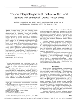
  • 1. ORIGINAL ARTICLE Proximal Interphalangeal Joint Fractures of the Hand Treatment With an External Dynamic Traction Device Kanthan Theivendran, BSc, MBBS, MRCS, Jonathan Pollock, BMBS, MRCS, and Vaikunthan Rajaratnam, FRCS, Dip Hand Surg(Eur) Intra-articular PIP joint fractures can be treated with Abstract: The authors present a series of 11 consecutive patients who sustained an intra-articular fracture of the proximal interpha- various techniques. This is dependent on the articular surface langeal (PIP) joint and 1 patient with a fracture of the interphalan- involvement of the base of the middle phalanx and the degree geal joint of the thumb treated with a Kirschner wire external of comminution. It is generally accepted that if there is less dynamic traction device. Range of motion and grip strength were than 40% involvement of the joint surface, then the fracture measured. We used the Michigan Hand Outcome Questionnaire is stable and can be treated nonoperatively.3 However, greater (MHQ) to obtain a subjective response to the treatment. Average than 40% of joint surface is unstable and requires surgical range of motion of the PIP joint was 64° and distal interphalangeal intervention.3 joint was 52°. The average grip strength was 86% with a mean MHQ Treatment modalities include extension block splint- score of 90. Two patients had pin site infections treated successfully ing,4 open reduction and internal fixation,5,6 closed reduction with antibiotics. All fractures had united at final follow up with and percutaneous fixation, and volar plate arthroplasty. There 1-mm articular step present. Our series highlights that these are are 2 main problems with comminuted fractures at the PIP difficult injuries to treat and that anatomic reduction is not always joint: 1) the articular bony fragments are too small to openly possible. However, this technique is simple and quick to perform reduce and internally fix to gain anatomic reduction; and 2) if while allowing early joint mobilization. The subjective response to the joint is immobilized, the fibrotic changes that occur after treatment is comparable with other studies using a similar device. injury will cause stiffness. These problems can be overcome by early mobilization Key Words: dynamic, external fixation, fracture, proximal after anatomic reduction of the fracture using the principle of interphalangeal joint ligamentotaxis.7,8 There have been various techniques de- (Ann Plast Surg 2007;58: 625– 629) scribed in the literature using these principles.4,7,9 –11 We report a series of 11 patients with intra-articular PIP joint fractures and 1 patient with an intra-articular interphalangeal (IP) joint fracture of the thumb treated with a Kirschner (K) wire dynamic external fixation device.10 P roximal interphalangeal joint (PIP) joint fractures are relatively rare but can be severely disabling for the patient and lead to stiffness and persistent pain if inadequately treated. METHODS The stability of the PIP joint is provided by the articular We reviewed 11 consecutive patients who sustained an congruency and soft tissue supports. Soft tissue stability is intra-articular fracture of the PIP joint and 1 patient of the IP provided by collateral ligaments, volar plate, joint capsule, joint of the thumb. All were treated with a dynamic external dorsal expansion, and extensor tendon as well as the flexor fixator frame by the senior author (V.R.) between 2004 and tendons.1 These fractures commonly occur when an axial 2005. We used the classification system by Seno et al (Fig. 1) load is applied to the finger tip causing hyperextension to describe the fracture configuration at the base of the middle leading to a dorsal fracture and/or dislocation.2 phalanx.12 Objective measurements of the active range of motion (AROM) of the distal interphalangeal (DIP) and PIP joints of the affected finger or thumb were measured using a standard goniometer. The grip strength was also measured Received June 27, 2006, and accepted for publication, after revision, August using the Jamar dynamometer and was compared with the 30, 2006. From The Birmingham Hand Centre, University Hospital Birmingham Selly uninjured side. We used the Michigan Hand Outcome Ques- Oak Hospital, Birmingham, U.K. tionnaire13 (MHQ) to obtain a subjective response to the Reprints: Kanthan Theivendran, BSc, MBBS, MRCS, 81 Pennine Way, treatment. Duration of the procedure was obtained from Ashby-de-la-Zouch, Leicestershire LE65 1EZ, United Kingdom. E-mail: operating room records. Bony union was confirmed by re- kanthan@hotmail.co.uk. Copyright © 2007 by Lippincott Williams & Wilkins view of the radiographs at the latest follow up. ISSN: 0148-7043/07/5806-0625 Statistical analysis was performed using the Pearson DOI: 10.1097/01.sap.0000245132.14908.9d correlation test to identify if there was any significant rela- Annals of Plastic Surgery • Volume 58, Number 6, June 2007 625
  • 2. Theivendran et al Annals of Plastic Surgery • Volume 58, Number 6, June 2007 FIGURE 1. Classification of base of middle phalanx fractures by Seno et al.12 Reproduced with permis- sion and copyright of the British Editorial Society of Bone and Joint Surgery. tionship between the time of delay to operation, the age of the and distraction across the fracture site. Early active move- patient, or the outcome of treatment. The SPSS 14.0 (SPSS ment was encouraged within 24 hours and all patients re- Inc., Chicago, IL) statistical software was used to perform the ceived hand therapist input. analysis with P values of less than 0.05 considered to be significant. RESULTS Surgical Technique The mean follow up was 24 weeks (range, 12–56 weeks) All of the patients had local anesthetic for the proce- and the average age of the patient was 35 years (range, 18 – 60 dure. A radiographic image intensifier (II) was used to visu- years). There were 10 men and 2 women who sustained intra- alize fracture manipulation and reduction. A 1.1-mm K-wire articular digital fractures. Table 1 outlines the demographic was inserted percutaneously and perpendicular to the long details. The device was left in situ for an average of 4 weeks and axis of the phalanx. This wire was directed to the center of the mean time from injury to operation was 8 days. The mean rotation of the head of the proximal phalanx while avoiding AROM of the injured PIP joint was 64° and DIP joint (including breaching the joint capsule. A second longer K-wire was the IP joint of the thumb) was 52°. The mean grip strength inserted distally and at right angles to the middle phalanx of the injured side compared with the uninjured hand was through the center of rotation of the head of the middle 86%. The average duration of the procedure was 12 minutes phalanx while avoiding breaching the joint capsule. Both (range, 10 –16 minutes). The mean MHQ score was 90. Eight wires were used to manipulate and reduce the fracture under patients were employed before their injury and all have II guidance. The proximal wire was bent into a hook approx- returned to their original occupations. imately 5 mm from the skin. The distal K-wire was bent The time delay to operation correlated negatively with parallel to the long axis of the phalanx pointing toward the the grip strength (r 0.556, P 0.061); however, this was proximal K-wire and shaped into a parabolic curve. This not significant. There was no significant correlation between preloaded curve was then made into a hook at its distal end time delay to operation and the AROM of the PIP joint (r and interlocked with the hook from the proximal wire while 0.176, P 0.583) or the delay to operation and the MHQ applying traction to the digit (Fig. 2). This provided tension score (r 0.084, P 0.795). No significant correlation was FIGURE 2. Operative technique: A, Proximal K-wire through the center of the head of proximal phalanx. B, Another distal K- wire parallel to proximal wire through center of the head of middle phalanx. C, The distal wire is bent backward and the proximal wire is bent forward. D, A preloaded dorsal curve is created from the distal wire. E, Wire ends are shaped into hooks that engage and maintain in-line traction. F, Fracture is reduced and maintained while allowing movement at the hooks of the K-wire. 626 © 2007 Lippincott Williams & Wilkins
  • 3. Annals of Plastic Surgery • Volume 58, Number 6, June 2007 External Fixation of PIP Joint Fractures found between age of the patient and the outcome (r Pin site infection Pin site infection Questionnaire Complications 0.04, P 0.903 for the grip strength; r 0.443, P 0.149 Disassembly for the AROM of the PIP joint; and r 0.292, P 0.357 for the MHQ). Two patients developed minor pin track infection suc- Nil Nil Nil Nil Nil Nil Nil Nil Nil cessfully treated with oral antibiotics. One patient knocked the device on the gearshift of a car. It became loose and Michigan Outcome Hand needed reapplication at 4 weeks. In this case, the disengage- 88 93 93 96 98 82 95 85 83 89 80 94 ment of the construct occurred as a result of a technical error of inadequate crimping of the ends of the wire. All patients were pain-free at the latest follow up, and there was no Joint Extensor Strength Grip clinical or radiologic evidence of osteomyelitis. All fractures (%) 100 100 77 75 80 80 92 85 86 95 75 90 were united at final follow up with an average extensor lag at the PIP joint of 11° with pain-free range of motion. However, Interphalangeal Lag (degrees) all our patients had less than a 1-mm intra-articular step present Proximal on radiographs with no evidence of reduction in joint space at 8 12 10 8 9 12 10 8 12 15 16 12 final follow up. Case no. 12 (Fig. 3) shows a Seno type 3 base of the middle phalanx fracture that shows bony union at 3 months postinjury with an intra-articular step present. Interphalangeal Interphalangeal Joint Active Range of (degrees) Motion Distal DISCUSSION 60 50 52 25 72 60 50 55 60 52 30 60 The treatment goals for intra-articular PIP joint frac- tures are to restore anatomic alignment of the joint and to allow early active movement to avoid stiffness. These chal- lenging goals have sparked renewed interest in the techniques Joint Active Proximal Range of (degrees) Motion for treating these difficult fractures. Previous attempts with 20 32 62 60 78 75 82 78 69 90 40 80 open reduction and internal fixation have proved difficult with small fragments, resulting in high complication rates and reduced ability to gain joint congruency.9,14 Stern et al performed a comparative study looking at splinting, skeletal Removal of Fracture Operative Operative (minutes) Time traction with early mobilization, and open reduction and 15 10 16 12 10 9 16 14 12 10 12 10 internal fixation (ORIF) of pilon fractures of PIP joint.9 They found that ORIF can achieve anatomic reduction in some cases, but concluded that severely comminuted subchondral (days) Delay 2 3 11 8 10 3 9 6 11 2 14 14 fragments are almost impossible to reduce and may be un- TABLE 1. Demographic Details and Results of Patients Treated necessary. ORIF can cause extensive soft tissue disruption leading to reduced blood supply to the fracture fragments (Seno et al) resulting in joint stiffness.9 To avoid these problems, many Type 1 1 2 5 1 2 3 3 2 1 1 3 authors have devised innovative techniques using the princi- ple of ligamentotaxis to provide stable reduction and early active movement of the PIP joint. Schenck used a dynamic circular frame, which allowed passive finger flexion and (weeks) Device extension at regular intervals.15 This device was large and 4 5 4 4 4 5 4 4 5 2 4 5 cumbersome and was worn for at least 7 weeks. Smaller fixators using springs and pulleys are often difficult to con- Follow Up struct.8,16 The Pins and Rubber Traction System (PRTS) by (weeks) 16 52 15 12 16 16 28 20 20 56 20 16 Suzuki et al11 used rubber bands that could be tensioned differentially to correct valgus and varus deformity. How- ever, although they reported excellent results, it was com- R middle L middle L middle Injured L thumb Finger monly known that rubber bands were subjected to plastic R little R little R little R little R ring R ring R ring L ring deformation and could become loose. The senior author found that rubber bands were difficult to manage postopera- (years) Sex M M M M M M M M M M tively and found it cumbersome to apply and maintain be- F F cause the elasticity was not constant. Simple external dy- Age namic fixators using K-wires only to provide ligamentotaxis 35 19 40 38 49 26 25 22 48 60 18 40 have been described by Hynes and Giddins10 and Gaul and Rosenberg.17 Our surgical technique is based on a similar Case No. 1 2 3 4 5 6 7 8 9 10 11 12 configuration of K-wires. This frame, if applied properly, © 2007 Lippincott Williams & Wilkins 627
  • 4. Theivendran et al Annals of Plastic Surgery • Volume 58, Number 6, June 2007 FIGURE 3. Case no. 12: A, Preoperative radiograph showing a Seno type 3 fracture. B, Seven-day postoperative radiograph with the frame in situ. C, Three-month postoperative radiograph with fracture union with articular step. D, E, Range of mo- tion at 3 months follow up. maintains constant distraction and, if overdistraction oc- sponse to treatment with other studies using a similar dy- curred, can be reduced by crimping the wire. A similar study namic external fixation device. by Deshmukh et al,18 who used a modified PRTS of Suzuki et al,11 produced an average AROM of the PIP joint of 85°, REFERENCES an average grip strength of 92%, and a mean normalized 1. Liss FE, Green SM. Capsular injuries of the proximal interphalangeal MHQ of 84 at an average follow up of 34 months. Our study joint. Hand Clin. 1992;8:755–768. showed a better average MHQ score of 90 as compared with 2. Glickel SZ, Barron OA. Proximal interphalangeal joint fracture disloca- that achieved in the study by Deshmukh et al. The mean tions. Hand Clin. 2000;16:333–344. AROM of the PIP joint from our study was lower compared 3. Glickel SZ, Barron OA, Eaton RG. Dislocations and Ligament Injuries in the Digits, 4th ed. New York: Churchill Livingstone; 1999:772– 808. with other studies using a similar K-wire external fixator.19,20 4. McElfresh EC, Dobyns JH, O’Brien ET. Management of fracture– This may be the result of the shorter follow-up period of 6 dislocation of the proximal interphalangeal joints by extension-block months in our study compared with similar fixators by Badia splinting. J Bone Joint Surg Am . 1972;54:1705–1711. et al (89° at 24 months) and Syed et al (79° at 2.2 years).19,20 5. Agee JM. Unstable fracture dislocations of the proximal interphalangeal joint of the fingers: a preliminary report of a new treatment technique. Importantly, all those patients who were employed before the J Hand Surg Am . 1978;3:386 –389. injury have now returned to their original jobs. All patients 6. Wilson JN, Rowland SA. Fracture– dislocation of the proximal interpha- were satisfied with the treatment and reported pain-free range langeal joint of the finger. J Bone Joint Surg Am . 1966;48:493–502. of motion. Fracture union had occurred in all patients at final 7. Agee JM. Unstable fracture dislocations of the proximal interphalangeal joint. Treatment with the force couple splint. Clin Orthop. 1987;214: follow up with less than a 1-mm intra-articular step present at 101–112. the PIP joint. Our series highlights that these are difficult 8. Allison DM. Fractures of the base of the middle phalanx treated by a injuries to treat and that anatomic reduction is not always dynamic external fixation device. J Hand Surg Br . 1996;21:305–310. possible as compared with open reduction with rigid internal 9. Stern PJ, Roman RJ, Kiefhaber TR, et al. Pilon fractures of the proximal fixation. However, internal fixation can be technically de- interphalangeal joint. J Hand Surg Am . 1991;16:844 – 850. 10. Hynes MC, Giddins GE. Dynamic external fixation for pilon fractures of manding and often require extensive soft tissue dissection the interphalangeal joints. J Hand Surg Br . 2001;26:122–124. that may contribute to complications and the need for sec- 11. Suzuki Y, Matsunaga T, Sato S, et al. The pins and rubbers traction ondary operative procedures.21 system for treatment of comminuted intraarticular fractures and frac- The main limitation to this study is the small sample ture– dislocations in the hand. J Hand Surg Br . 1994;19:98 –107. 12. Seno N, Hashizume H, Inoue H, et al. Fractures of the base of the middle size and short follow-up period. Long-term follow up will be phalanx of the finger. Classification, management and long-term results. required to identify if patients develop post-traumatic arthritis J Bone Joint Surg Br . 1997;79:758 –763. within these joints. Despite the current limitations, we believe 13. Chung KC, Pillsbury MS, Walters MR, et al. Reliability and validity that this system provides a simple and cost-effective method testing of the Michigan Hand Outcomes Questionnaire. J Hand Surg of treatment of these difficult fractures. The application of the Am . 1998;23:575–587. 14. Hastings H 2nd, Carroll CT. Treatment of closed articular fractures of external fixator is quick (average operative time of 12 min- the metacarpophalangeal and proximal interphalangeal joints. Hand utes), is done under local anesthetic, and is acceptable to the Clin. 1988;4:503–527. patient. Our case series shows a comparable subjective re- 15. Schenck RR. Dynamic traction and early passive movement for fractures 628 © 2007 Lippincott Williams & Wilkins
  • 5. Annals of Plastic Surgery • Volume 58, Number 6, June 2007 External Fixation of PIP Joint Fractures of the proximal interphalangeal joint. J Hand Surg Am . 1986;11:850 – pins and rubbers traction system. J Bone Joint Surg Br . 2004;86:406 – 858. 412. 16. Inanami H, Ninomiya S, Okutsu I, et al. Dynamic external finger fixator 19. Syed AA, Agarwal M, Boome R. Dynamic external fixator for pilon for fracture dislocation of the proximal interphalangeal joint. J Hand fractures of the proximal interphalangeal joints: a simple fixator for a Surg Am . 1993;18:160 –164. complex fracture. J Hand Surg Br . 2003;28:137–141. 17. Gaul JS Jr, Rosenberg SN. Fracture– dislocation of the middle phalanx at 20. Badia A, Riano F, Ravikoff J, et al. Dynamic intradigital external the proximal interphalangeal joint: repair with a simple intradigital fixation for proximal interphalangeal joint fracture dislocations. J Hand traction-fixation device. Am J Orthop. 1998;27:682– 688. Surg Am . 2005;30:154 –160. 18. Deshmukh SC, Kumar D, Mathur K, et al. Complex fracture– dislocation 21. Stern PJ, Wieser MJ, Reilly DG. Complications of plate fixation in the of the proximal interphalangeal joint of the hand. Results of a modified hand skeleton. Clin Orthop. 1987;214:59 – 65. © 2007 Lippincott Williams & Wilkins 629