Weitere ähnliche Inhalte
Ähnlich wie 4 cd1 nature (20)
Mehr von ufamimunologia (18)
Kürzlich hochgeladen (20)
4 cd1 nature
- 1. REVIEWS
CD1 antigen presentation:
how it works
Duarte C. Barral and Michael B. Brenner
Abstract | The classic concept of self–non-self discrimination by the immune system focused
on the recognition of fragments from proteins presented by classical MHC molecules.
However, the discovery of MHC-class-I-like CD1 antigen-presentation molecules now
explains how the immune system also recognizes the abundant and diverse universe of lipid-
containing antigens. The CD1 molecules bind and present amphipathic lipid antigens for
recognition by T-cell receptors. Here, we outline the recent advances in our understanding of
how the processes of CD1 assembly, trafficking, lipid-antigen binding and T-cell activation
are achieved and the new insights into how lipid antigens differentially elicit CD1-restricted
innate and adaptive T-cell responses.
Endocytic pathway
The CD1 family of MHC-class-I-like glycoproteins strategies by which antigen-presenting cells (APCs)
A trafficking pathway used by (CD1a, CD1b, CD1c, CD1d and CD1e) present both stimulate CD1-restricted T-cell responses and how
all cells for the internalization foreign and self lipids as cognate antigens to T cells. these responses are linked to the types of lipid antigen
of molecules from the plasma The CD1 proteins share sequence homology and (self versus foreign), the CD1 isoform (group 1 versus
membrane to lysosomes.
overall domain structure with MHC class I molecules, group 2) and the responding T-cell population (clonally
Capping motifs being comprised of a heavy chain with three extracell specific versus population en masse). The mechanisms
Carbohydrates that are ular domains that are non-covalently associated with by which CD1-restricted T cells affect a broad range
attached to the branches of β2-microglobulin (β2m). On the basis of sequence analy- of immune responses can now be understood. For a
the arabinan domain in sis, the CD1 isoforms can be classified into three groups: detailed overview on the roles of NKT cells in disease,
lipoarabinomannan (LAM).
In the case of Man-LAM, which
group 1 comprises CD1a, CD1b and CD1c; group 2 the reader is referred to other excellent reviews on this
is found in Mycobacterium comprises CD1d; and group 3 comprises CD1e. All subject1,2.
tuberculosis and other mammals studied so far have been found to express CD1
pathogenic species of molecules. Interestingly, however, humans express all Diversity of lipids presented by CD1 molecules
mycobacteria, the
CD1 isoforms (CD1a–CD1e), whereas muroid rodents The lipid antigens presented by CD1 molecules include
carbohydrates are mannose
groups. Ara-LAM is not
express only CD1d. Group 1 CD1 molecules mainly a broad array of classes, ranging from foreign lipids
capped and it is found in present lipid antigens to clonally diverse T cells that that are unique to specific microorganisms to common
non-pathogenic, fast growing mediate adaptive immunity to the vast range of micro- mammalian self lipids.
strains of mycobacteria. bial lipid antigens. By contrast, CD1d (group 2) mol-
ecules present lipid antigens to natural killer T (NKT) Foreign lipid antigens. Many of the unique lipids found
cells, a subset of which, the invariant NKT (iNKT) cells, in Mycobacterium tuberculosis can be presented by CD1a,
expresses an invariant T-cell receptor (TCR) α-chain, CD1b and CD1c to activate clonally diverse T cells. These
responds rapidly en masse following antigen recognition include the abundant mycolates, such as the free fatty
Division of Rheumatology, and is a potent effector of innate immunity. acid mycolic acid, and mycolates with esterified glycans,
Immunology and Allergy, CD1 proteins survey the endocytic pathway to intersect such as glucose monomycolate. Mycolic acid (BOX 1) was
Department of Medicine,
and bind lipid antigens. Several molecules involved in the first CD1-presented lipid antigen to be identified3.
Brigham and Women’s
Hospital, Harvard Medical lipid metabolism have recently been shown to also func- M. tuberculosis-derived lipoglycans (such as lipoarabino
School, 1 Jimmy Fund Way, tion in CD1-mediated lipid-antigen presentation, such mannan), which are composed of a phosphatidylinositol
Boston, Massachusetts as saposins, which mediate the loading of lipids onto anchor, a macromolecular polysaccharide backbone and
02115, USA. CD1 molecules in lysosomes. In this Review, we describe capping motifs, or their component phosphatidylinositol
Correspondence to M.B.B.
e-mail: mbrenner@rics.bwh.
the pathway of CD1 antigen presentation, including the mannosides4, were found to activate CD1b-restricted
harvard.edu delivery, processing, loading and presentation of lipid T cells5. By contrast, Mycobacterium spp. lipids that are
doi:10.1038/nri2191 antigens to T cells. New insights now delineate the closely related to mannosyl β-1-phosphomycoketides are
nature reviews | immunology volume 7 | december 2007 | 929
© 2007 Nature Publishing Group
- 2. REVIEWS
Box 1 | Structures of microbial and self lipids presented by CD1 molecules
Mycobacterial lipids (see figure, part a), such as sulpholipids from Mycobacterium tuberculosis, have a sulphate
group on a trehalose moiety, which is acylated by two to four very long chain branched fatty acids. The diacylated
sulpholipid has been shown to be recognized by CD1b-restricted T cells8. Lipopeptides, such as didehydroxymyco-
bactin, an intermediate in the biosynthesis of the mycobacterial iron scavenger mycobactin siderophores, can also
be presented by CD1a molecules124. Mycobacterial mycolates contain α-alkyl branched β-hydroxy fatty acids in
which the main meromycolate chain (C20–C90) may contain R-substitutions such as the keto function group that is
shown3. Diacylglycerols, such as the α-galactosyldiacylglycerol from the spirochete Borrelia burgdorferi, and the
common mammalian phosphoglycerides, such as phosphatidylinositol (part b), can be presented by CD1d to
stimulate invariant natural killer T (iNKT) cells11,18. Glycosphingolipids (part c) are among the most studied lipids
presented by CD1 molecules. α-galactosylceramide is a synthetic antigen mimic that, in contrast to mammalian
glycosphingolipids, contains an α-linkage between the 1′ carbon of the sugar and the sphingosine base125. A similar
α-linkage glycosphingolipid (α-glucuronosylceramide) found in α-proteobacteria activates iNKT cells9,10.
By contrast, abundant mammalian β-linked ceramides, such as sulphatide from myelin, and isoglobo-series
glycosphingolipids, such as isoglobotrihexosylceramide, can be presented by CD1 molecules19,23.
a O OH
HO OH
O
NH
O
O O
O
O OSO3H
NH
O O
Didehydroxymycobactin: Diacylated sulpholipid: M. tuberculosis
M. tuberculosis HO OH
OH
O O
O
NH
HN
O O
N HO
O
OH HOOC Keto mycolic acid: M. tuberculosis
HO
b O O O O
HO OH
O O
HO
–O O O O
P
OH
O O O Phosphatidylinositol: self O
OH
α-Galactosyldiacylglycerol: B. burgdorferi
HO OH
HO
O
Trehalose c O
A disaccharide formed by two HN
glucose units. HN
O OH
OH OH O
Siderophores O O
Low molecular-weight OH
OH OH
HO OH
compounds that are secreted
by numerous types of bacteria O HO Isoglobotrihexosylceramide: self HO α-Galactosylceramide: synthetic or
and that have a high affinity for OH marine sponge
O
iron and other metal ions.
These molecules chelate metal HO OH O
ions and carry them into the O
OH O
cell through specific receptors. O
HN
They are bacterial virulence OH HN
HO OH OH
factors. O O
O
HO OH
Sphingosine O OH O
OH OH
An amino alcohol that can be HO OH Sulphatide: self
OH
linked to a fatty acid via the HO
amino group to form the basic OSO3–
structure of sphingolipids. α-Glucuronosylceramide: Sphingomonas spp.
Nature Reviews | Immunology
930 | december 2007 | volume 7 www.nature.com/reviews/immunol
© 2007 Nature Publishing Group
- 3. REVIEWS
Purified protein derivative presented by CD1c to human T-cell clones6. Among the and B. burgdorferi infection11,12. So, microbial lipids that
A protein from Mycobacterium other unique mycobacterial lipids are lipopeptides and are distinct to those derived from M. tuberculosis and
tuberculosis that is used in the sulpholipids (BOX 1), which can be presented by CD1a that are presented by CD1a, CD1b and CD1c can be
tuberculin sensitivity test, and CD1b, respectively. Importantly, reactivity against presented by CD1d and can stimulate iNKT cells.
which determines previous
interactions of the host with
several of these lipids has been observed with increased
the bacterium. A positive frequency in patients infected with M. tuberculosis or Mammalian self-lipid antigens. The first reported
tuberculin test is generally in healthy individuals who are positive for the purified examples of group-1-CD1-restricted human T cells13
taken as an indication of protein derivative6–8, suggesting a role for these lipids in and CD1d-restricted mouse T cells14,15 were examples
previous exposure to
host defence against tuberculosis infection. Although all of self-reactive responses. It is widely assumed that
M. tuberculosis.
of the mycobacterial lipids noted above are recognized by iNKT cells recognize self lipids during selection in the
α-Galactosylceramide a clonal population of CD1a-, CD1b- and CD1c-restricted thymus16. Yet, it is also clear that the same iNKT cells
(α-GalCer). A synthetic or T cells, so far, similar reactivity for individual iNKT cells bearing self-lipid–CD1d-reactive TCRs also recognize
marine-sponge-derived has not been observed. Instead, different microorganism- foreign-lipid antigens in the periphery1. Similarly,
glycolipid containing an
α-anomeric glycosidic linkage
derived lipids of several chemical classes have been shown group-1-CD1-restricted human T cells are weakly self
of the galactose residue to the to activate iNKT cells. reactive and display stronger reactivity to foreign-lipid
sphingosine base. This lipid, Studies of iNKT cells for many years focused on their antigens17. So, the combination of self- and foreign-
and structurally related ones, activation by the synthetic mimic α-galactosylceramide antigen reactivity is likely to be an inherent feature
potently activates CD1d-
(α-GalCer)(BOX 1). This raised the question of whether of CD1-restricted T cells, as it is for MHC-restricted
restricted natural killer T cells
that express the semi-invariant
similar structures existed in microorganisms in nature. T cells.
Vα14–Jα18 T-cell receptor in Although their occurrence among pathogens remains In cell-free assays, it was shown that CD1d-restricted
mice (and the Vα24–Jα18 unclear, non-pathogenic, soil and water α-proteobacteria T cells depend on loading of the CD1 molecules with
equivalent receptor in have now been found to contain iNKT-cell stimulating mammalian self lipids to activate NKT-cell hybri-
humans).
α-linked sphingolipids9,10 (BOX 1). Instead of α-linked domas18. These studies revealed that small mammalian
sphingolipids, another class of lipids, the diacylglycerols phosphoglycerides, such as phosphatidylinositol (BOX 1),
(BOX 1) that are present in the spirochete Borrelia burg- phosphatidylglycerol and phosphatidylethanolamine,
dorferi (the aetiological agent of lyme disease in humans), could stimulate various NKT-cell populations. One fea-
were found to stimulate iNKT cells11. Further, the lipo- ture common to these self antigens was that they were
phosphoglycan of the protozoan parasite Leishmania weak agonists compared with the potent α-GalCer
donovani was found to bind CD1d and stimulate mimic. Several glycosphingolipids have also been
iNKT cells in vitro12. Importantly, iNKT-cell-deficient shown to be presented by CD1 molecules (TABLE 1).
mice showed increased susceptibility to L. donovani Sulphatide, a major component of myelin (BOX 1), was
found to be a promiscuous self-lipid antigen that could
be presented by CD1a, CD1b and CD1c and activate
Table 1 | CD1-restricted lipid antigens clonally restricted human T cells19. Further, GM1 and
Source Antigen CD1 isoform Refs related gangliosides were also found to be presented
by CD1b, suggesting that self-lipid antigens may be
Mycobacterium Mycolic acids CD1b 3
tuberculosis and other important targets for autoimmune T cells in condi-
mycobacteria Glucose monomycolate CD1b 132 tions such as multiple sclerosis20,21. Glycosphingolipids
Sulpholipid (diacylated CD1b 8 have also been reported to stimulate iNKT cells. The
sulphoglycolipid) ganglioside GD3, which is markedly overexpressed
Phosphatidylinositol mannosides CD1b, CD1d 5,133 in human melanomas, is a major target for ‘autoanti
Mannosylated lipoarabinomannan CD1b 5 bodies’. The injection of mice with human melanoma
cells or GD3-loaded dendritic cells (DCs) also stimu-
Mannosyl-β1-phosphomycoketides CD1c 6,134 lates a CD1d-restricted NKT-cell response22. Recently,
Didehydroxymycobactin CD1a 124 iGb3 (isoglobotrihexosylceramide), a member of the
Sphingomonas spp. α-Glucuronosylceramide CD1d 9,10 globo/isoglobo-series glycosphingolipids (BOX 1), was found
to activate iNKT cells in vitro10,23. However, the sug-
Borrelia burgdorferi α-Galactosyldiacylglycerol CD1d 11
gestion that iGb3, as a single glycosphingolipid, is the
Leishmania donovani Lipophosphoglycan CD1d 12 critical self-lipid antigen controlling both iNKT-cell
Mammalian (self) Phosphatidylinositol CD1d 18 development in the thymus and activation in the periph-
Phosphatidylglycerol CD1d 18
ery in vivo has been challenged by the failure to detect
iGb3 in mouse or human thymi using sensitive high-
Phosphatidylethanolamine CD1d 18 performance liquid chromatography methods24 and by
GM1 CD1b 20,21 the finding that iGb3-synthase-deficient mice, which
GD3 CD1d 22 lack the entire family of isoglobo-series glycosphingo
lipids, have normal numbers of iNKT cells and iNKT-
Sulphatide CD1a, CD1b, 19
CD1c cell development25. So, it seems likely that there may
be several self-lipid antigens that collectively provide a
Isoglobotrihexosylceramide CD1d 10,23
range of ligands for positive and negative selection of
Synthetic or marine α-Galactosylceramide CD1d 125 NKT cells in the thymus and the activation of NKT cells
sponge in the periphery.
nature reviews | immunology volume 7 | december 2007 | 931
© 2007 Nature Publishing Group
- 4. REVIEWS
a b O
α-GalCer C16 spacer lipid
HN
OH
OH O
O
α1 OH Phe70
OH
α-GalCer variant
HO HO
Cys12
α2 A' F'
c O
O
HO
HO
HO
β2-microglobulin O
HO Glucose monomycolate (C60)
OH Phe70
α3
Val12
F'
A' T'
C'
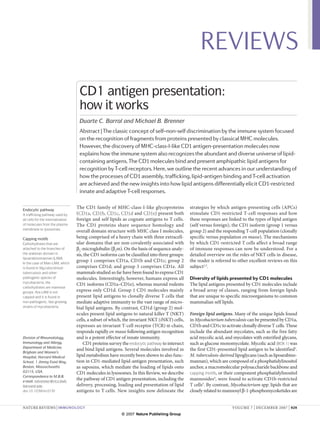
Figure 1 | Crystal structures of CD1b and mouse CD1d loaded with lipids. a | A ribbon diagram of the mouse CD1d
crystal structure loaded with a synthetic variant of α-galactosylceramide (α-GalCer) that contains a shorter fatty acid
Nature Reviews | Immunology
chain (the α-GalCer variant is shown in stick representation with the carbon backbone in yellow and oxygen atoms in
red)130. A C16 spacer lipid was found to fill the empty space in the A′ channel of CD1d, which would be occupied by the
longer fatty acid of the non-variant α-GalCer. b | Surface representation of the mouse CD1d antigen-binding groove
loaded with the same lipids as in a. The A′ and F′ hydrophobic channels are shown, as well as two amino-acid residues that
form the A′ pole (Cys12 and Phe70) and divert the channel containing the lipid alkyl chain around them. The short fatty
acid chain of the lipid inserts into the A′ channel, whereas the sphingosine chain is inserted into the F’ channel. c | Surface
representation of CD1b loaded with the C60 species of glucose monomycolate47. The long fatty acid chain sequentially
traverses the A′, T′ and F′ superchannel and the α-branched chain inserts into the C′ channel. The diagrams were
generated using CCP4MG software131.
Structure of CD1 proteins F′ and the A′ channels. There are similar channels in
Similar to MHC class I molecules, CD1 heavy chains CD1a, with the A′ channel having a fixed length, in which
consist of α1 and α2 domains that form the antigen- a closed terminus limits the length of the alkyl chains
binding region, contained within two antiparallel that can be accommodated, like a ‘molecular ruler’, to
α-helical structures that are situated on a β-pleated approximately 18–23 carbon atoms 27. Interestingly,
sheet (FIG. 1a). The α1 and α2 antigen-binding region the F′ channel of CD1a can bind either an alkyl chain
Lyme disease
A disease caused by the
is linked to an immunoglobulin-like α3 domain, which or a peptide, which enables the molecule to bind and
bacterium Borrelia burgdorferi is attached to the membrane by a transmembrane seg- present antigens to T cells that have one or two alkyl
or other Borrelia spp. that is ment, followed by a short cytoplasmic tail. However, chains, such as the lipopeptide didehydroxymycobac-
transmitted to humans via the unlike MHC class I molecules, CD1 proteins bind alkyl tin and sulphatide, respectively27,28. By contrast, CD1b
bites of infected blacklegged
chains in hydrophobic channels that reside beneath the can accommodate longer alkyl chains and contains
ticks. Symptoms can include
skin rash, fever, fatigue, surface of CD1 molecules, whereas the hydrophilic head four channels: the A′, F′, C′ and T′ channels29 (FIG. 1c).
headache, muscle pain, stiff groups of the lipid antigens protrude where the hydro- In CD1b, the A′ and F′ channels are connected via the
neck and swelling of the knee phobic channels open to the membrane distal surface T′ tunnel, which forms a superchannel that can accom-
and other large joints. Most of the CD1 molecule (FIG. 1a). These head moieties are modate multiple alkyl chains or a single alkyl chain
cases can be successfully
treated with antibiotics.
stabilized by hydrogen bonds, which also contribute of up to 60 carbons. Moreover, besides the surface
to the correct positioning of the lipid antigens. CD1 region where the A′ and F′ channels converge, CD1b
Globo/isoglobo-series proteins have deeper and more voluminous antigen- has an extra portal where the C′ channel contacts the
glycosphingolipids binding compartments than MHC class I molecules. molecular surface. This allows egress of the alkyl chain
One arm of the glycosphingo-
Moreover, the different architecture of the antigen- under the α2 helix of CD1b such that chains longer
lipid family, which is
characterized by an α-linked binding compartment of different CD1 isoforms allows than 16 carbon atoms might extend out of this chan-
galactose sugar in the third them to bind distinct lipid antigens. The antigen- nel. Together, these features enable the CD1b groove to
sugar position. Globotrihexo- binding groove of mouse CD1d can be divided into two accommodate very long fatty acid chains and, poten-
sylceramide (Gb3) has an channels: the A′ channel and the F′ channel, classified tially, lipids with three alkyl chains29.
α1–4 linked galactose sugar,
whereas isoglobotrihexosyl
by comparison with the pockets of the MHC class I Recently, the crystal structure of the TCR–α-GalCer–
ceramide (iGb3) has an α1–3 peptide-binding groove26 (FIG. 1b). A narrow entrance CD1d ternary complex was elucidated30. As predicted,
linked galactose sugar. into the groove is found at the junction between the the TCR of the NKT cell contacts the protruding head
932 | december 2007 | volume 7 www.nature.com/reviews/immunol
© 2007 Nature Publishing Group
- 5. REVIEWS
group of α-GalCer and the CD1 α-helices. Interestingly, to MHC class I molecules, CD1d also associates with the
the footprint of the NKT-cell TCR on the surface of the thiol oxidoreductase ERp57, which is involved in the
α-GalCer–CD1d complex is substantially different from formation of disulphide bonds33–35 (FIG. 2a). In general,
the footprint of MHC-class-I-restricted TCRs, being CD1 proteins depend on the association with β2m for
parallel to the long axis of the CD1d binding groove their exit from the ER and for trafficking through the
and positioned in the extreme end of CD1d, above the secretory pathway to the cell surface31,36,37, although CD1d
F′ pocket; and with the invariant TCR α-chain regions can also be expressed at the cell surface independently
CDR1 (complementarity-determining region 1) and of β2m and might still be functional38–41.
CDR3 dominating the recognition of the antigen.
Loading of endogenous lipids onto CD1 in the ER.
CD1 trafficking in the secretory pathway Studies in which natural ligands have been eluted from
Assembly of CD1 molecules. Newly synthesized CD1 CD1 have revealed that CD1b and CD1d associate with
molecules have signal sequences for translocation into endogenous lipids, including phosphatidylinositol and
the lumen of the endoplasmic reticulum (ER). Following glycosylphosphatidylinositols42–44. The use of native
synthesis, they rapidly become glycosylated by the mass spectrometry to study lipid–CD1 complexes
addition of N-linked oligosaccharides. This enables the revealed that CD1b was associated with phosphati-
newly synthesized heavy chains to bind the ER chaper- dylcholine45. Another study that used a recombinant
ones calnexin and calreticulin, which have specificity for form of CD1d containing a KDEL ER-retention signal
monoglucosylated immature glycans31–33 (FIG. 2a). Similar showed that phosphatidylinositol associated with the
Plasma
membrane
c
Clathrin-coated pit
AP2 d
b
Trans-Golgi
network Sorting
endosome e
Golgi AP3
Endocytic recycling compartment
Peptide
Ii
ER
MHC MHC class II
class I
Self lipid f
ERp57
Signal sequences
Short peptide sequences Nucleus a
involved in the post- β2m
translational targeting of
proteins. In particular, the
Calnexin Calreticulin
endoplasmic reticulum (ER)
signal sequence is recognized
CD1 (heavy chain) MHC class II compartment or lysosome
by the signal-recognition
peptide after synthesis of the
signal and the protein is Figure 2 | Intracellular trafficking of CD1 molecules. a | CD1 heavy chains are assembled in the endoplasmic
co-translationally inserted into Nature Reviews | Immunology
reticulum (ER), where they bind the chaperones calnexin, calreticulin and ERp57. They also bind β2-microglobulin (β2m)
the ER lumen. The signal non-covalently in the ER. b | CD1 molecules then follow the secretory route through the Golgi apparatus to the plasma
sequence is normally removed membrane. MHC class I and II molecules also assemble in the ER and follow a similar route, with MHC class II molecules
by a signal peptidase. (in complex with invariant chain (Ii)) being diverted from the trans-Golgi network to the endosomes. c | CD1 molecules are
internalized in clathrin-coated pits via the interaction of the adaptor complex AP2 with tyrosine-based sorting motifs
Secretory pathway
present in the cytoplasmic tails of CD1. From the sorting endosome, CD1 molecules can follow two main routes. d | CD1
A trafficking pathway from the
endoplasmic reticulum to
molecules such as CD1a and CD1c can follow the slow recycling pathway, back to the plasma membrane, through the
the plasma membrane that is endocytic recycling compartment. e | CD1 molecules such as CD1b and mouse CD1d can traffic to late endosomal and
taken by newly synthesized lysosomal compartments via the interaction of AP3 also with tyrosine-based motifs contained in the cytoplasmic tails of
molecules that are destined these CD1 molecules. f | CD1 and MHC class II molecules recycle from lysosomal compartments to the plasma membrane.
to be secreted. During their trafficking, CD1 molecules are thought to be loaded with a lipid molecule.
nature reviews | immunology volume 7 | december 2007 | 933
© 2007 Nature Publishing Group
- 6. REVIEWS
recombinant CD1d, which supports the idea that self proteins into clathrin-coated pits56. Using surface plasmon
lipids are loaded into CD1 molecules during their resonance, the cytoplasmic tails of CD1b, CD1c and
assembly in the ER43,44. Such ER-loaded self lipids might mouse CD1d were shown to bind AP2 or one of its
be antigenic or alternatively they might function as subunits, and immunoelectron microscopy studies of
chaperones to facilitate the assembly and to stabilize CD1b and CD1c showed that these molecules promi-
CD1 molecules in the ER. Consistent with this, no nently localize to clathrin-coated pits and vesicles53,57–59.
CD1b crystal structures have been obtained without Accordingly, a marked reduction in CD1b and CD1d
a lipid or other molecule filling the entire antigen- internalization, and consequent accumulation on the
binding groove of the molecule, suggesting that the cell surface, occurred when the respective cytoplasmic
hydrophobic groove is stable only when occupied by tails of these molecules were deleted, demonstrating
a bound lipid45–47. the importance of these domains for internalization
of the proteins from the plasma membrane53,54,57,59–61.
Chylomicrons Role for MTP in CD1 antigen presentation. Micro- In contrast to the other isoforms, CD1a does not contain
Large lipoprotein particles
primarily composed of
somal triglyceride transfer protein (MTP; also known any sorting motifs in its cytoplasmic tail, and yet it is also
triglycerides, secreted by the as MTTP) is an ER-resident lipid-transfer protein that internalized from the plasma membrane into endosomal
intestine into the lymphatic is required for the proper assembly and secretion of compartments by an unknown mechanism.
system and degraded by apolipoprotein-B-containing lipoproteins, namely
lipoprotein lipase.
very low-density lipoproteins (VLDLs) by the liver and CD1 trafficking through the endocytic pathway. After
Endoglycosidase-H chylomicrons by the intestine48,49. Interestingly, the inhi- internalization into the early or sorting endosomes, the
resistance bition of MTP in APCs causes a defect in the presenta- different CD1 isoforms follow different trafficking path-
Endoglycosidase H cleaves tion of lipid antigens by CD1d, and mice lacking MTP ways. CD1a and CD1c, but not CD1b, co-localize with
high-mannose oligosaccharides are also deficient in CD1d antigen presentation50,51. a dominant-negative form of the small GTPase ARF6
from N-linked glycoproteins.
The acquisition of resistance to
Although the precise mechanism of action of MTP (ARF6-T27N) in the endocytic recycling compartment,
this enzyme is related to the has not been delineated, MTP was shown to transfer indicating that CD1a and CD1c follow the slow recycling
processing of the high a phospholipid (phosphatidylethanolamine) to mouse pathway back to the plasma membrane58,62 (FIG. 2d). Indeed,
mannose into complex CD1d in vitro51, and therefore could have a role in the CD1a was shown to co-localize with RAB11, another
oligosaccharides in the medial
loading of CD1d with endogenous lipids that are marker for the endocytic recycling compartment, and
Golgi apparatus and therefore
indicates progression through important both for NKT-cell selection in the thymus to recycle back to the plasma membrane from an intra
the secretory pathway. and antigen presentation by APCs in the periphery. cellular pool63.
Unexpectedly, MTP deficiency was also shown to Some of the tyrosine-based sorting motifs, such
Invariant chain inhibit the recycling of mouse CD1d from lysosomes as those in CD1b and mouse CD1d molecules, can
(Ii). A non-polymorphic
molecule that associates with
to the cell surface, suggesting possible effects of MTP bind both AP2, which mediates CD1 internalization
MHC class II proteins. By that are distal from its location in the ER52. from the plasma membrane, and AP3, which diverts
occupying the antigen-binding these CD1 molecules from the early recycling path-
cleft, the invariant chain CD1 trafficking from the ER to the plasma membrane. way to late endosomes and lysosomes 59,64–66 (FIG. 2e).
stabilizes newly synthesized
Studies of the time-course of acquisition of endoglyco- Accordingly, AP3-deficient cells have increased cell-
MHC class II molecules in the
endoplasmic reticulum and sidase-H resistance of CD1b and the appearance of CD1b at surface expression of CD1b, with an accumulation of
directs the mature molecules the cell surface suggest that, following assembly in the ER, molecules in the early recycling pathway and a near
to compartments in which this isoform follows the secretory pathway through the absence in lysosomes 64. The functional importance
binding with antigenic peptides Golgi directly to the plasma membrane53 (FIG. 2b). CD1d of AP3-dependent CD1b trafficking is highlighted
occurs.
has also been observed to associate with MHC class II by the defects displayed by the AP3-deficient cells in
Surface plasmon resonance molecules and the invariant chain (Ii), which can direct the CD1b-restricted presentation of a microbial-lipid
A technique used to measure CD1 complexes with these proteins from the trans-Golgi antigen, such as glucose monomycolate64. In the case of
molecular interactions by network to endosomal compartments without first reach- mouse CD1d, AP3-deficient mice show a similar defect
observing how much of an
ing the plasma membrane54,55. However, the functional in CD1d localization to lysosomes and CD1d-mediated
input molecule (for example,
a protein) is bound to a chip significance of this alternative CD1 trafficking pathway antigen presentation, which is reflected in a significant
immobilized with another is not known. reduction in the number of NKT cells in these mice65,66.
molecule. The amount of Interestingly, unlike CD1b and mouse CD1d, the
bound input is directly CD1 trafficking in the endocytic pathway tyrosine-based motifs of CD1c and CD1d (human) are
proportional to the change
in the light reflected off the
CD1 internalization from the plasma membrane. The not capable of binding AP3, as assessed using yeast two-
immobilized chip, which is internalization of CD1 from the plasma membrane is hybrid assays64. As a result, AP3-deficient human cells
specifically measured. This essential for its ability to sample antigens in the endo- do not display any defects in the presentation of CD1c-
technique can be used to cytic system. After trafficking to the plasma membrane, mediated lipid antigens. The importance of CD1 traf-
calculate single-molecule
CD1 molecules appear to follow a dominant, pre- ficking through the endocytic pathway for lipid loading
affinities, as well as binding
on and off rates. sumably constitutive, pathway of internalization into is highlighted by studies that examined the function of
endosomes (FIG. 2c). This clathrin-dependent pathway CD1 molecules that do not internalize properly from
Slow recycling pathway is common to proteins that contain well character- the plasma membrane owing to mutations in the cyto-
One arm of the early recycling ized tyrosine-based sorting motifs of the YXXφ type plasmic tail. Indeed, mutant mouse CD1d and CD1b
pathway that involves
trafficking through early
(where Y is a tyrosine, X is any amino acid and φ is a molecules that lack the cytoplasmic tail are defective
endosomes and the endocytic bulky hydrophobic residue), which bind the adaptor in the presentation of lipid antigens to iNKT cells and
recycling compartment. protein complex 2 (AP2) and allow sorting of cargo T cells, respectively60,61,67.
934 | december 2007 | volume 7 www.nature.com/reviews/immunol
© 2007 Nature Publishing Group
- 7. REVIEWS
So, once internalized from the plasma membrane, and CD1 trafficking are differentially regulated during
CD1a molecules traffic mainly through the endocytic DC maturation.
recycling compartment, whereas CD1b and mouse
CD1d molecules traffic mainly through late endosomes Lipid trafficking
and lysosomes. These distinct trafficking routes taken As described, CD1 molecules can survey different cell
by the different CD1 isoforms presumably allow each ular compartments by virtue of differential endosomal
isoform to survey distinct intracellular compartments trafficking. Collectively, CD1 molecules ensure a broad
for lipid antigens. Of the CD1 isoforms, CD1c appears survey of endocytic compartments for endogenous and
to have the most promiscuous localization, as it can be exogenous lipid antigens, which are ultimately presented
found in both early and late endocytic compartments58. to T cells. Therefore, CD1 molecules must intersect rele
vant lipid antigens in endocytic compartments for lipid
CD1 recycling from endosomes to the plasma membrane. loading to occur.
The sequence of antigen loading onto CD1 molecules
probably involves exchanging self lipids that are bound to Delivery of lipid antigens to APCs. Cells continuously
CD1 during assembly in the ER or delivery to the plasma take up lipids for their metabolic needs. Most of these
membrane with other self or foreign lipids that are are transported in the blood in complex with apoli-
encountered within endosomes. Once this has occurred, poproteins, forming lipid-transport particles76. One
CD1 molecules must return to the plasma membrane mechanism of uptake of lipid-transport particles is the
for presentation of the antigens to T cells. For CD1a LDL receptor (LDLR)-dependent internalization of
molecules, return to the plasma membrane can readily the VLDL particles into endosomes. The LDLR binds
be accounted for because of their extensive localization apolipoprotein E (apoE) on VLDL particles and deliv-
in the endocytic recycling compartment. Like other cell- ers it to the endocytic system 77 (FIG. 3b). The low pH
surface molecules that are internalized into the endocytic in endosomes facilitates the release of bound apolipo-
recycling compartment from the plasma membrane, after proteins from the receptor. The LDLR is then recycled
a period of retention, they correspondingly recycle back to the plasma membrane, whereas the lipoproteins
to the plasma membrane. In recent years, it has become are delivered to late endosomes and lysosomes for
well recognized that recycling from late endosomes degradation78. Interestingly, apoE can bind exogenous
and lysosomes back to the plasma membrane is also a CD1 lipid antigens and dramatically enhances the
functionally significant trafficking route for molecules CD1d-dependent presentation of a lipid antigen that
that are not degraded in these compartments, such as requires lysosomal processing79. It has also been shown
MHC class II molecules68,69 (FIG. 2f). Although the route that APCs secrete large amounts of apoE, suggesting
followed by CD1 molecules from lysosomes to the a mechanism for how APCs sample the extracellular
plasma membrane has not been clearly delineated, this milieu for lipid antigens by secreting and taking up
pathway is the target for immune evasion by herpes apoE-bound lipids from their local environment79.
simplex virus 1 (HSV1), as it was shown that this virus Other families of receptors may also be involved in
can inhibit the recycling of CD1d to the plasma mem- the uptake of lipid antigens by APCs. Scavenger recep-
brane, with consequent accumulation of the protein in tors, including SR-A, SR-BI, LOX1 (lectin-type oxidized
lysosomes70. LDL receptor 1; also known as OLR1) and CD36, bind
modified forms of LDL, such as acetylated LDL and
CD1 trafficking in mature DCs. The recycling of oxidized LDL, and also apoptotic cells80, and may there-
molecules from lysosomes to the plasma membrane fore be involved in the uptake of exogenous lipids from
is influenced by the maturation of DCs. Strikingly, infected cells (FIG. 3b).
MHC class II molecules are rapidly delivered from C-type lectins bind carbohydrate moieties via their
lysosomes to the plasma membrane within hours carbohydrate-recognition domains (CRDs) in a cal-
of receiving DC maturation signals71. This is one of cium-dependent manner81 (FIG. 3b). Most C-type lectins
Pinocytosis the several changes DCs undergo when exposed to bind mannosylated moieties, which are present in
A type of endocytosis in which maturation stimuli, which also include decreased several CD1 lipid antigens. Indeed, lipoarabinoman-
fluid is taken up by cells. pinocytosis and increased half-life of MHC class II nan has been shown to be internalized and delivered
molecules on the cell surface71–74. By contrast, it is not to late endosomal and lysosomal compartments by the
C-type lectins
Receptor proteins that bind
clear whether the recycling of CD1 molecules shows macrophage mannose receptor82. Another C-type lectin,
carbohydrates in a calcium- similar changes in response to DC maturation. Indeed, langerin, is expressed by Langerhans cells and localizes to
dependent manner. The cell-surface expression of CD1a, CD1b, CD1c and Birbeck granules, an endosomal recycling compartment
binding activity of C-type CD1d only shows minor changes on DC maturation, in which CD1a is also found83. The failure of APCs to
lectins is based on the
and the presentation of glucose monomycolate is not present CD1a-dependent lipid antigens when langerin
structure of the carbohydrate-
recognition domain, which is affected by DC maturation, whereas the presentation is blocked by antibodies suggests that langerin might be
highly conserved between of an MHC-class-II-presented peptide is markedly involved in the uptake of glycolipid antigens84.
members of this family. enhanced in mature DCs74. Another difference between Finally, the uptake of particulate material or the
CD1 and MHC class II molecules is the continued active pathogens themselves by phagocytosis is also an impor-
Langerhans cells
A type of dendritic cell that is
internalization of CD1b after DC maturation, whereas tant route of delivery of exogenous and microbial lipid
resident in the epidermal layer the internalization of MHC class II molecules is strongly antigens into the endocytic system, where CD1 isoforms
of the skin. reduced in mature DCs72,75. Therefore, MHC class II can bind them (FIG. 3b).
nature reviews | immunology volume 7 | december 2007 | 935
© 2007 Nature Publishing Group
- 8. REVIEWS
Pathogen
ApoE Plasma
b membrane
Scavenger receptor
Phagocytosis C-type lectin
Clathrin
c
CD1e
LDLR
Clathrin-coated pit
ER
MHC class II compartment
or lysosome
?
a Saposin B
MTP Microbial
CD1 lipid
Nucleus Self lipid
Figure 3 | Lipid loading and exchange in the endocytic pathway. a | Self lipids (shown in orange) from the
endoplasmic reticulum (ER) are loaded onto CD1 molecules. In the case of CD1d, this process is facilitated by a poorly
Nature Reviews | Immunology
characterized mechanism that involves microsomal triglyceride transfer protein (MTP). b | Four possible mechanisms
for the uptake of foreign lipid antigens are shown: clathrin-dependent internalization of apolipoprotein E (apoE)–lipid
complexes bound to the low-density lipoprotein receptor (LDLR); phagocytosis of particulate material or whole
pathogens; C-type lectins, which can bind mannose residues on glycolipids; and internalization through scavenger
receptors, which can bind modified forms of LDL and apoptotic cells. c | The exchange of endogenous lipids, loaded in
the ER or the secretory pathway, by foreign lipids or different endogenous lipid antigens (in blue), takes place in
endocytic compartments, such as lysosomes. Several accessory molecules, such as saposins and CD1e, have been
implicated in the loading of lipids in these compartments. In the case of saposin B, the protein probably binds lipids,
extracts them from membranes and transfers them onto CD1d molecules.
Intracellular trafficking of lipids. Our understand- Lipid exchange in the endocytic pathway
ing of intracellular lipid trafficking is very incomplete. Several lines of evidence indicate that microbial and
Nevertheless, it is now appreciated that lipids are distrib- self antigens may be predominantly loaded onto CD1
uted differently among compartments and, consequently, molecules in the late endosomal or lysosomal compart-
different compartments of the endocytic pathway have ments. This would explain why the steady state distribu-
distinct lipid compositions. For example, early endo- tion of many CD1 molecules (such as CD1b and mouse
somes have been shown to be enriched in PtdIns3P CD1d) is mainly localized to these compartments. The
(phosphatidylinositol-3-phosphate) and cholesterol, many examples of defects in presentation that occur
whereas late endosomes are enriched in PtdIns(3,5)P2 when CD1 molecules fail to be internalized from the
(phosphatidylinositol-3,5-bisphosphate) and bis(mono- plasma membrane and/or delivered to late endosomes
acylglycero)phosphate (also known as lysobisphosphatidic and lysosomes are consistent with the acquisition of lipid
acid)85,86. Furthermore, it has been shown that some lipids antigens in endosomal compartments. Some self lipids
traffic intracellularly according to their biophysical prop- are loaded onto CD1 molecules in the ER, which sug-
erties. Lipid analogues that contain long and saturated gests that the nascent CD1 molecules internalized from
lipid tails traffic to late endocytic compartments, whereas the plasma membrane contain bound lipids that can be
lipids with short or unsaturated tails, and the same head exchanged for others in the endocytic compartments,
group, follow the endocytic recycling pathway87. Indeed, such as late endosomes and lysosomes. Given that the
the long-chain version of the CD1b-restricted antigen lipids in endocytic compartments are probably inserted
glucose monomycolate (containing 80 carbons) prefer- into endosomal membranes, these lipid antigens must be
entially accumulates in late endocytic compartments, transferred from the internal or limiting membrane of an
when compared with a shorter version (containing 32 endosome into the CD1 antigen-binding groove. This
carbons)88. The stereochemistry of the lipid has also been process is facilitated by accessory molecules that have
shown to influence lipid internalization and trafficking89. been recently identified.
Given that lipids are sorted into different compartments
and traffic according to their biophysical properties, it has Saposins. Saposins are membrane-perturbing sphingo
been proposed that CD1 trafficking evolved to sample the lipid activator proteins that were previously charac-
compartments to which the lipid antigens traffic90. terized for their role in the lysosomal degradation of
936 | december 2007 | volume 7 www.nature.com/reviews/immunol
© 2007 Nature Publishing Group
- 9. REVIEWS
glycosphingolipids. Although not enzymatically active, of lipid antigens onto CD1, in lysosomes, as recently
deficiencies in these glycoproteins lead to sphingolipid- shown for Niemann-Pick type C2 protein98.
storage disorders by failing to activate the enzymatic deg-
radation of glycosphingolipids by glycosidic enzymes91 CD1e. CD1e is the only CD1 isoform that is not expressed
(BOX 2). Saposins are generated from a single precursor on the cell surface of DCs99,100. In immature DCs, CD1e
protein, prosaposin, that yields saposins A–D upon its is mainly localized in the Golgi apparatus, whereas in
endosomal proteolytic cleavage91. Prosaposin-deficient mature DCs it is nearly exclusively detected in late endo-
fibroblasts were found to have impaired presentation of somes and lysosomes, where it is cleaved into a functional
CD1b- and mouse CD1d-restricted exogenous antigens soluble form99–101. It has been suggested that CD1e could
in vitro92,93. Interestingly, the CD1b presentation defect be involved in antigen processing as it was found that it
could be rescued with the addition of saposin C but facilitates the α-mannosidase-dependent processing of
not other saposins92. Such selectivity for saposin–CD1 PIM6 into PIM2 (Ref. 101).
combinations is further supported by the finding that In the case of peptide loading onto MHC class II mol-
presentation of α-GalCer by CD1d is preferentially ecules, an accessory protein, HLA-DM (known as H-2M
restored by saposin B 94. In addition, prosaposin- in mice) facilitates the displacement of class-II-associated
deficient mice exhibit defects in CD1d-mediated antigen invariant chain peptide (CLIP) from the peptide-binding
presentation to NKT cells, as well as in the thymic devel- groove to allow new peptides to bind to MHC class II
opment of NKT cells95. Saposins are known to mobilize molecules102,103. So far, no specific mechanism for displac-
sphingolipids from donor membranes and transfer ing the ER-bound lipids to favour loading of other lipids
them to acceptor membranes (FIG. 3c). Indeed, saposin B onto CD1 molecules has been identified. Future studies
was shown to transfer GM1, phosphatidylinositol and will be needed to fully understand the specific molecules
phosphatidylcholine between membranes96,97. The exact and topologies of lipid-antigen loading onto CD1 mol-
mechanism by which saposins mediate the transfer of ecules. Interestingly, it is clear that some lipids may load
lipid antigens onto CD1 is not known, but it was found onto CD1 molecules on the cell surface88,104 or in early
by co-immunoprecipitation that saposin C and, to a endosomes, where saposins have not been detected.
lesser extent, saposin D interact directly with CD1b
and that saposin A and lipid-loaded CD1d can form Functions of CD1-restricted T cells
a complex, as analysed by western blotting after native Characteristics of group-1-CD1-restricted T cells. Group-
isoelectric focusing 92,95. These studies suggest that 1-CD1-restricted T cells specific for microbial-lipid anti-
saposins may directly bind lipid antigens, extract them gens have highly diverse TCR α- and β-chains, which
from endosomal membranes and then transfer the lipids are indistinguishable from comparable peptide-specific
while bound to CD1. However, this mechanism would TCRs105. CD1a-, CD1b- and CD1c-restricted T cells
require that saposins be capable of binding every lipid were initially identified among the CD4–CD8– double-
that CD1 molecules present. Although this is conceiv- negative T-cell subset but have now been recognized to
able, it is likely that some saposins have a more general be even more common in the CD4+ and CD8+ αβ TCR
role as membrane disruptors that facilitate access to T-cell pool3,7,106–109. Most microbe-specific group-1-
Class-II-associated invariant lipids or destabilize lipid topologies in membranes, CD1-restricted T cells appear to be T helper 1 (TH1)-like
chain peptide rendering them more available for loading onto CD1 cells7,107–109. The clonal heterogeneity of group-1-CD1-
(CLIP). A fragment of invariant molecules without directly transferring them (BOX 2). restricted T cells and their expansion in infection suggests
chain that occupies the MHC Other molecules might also be involved in the loading that they provide adaptive immunity to microbial-lipid
class II antigen-binding groove
and prevents loading of
antigenic peptides.
Box 2 | Saposins
Metachromatic
The saposins (saposin A to saposin D) are small, non-enzymatic proteins that localize to lysosomes and facilitate the
leukodystrophy
An inherited lipid storage
hydrolysis of different glycosphingolipids in these compartments. For example, saposin B ‘activates’ the degradation of
disorder that affects the growth sulphatide by arylsulphatase A, and saposin C promotes the degradation of glucosylceramide by glucosylceramide-β-
and/or development of the glucosidase. Consequently, defects in these saposins lead to the accumulation of the referred substrates in lysosomes
myelin sheet. Although several and result in a variant form of metachromatic leukodystrophy and in a juvenile variant of Gaucher’s disease, respectively.
cell types can be affected, the Saposin C and saposin D bind and destabilize phospholipid membranes in a pH-dependent manner126. The neutralization
pathology is essentially of the highly negatively charged surface of saposin C, by protonation of acidic residues upon a decrease in endosomal
associated with the nervous pH, is thought to facilitate the interaction with membranes, as the negatively charged surface might hinder the insertion
system. This disease has into the apolar interior of membranes or cause electrostatic repulsion from the negatively charged groups of the
infantile, juvenile and adult
membrane127. This destabilizes the membrane bilayer architecture and may make glycolipid substrates more accessible
forms.
to hydrolases. By contrast, saposin B binds poorly to phospholipid membranes and does not destabilize them
Gaucher’s disease significantly, even at low pH126. Saposin B forms a dimer in solution and contains a large hydrophobic cavity where lipids
A heterogeneous genetic lipid can bind128. Indeed, a phospholipid was shown to co-purify and crystallize with saposin B and could be modelled in the
storage disorder. The most dimer structure, in which the acyl chains are buried inside the hydrophobic cavity of saposin B, whereas the hydrophilic
common form, type 1, is head group is exposed to the solvent in a manner similar to lipid-loaded CD1 molecules. So, saposin B can extract and
characterized by anaemia, low transfer sphingolipids and phospholipids between membranes and does so more efficiently at low pH97,129. Therefore, this
platelet counts, enlarged liver saposin is considered to be a lipid ‘solubilizer’ that forms a complex with the lipid after extracting it from the membrane,
and spleen and skeletal whereas saposin C is considered to be a membrane disruptor, increasing exposure of lipid head groups to enzymes. Both
disorders and may involve lung
of these mechanisms could facilitate the transfer of lipids from lysosomal membranes to CD1 molecules.
and kidney impairment.
nature reviews | immunology volume 7 | december 2007 | 937
© 2007 Nature Publishing Group
- 10. REVIEWS
antigens, analogous to the adaptive immunity of MHC- the innate phase of the immune response, it is also clear
restricted T cells against microbial-protein antigens that their early activation can provide help to B cells,
(TABLE 2). influencing antibody production122, as well as enhancing
MHC-restricted T-cell responses to peptide antigens123.
Innate-like function of iNKT cells. CD1d-restricted
iNKT cells comprise a large pool of up to several percent Activation of CD1-restricted T cells by microorganisms.
of all T cells that respond rapidly and display the fea- The strategy for the activation of group-1-CD1-restricted
tures of innate immune cells (TABLE 2). Resting NKT cells T cells by microbial antigens appears to be similar to that
have a memory or partially activated phenotype and used for the recognition of microbial peptides presented
respond rapidly following TCR stimulation to produce by classical MHC molecules. A clonally restricted T-cell
cytokines, such as interferon-γ (IFNγ) and interleukin-4 response occurs that is rigorously determined by recog-
(IL-4), and to become cytotoxic T lymphocytes (CTLs). nition of the lipid-antigen–CD1 complex by the TCR
Although all iNKT cells use nearly the same TCR (FIG. 4a). As discussed earlier, microbial lipids presented
α-chain sequence, they include both CD4 + and as cognate antigens that activate iNKT cells have also
CD4– subsets that are distinct in terms of various func- been recently identified. Sphingolipids in α-proteobacte-
tions. For example, human CD8+ and double-negative ria, diacylglycerols in spirochetes and lipophosphoglycans
iNKT cells are more likely to be cytotoxic and produce in Leishmania spp. parasites stimulate iNKT cells and are
TH1-type cytokines, whereas CD4+ iNKT cells are more recognized directly by the invariant Vα14–Jα18 (mouse)
likely to produce both TH1-type and TH2-type cytokines or Vα24–Jα18 (human) TCRs (FIG. 4b) . Currently,
after stimulation110,111, and these subsets also have some- α-GalCer-loaded CD1d tetramers are considered the
what different patterns of chemokine and chemokine gold standard for identifying iNKT cells. By compari-
receptor expression112,113. Moreover, the CD4– subset of son, tetramers of CD1d loaded with Sphingomonas spp.
NKT cells has the main capacity for tumour rejection in α-glucuronosylceramide and B. burgdorferi α‑galactosyl
several mouse models114. diacylglycerol stain 25% and 12% of iNKT cells, respec-
Once activated, iNKT cells rapidly stimulate DCs, tively9–11. These subpopulations of iNKT cells are activated
macrophages and NK cells and recruit neutrophils to en masse by such antigens.
expand the immediate innate immune response and So far, no cognate lipid antigens that are recognized by
have an impact on the subsequent adaptive T-cell and the iNKT-cell TCR have been found in the main Gram-
B-cell responses. There is a striking reciprocal stimulation negative and Gram-positive bacterial pathogens that are
axis between DCs and NKT cells, such that activation of prominent in human disease, and viruses do not encode
iNKT cells by DCs, using either α-GalCer or CD1- enzymatic machinery for lipid synthesis. In the search for
restricted self reactivity, results in upregulation of CD40 a mechanism by which a wider range of microorganisms
ligand expression and IFNγ secretion by the NKT cells, might be able to stimulate iNKT cells, even in the absence
which in turn stimulates DC maturation115–117. The cycle of specific cognate foreign lipid antigens, an alternative
is self-amplifying as IL-12 production by the DCs in this strategy was found that depends on cytokines (such as IL-
context further activates the iNKT cells118. The secretion 12) in addition to TCR stimulation by CD1d-presented
of IFNγ by iNKT cells activates macrophages to enhance self antigens118 (FIG. 4c). Indeed, the stimulation by an array
their capacity for killing intracellular microorganisms119,120. of Gram-negative bacteria (such as Salmonella typhimu-
Therefore, NKT cells contribute to the innate phase of rium) or Gram-positive bacteria (such as Staphylococcus
immunity to microorganisms through their role at the aureus) cultured with myeloid DCs could be blocked by
centre of a broad immune response (TABLE 2). In fact, the either CD1d-specific or IL-12-specific monoclonal anti-
full innate immune response to the Toll-like receptor (TLR) bodies in vitro and in vivo. So, TLR-stimulated cytokines
agonist lipopolysaccharide is blunted in NKT-cell-deficient from APCs can drive iNKT cells interacting with the same
mice121. Although iNKT cells are mainly associated with or nearby APC, enabling them to respond to virtually any
Table 2 | Characteristics of CD1-restricted T cells
Group-1-CD1-restricted CD1d-restricted iNKT cells CD1d-restricted
T cells diverse NKT cells
Antigens Microbial and self lipids Microbial and self lipids Unknown
Toll-like receptor T-cell population Clonally diverse Canonical TCRα but polyclonal Clonally diverse
(TLR). A family of pattern-
recognition receptors that TCR TCRα: diverse; TCRβ: diverse TCRα: invariant Vα14 or Vα24 and Jα18; TCRα: diverse;
recognize conserved molecules TCRβ: limited Vβ repertoire with diverse TCRβ:diverse
from pathogens, such as CDR3
lipopolysaccharide or
Precursor One per thousands, unique <1% of T cells in humans; 2–50% of T cells Unknown
endotoxin, initiating innate
frequency specificity for single antigen in mice; pool of cells that responds
immune responses.
en masse to a single antigen
Spirochetes Memmory Yes No Unknown
Phylum of flagellated helical-
Immunity Adaptive, slow Innate-like, rapid (hours to few days) Unknown
shaped, Gram-negative
bacteria. CDR3, complementarity-determining region 3; iNKT cell, invariant natural killer T cell; TCR, T-cell receptor.
938 | december 2007 | volume 7 www.nature.com/reviews/immunol
© 2007 Nature Publishing Group
- 11. REVIEWS
a Cognate microbial-antigen b Cognate microbial-antigen c Cytokine-driven self-antigen
activation of T cells restricted activation of CD1d-restricted activation of CD1d-restricted
by CD1a, CD1b or CD1c iNKT cells iNKT cells
Clonally unique T cells iNKT cells
TCR Vα14(Vα24)–Jα18 TCR
CD1a, Lipid Plasma TLR
CD1b IL-12
or CD1c membrane CD1d CD1d
Antigen- Microorganism
presenting
cell
Phagosome
Self-lipid antigen
Saposin Microbial-lipid
Lysosome antigen
Figure 4 | Strategies for activation mechanisms of CD1-restricted T cells. a | Activation of CD1a-, CD1b- and CD1c-
restricted T cells. Distinct microbial lipid antigens presented by CD1a, CD1b and CD1c are recognized by low Immunology
Nature Reviews | precursor
frequency T cells bearing individual diverse αβ T-cell receptors (TCRs). b | Cognate microbial-antigen activation of
invariant natural killer T (iNKT) cells. Certain bacteria contain lipid antigens recognized by the Vα14–Jα18 (mouse) or
Vα24–Jα18 (human) invariant TCR that stimulate a large pool of iNKT cells en masse. c | Cytokine-driven self-antigen
activation of iNKT cells. Even in the absence of a cognate microbial lipid antigen for the iNKT-cell TCR, most microorganisms
can activate iNKT cells by stimulating antigen-presenting cells to produce interleukin-12 (IL-12), which in combination
with the self-lipid antigens presented by CD1d can stimulate potent iNKT-cell responses. This allows rapid activation of a
large pool of iNKT cells without direct microbial lipid antigen recognition by the TCR.
microorganism that can stimulate the APC accordingly118. they circulate through various endocytic compartments
That this cytokine-dependent mechanism is depend- before returning to the plasma membrane bearing lipid
ent on TLR engagement of the APC was confirmed in antigens. CD1 molecules first bind lipid antigens in
studies showing that S. typhimurium-exposed wild-type the ER and then may exchange these for other self- or
bone-marrow-derived DCs, but not cells deficient in microbial-lipid antigens that are encountered along the
the TLR-adaptor proteins MyD88 (myeloid differen- endocytic pathway. The ER loading may involve MTP,
tiation primary-response gene 88) or TRIF (Toll/IL-1- and ER-loaded lipids may be exchanged in lysosomes
receptor (TIR)-domain-containing adaptor protein by saposins. Foreign lipid antigens may be delivered to
inducing IFNβ), were able to stimulate iNKT cells APCs in phagocytosed microorganisms or in lipoprotein
in vitro10. So, there are two major mechanisms for the particles. Lipid-antigen–CD1 complexes expressed on the
activation of iNKT cells, either via cognate microbial cell surface then can activate both adaptive and innate-like
antigens (for those organisms that have them) or via self lymphocyte responses. Group-1-CD1-restricted T cells
antigens and APC-derived cytokines (for organisms with appear to function as clonally diverse adaptive immune
potent TLR agonists)118,121 (FIG. 4). cells that expand following infection. By contrast, CD1d-
restricted iNKT cells are innate-like lymphocytes that
Concluding remarks respond within hours after activation as a pool of charged
The structure of CD1 molecules enables the hydrocarbon cells that are potent sources of cytokines and elicit broad
chains of lipids of a wide variety of types to embed them- responses. Recent studies of lipid antigens provide insight
selves in the hydrophobic channels of the CD1 antigen- into this surprising range of T-cell responses, which are
binding groove, below the surface of the molecule. The closely linked to the distinct CD1 isoforms and the invari-
TCR then recognizes the surface-exposed polar moieties ant and diverse T-cell responses they elicit. Even more
of the lipids along with CD1 surface determinants, in a remarkably, the ability of iNKT cells to be activated by
manner similar to that of peptide recognition by MHC self antigens and APC-derived cytokines in the absence
molecules. To survey the intracellular compartments of of cognate foreign antigen recognition appears an entirely
APCs for lipid antigens, CD1 molecules traffic from the unexpected mechanism of T-cell response that has no
ER to the cell surface and then back into the cell, where parallel among MHC-restricted responses.
nature reviews | immunology volume 7 | december 2007 | 939
© 2007 Nature Publishing Group