2. 68 R.G. Atnip
The ideal flaps will be just long enough to coapt without
tension, but “too long” is always preferable to “too short.”
When faced with inadequate soft tissue for closure, the sur-
geon can use standard plastic techniques to mobilize the flaps
further, or can attempt to shorten the bone, even to the point
of excising the entire base of the phalanx. The options in that
case are to convert to a transmetatarsal amputation (see next
section), or to leave the metatarsal head intact. In the latter
case, it is imperative to remove the articular cartilage to avoid
necrosis and infection of this nonvascular tissue layer.
As described in a previous section, closure of the skin can
be accomplished with the suture method and material of
choice, provided that the technique is as gentle and atrau-
matic as possible. A minimal number of sutures. combined
with interspersed thin adhesive strips, provide a secure clo-
sure with minimal tissue injury.
Transmetatarsal
Amputation (TMA)
This procedure consists of amputation of one or more toes
along with a portion of the corresponding metatarsal bone(s).
The success of the procedure depends heavily on the health
and integrity of the plantar skin and soft tissues that will
provide coverage of the bone stump and ultimately form the
weight bearing surface. Transmetatarsal amputation is a very
useful and effective method for treating ischemic necrosis of
the forefoot, and often represents the patient’s last hope for
salvage of a functional foot. In cases where the plantar tissues
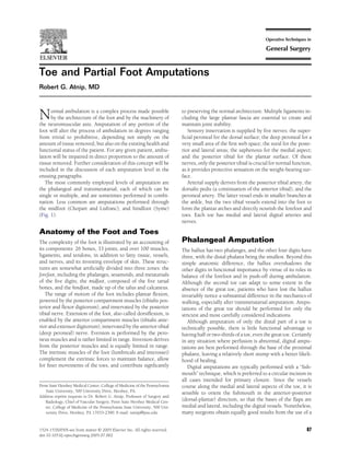
Figure 1 The skeleton of the foot, showing the level of bony transec-
tion for each of the four standard toe or partial foot amputations.
Creation of the soft-tissue flaps for each of these procedures is de-
scribed in more detail in the text.
medial-lateral fishmouth with anterior and posterior flaps. In
either case, the incisions are arc shaped and symmetric, each
encompassing a hemi-circumference of the toe. It is often
necessary for the apex of the flap to extend rather close to the
margin of necrosis, but the surgeon must visually verify that
the skin margins of the flap are viable and not grossly in-
fected. If the surgeon has any doubt regarding the skin mar-
gins, the wound might be better left open temporarily.
The soft tissue of the toes is sparse, consisting of skin,
minimal subcutaneous fat with nerves and vessels, investing
fascia, and tendons within their sheaths. Flaps must therefore
be incised perpendicular to the skin, full thickness down to
the bone, preserving all soft tissue with the flap. The flaps
should initially be generously long (as the distal pathology
permits), with the intent of shortening them to optimal
length for a tension-free closure. After stripping of the peri-
osteum, the bone should be amputated through the mid-
shaft, and then shortened and smoothed with a rongeur
down to the base, taking care not to violate the metatarso- Figure 2 A simple amputation through the proximal phalanx of the
phalangeal joint. The large flexor and extensor tendons left great toe. Symmetric medial and lateral flaps have been created,
should be then be distracted, amputated sharply, and al- based on the digital arteries. The stump of the phalanx is visible in
lowed to retract into the deeper soft tissues. Any final de- the base of the wound, along with the cut ends of the extensor and
bridement of the flaps can then be performed (Fig. 2). flexor tendons.
3. Toe and partial foot amputations 69
of the forefoot are extensively compromised, however, TMA
is unlikely to be a realistic option.
It is important to note that TMA includes resection of the
metatarsal head. Although sometimes tempting, amputation
of a toe through the metatarso-phalangeal (MTP) joint should
be avoided for several reasons. Leaving the metatarsal head
does not improve function, and instead creates a potential
pressure point that may predispose to recurrent ulceration
and infection. The bulk of the metatarsal head can make skin
closure more difficult. Since articular cartilage depends on
synovial fluid for its nutrient supply, the cartilage may die
once the joint has been disrupted. Removing the cartilage but
leaving the bony head offers no advantage over amputation of
the entire distal metatarsal.
Transmetatarsal amputation is indicated primarily in two
situations: necrosis or ulceration of the toe(s) at or proximal
to the level of the MTP joint; and/or plantar pressure ulcer-
ation over the metatarsal heads. The extent of the amputation
is dictated by the extent of necrosis, and can encompass a
single toe, two or three toes, or the entire forefoot. These
variations will be considered separately in the following para-
graphs.
Single Outer-Toe TMA
The toe and its metatarsal are sometimes called a “ray,” and
the corresponding surgery can be called a “ray” amputation.
The most commonly performed single ray amputations are
those of the first or fifth toes. Each is performed by the use of Figure 3 An example of the “racket-handle” type of incision used for
a “racket-handle” incision consisting of an elliptical cut transmetatarsal amputation of the great toe. The racket joins the
around the base of the affected toe, and a straight incision handle over the medial aspect of the metatarso-phalangeal joint, and
starting at the proximal end of the ellipse and continuing the handle extends along the metatarsal shaft. This incision can be
along the outer edge of the metatarsal shaft (Fig. 3). The exact modified for combined amputations of the first and second toes, and
contour of the incision must often be modified by the pattern can also be used for amputation of the fifth toe, or of the fourth and
fifth toes together.
of ulceration or necrosis of the toe, but must be designed to
preserve as much plantar skin and soft tissue as possible. It is
often convenient to use the elliptical incision to disarticulate
the toe at the MTP joint, and thus remove this ulcerated or then assess the closure potential of the dorsal and plantar
dead tissue from the surgical field before proceeding with the flaps. If at all possible, any redundancy should be trimmed
deeper dissection. This technique has the added advantage from the dorsal flap rather than the plantar, unless the plantar
that the metatarsal is easier to visualize and isolate after the tissue appears to be of poor quality. In cases where the flaps
toe itself has been removed (Fig. 4). will not approximate without tension, the surgeon has the
After disarticulation of the MTP joint, the joint capsule choices of resecting more bone, debulking the flaps, leaving
must be sharply and completely separated from the metatar- part of the wound open, or amputating the adjacent ray to
sal head. Great care must be taken in avoiding entry into the mobilize more soft tissue. When all is satisfactory, closure is
MTP joint of the adjacent ray, and in avoiding injury to the then performed as described in the preceding section.
plantar soft tissues abutting the shaft of the metatarsal. (In
these tissues are located the arterial supply to the plantar Single Inner-Toe TMA
flap.) Once the head is free, one then proceeds with stripping Transmetatarsal amputation of an inner toe (toes 2, 3, or 4)
of the periosteum of the metatarsal shaft to the desired level can be a useful procedure, but requires modifications in tech-
using a small elevator. The shaft is then divided with a bone nique. Because of the constraints imposed by the adjacent
cutter and recessed with a rongeur so that the stump is bev- rays, it is more difficult to perform isolated TMA of an inner
eled with the shorter edge on the plantar surface (to avoid a toe, and more difficult to obtain good closure. If the plantar
pressure point) (Fig. 5). tissues are relatively normal, the amputation can be done
The next step is to excise the remnants of the joint capsule, using the racket-handle technique, with the handle extend-
which in the case of the first toe, will include the sesamoid ing from the dorsal end of the ellipse along the dorsal surface
bone. These structures are virtually avascular and heal of the metatarsal shaft. Added difficulties occur when the
poorly. The dissection is best done with a very sharp #15 plantar skin is ulcerated or ischemic, in which case, it is
scalpel blade, taking only the ligamentous and bony compo- impossible to avoid an incision on the plantar weight-bearing
nents, and sparing the plantar fascia and other soft tissues. surface. In either case, the operation proceeds best by disar-
Once the tissue resection has been completed, one must ticulating and removing the toe at the MTP joint, freeing the
4. 70 R.G. Atnip
Figure 4 Transmetatarsal amputation
of the great toe. The specimen has
been removed after disarticulation of
the metatarso-phalangeal joint. The
sesamoid bone has been carefully ex-
cised from the plantar flap. The
transected flexor hallucis longus ten-
don can be seen posterior to the shaft
of the metatarsal. The plantar flap is
redundant, and will need to be
sculpted and trimmed before closure.
head from the joint capsule (while not entering the adjacent Multiple TMA
joints), stripping and resecting the desired length of shaft, Although in theory any combination of toes could be ampu-
and excising the remnants of joint capsule before closing. The tated at the TMA level, such a decision should take into
essentially fixed position of the adjacent metatarsal rays can account the relative importance of the various toes in the
make it rather difficult to close an inner-toe TMA without stability of the foot and the mechanics of walking. Significant
skin tension. The foot can be wrapped to compress the meta- stability and function are lost with amputation of the great
tarsals and reduce tension on the suture line, but only if toe, especially at the TMA level, and the loss is even greater if
precautions are taken to avoid pressure ulceration from the the second toe is also taken. To perform TMA of the first three
bandage itself. toes would likely be a disservice to the patient, leaving him/
Figure 5 Transmetatarsal amputation
of the great toe. The metatarsal shaft
has been cut on a posterior bevel, and
the plantar flap has been trimmed of
excess soft tissue. The flexor tendon
has been cut shorter than the bone.
The joint capsule of the adjacent sec-
ond MTP joint is intact, and has not
been entered or disrupted.
5. Toe and partial foot amputations 71
her with a narrow, tapered, and dysfunctional forefoot. Sim-
ilarly, the more toes removed from the lateral aspect of the
foot, the greater the asymmetry and imbalance of forces on
the remaining rays.
The technique for multiple TMA is a simple modification
of that for first or fifth ray amputation. An ellipitical incision
is created to encompass the base of the affected toes, modified
as needed to incorporate any areas of dorsal or plantar necro-
sis. The racket handle then extends along the outer aspect of
the metatarsal shaft. Flaps are created in identical fashion to
standard TMA. The MTP joints are disarticulated, the meta-
tarsal shafts amputated, recessed, and beveled appropriately.
The flaps are then sculpted and closed without tension.
Although preservation of the medial toes is more advanta-
geous than saving the lateral toes, it is questionable whether
TMA of more than two adjacent rays should ever be per-
formed. In patients with diabetic or other polyneuropathies,
amputations that create gross asymmetry of the forefoot are
associated with a notoriously high incidence of subsequent
breakdown and re-amputation. As a general rule, balance,
function, and stump integrity will be better with a complete
(full-foot) transmetatarsal amputation.
Full-Foot TMA
Amputation of the entire forefoot at the transmetatarsal level Figure 6 Flaps outlined for a “full foot” transmetatarsal amputation.
is one of the most useful procedures in the surgical armamen- The plantar flap is long, and the plantar incision extends along the
tarium. When properly performed, full-foot TMA results in a base of the toes. The dorsal incision crosses transversely over the
symmetric stump with favorable weight distribution. Al- mid- to distal level of the metatarsal shafts. Either the dorsal or
though there is no question that patients with TMA must plantar incisions may need to be modified if there is ulceration or
learn to adapt their balance, gait, and stride after loss of the necrosis of the forefoot.
forefoot, most patients will be able to walk, either indepen-
dently or with simple supportive devices. Foot orthoses or
custom shoes can be useful to facilitate walking, but prosthe- sesamoid bones and portions of the joint capsules, which
ses are not necessary. should be carefully excised, leaving adjacent muscle and ves-
If the plantar tissues are intact, the plantar incision for sels intact. All potentially viable skin and soft tissue of both
TMA crosses the foot as close to the base of the toes as pos- dorsal and plantar flaps should be spared until the final stage
sible. The dorsal incision is made across the mid- to distal of the procedure. Excess tissue can be removed and flaps
level of the metatarsal shafts, as dictated by the pattern of trimmed during closure, once it is known how the flaps can
forefoot necrosis (Fig. 6). The dorsal and plantar incisions are best be re-approximated.
then connected by axial incisions made along the shafts of the In the presence of ulceration or necrosis on the plantar
first and fifth metatarsals. The result will be a plantar flap of surface, the placement of the plantar incision and the creation
variable length. In developing the plantar flap, the incision of the plantar flap will need to be individualized. In the
should be carried down to the MTP joints, which should all common case of a neuropathic ulcer penetrating to the meta-
then be disarticulated. This allows the surgeon to find the tarsal head, the ulcer can be excised in elliptical or V-shaped
proper plane along the plantar surface of the metatarsal head fashion, which in essence will create two plantar flaps and
and shaft. From the plantar approach, the metatarsal shafts hence require a final T-shaped suture line. If the plantar
angle toward the dorsum of the foot as they traverse proxi- necrosis is more medial or lateral than central, the remaining
mally, and it is imperative that the surgeon adhere closely to plantar tissue can often be rotated to achieve final closure. In
the shafts to preserve the muscles and vessels of the plantar such situations, some of the metatarsal shafts may need to be
flap. amputated shorter than others to enable closure of the flaps
The dorsal incision is carried directly down through the without tension. It is in these cases that the imagination and
soft tissues, extensor tendons, and dorsal vessels to the ante- reconstructive skill of the surgeon become especially impor-
rior surface of the metatarsal shafts. At the desired level, these tant.
shafts are stripped of periosteum and divided with bone cut- Like most amputations below the ankle, a full-foot TMA
ter or rongeur. Working simultaneously from the plantar lends itself to only one layer of closure, the skin. In essence,
surface, the interosseus muscles are divided along with any the dorsal surface consists of skin, virtually no subcutaneous
remaining ligaments and tendons, and the specimen re- fat, and a very thin layer of fascia. If the plantar flap is too
moved. The metatarsal stumps should be recessed and bev- long, it should be shortened to eliminate redundancy and
eled, shorter on the plantar aspect. dead space (and thereby minimize the chance of hematoma).
Remaining on the plantar flap at this point will be the The optimal length is that which brings the plantar tissues up
6. 72 R.G. Atnip
Figure 7 Closure of the transmetatar-
sal amputation with simple inter-
rupted sutures. The metatarsal shafts
have been cut with a posterior bevel,
essentially flush with the dorsal inci-
sion. The plantar flap has been
sculpted to approximate the dorsal
tissue without tension or redundancy.
to abut and securely cover the bony stumps with minimal employed in America by battlefield surgeons in the Civil
dead space, while allowing the plantar and dorsal skin to be War. They hold out the prospect of saving part of the foot in
sutured without tension (Fig. 7). patients who fail or are not eligible for TMA, but they are
Given that the success and functionality of forefoot am- seldom used in modern amputation surgery. The chief dis-
putation are much superior to that of mid- or hindfoot advantage of the Lisfranc and Chopart procedures is that they
amputations, there can be a role for a certain surgical disrupt the tendinous attachments of the midfoot and predis-
“license” in performing modified TMA for patients with pose to stump deformities associated with dysfunctional am-
extensive forefoot necrosis. One option is to amputate the bulation. The loss of foot length and loss of tendon insertions
metatarsal shafts very short, provided that the surgeon is leaves the plantar flexors almost unopposed, resulting in an
aware of the dangers inherent in violating the tarso-meta- equinus deformity, with a consequent shift of weight bearing
tarsal joints. Removal of the first and/or fifth metatarsal from the calcaneus onto the stump itself. Although technical
bases will result in loss of part of the insertion of the modifications have been introduced that partly compensate
tibialis posterior and peroneus tendons, respectively. The for this imbalance of forces, midfoot amputation has still not
ensuing imbalance of forces on the TMA stump leads to gained wide acceptance as an alternative to below-knee am-
deformity, pressure ulceration, and impaired walking. putation. Braces and prostheses are usually required for
Wholesale entry into the tarso-metatarsal joints is tanta- walking, and there is a relatively high incidence of conversion
mount to performing a Lisfranc amputation, which is dis- to BKA.
cussed in the following section. The Lisfranc amputation is essentially a disarticulation of
If the bone and deeper tissues are viable but local coverage the tarso-metatarsal joints, using a plantar flap for coverage
is inadequate, vacuum-assisted closure and/or skin grafting with a technique virtually identical to transmetatarsal ampu-
may allow an “open” TMA to eventually heal. In rare cases, tation. The important technical point is to remove as much
the surgeon may wish to consider a free tissue transfer to articular cartilage as possible from the cuneiform and cuboid
salvage the foot, but an almost ideal set of conditions must surfaces to circumvent cartilaginous necrosis. Various ten-
pertain to justify such a complex undertaking. The indica- don transfers, reattachments, and tendo-Achilles lengthening
tions, techniques, risks, and outcomes of free-tissue transfer (TAL) have been proposed to prevent equinus deformity, but
are beyond the scope of this monograph. results are often suboptimal.
The Chopart amputation shortens the foot even further
Midfoot Amputations by removing the entire mid- and forefoot through the
talo-navicular and calcaneo-cuboid joints. Once again, a
(Lisfranc and Chopart) plantar flap is used for coverage, but problems with stump
These two surgical procedures were introduced by French deformity tend to be even more common than with the
surgeons in the 19th century, and they were supposedly first Lisfranc.
7. Toe and partial foot amputations 73
Conclusions ing amputations must approach each procedure with the
finest exacting technique and attention to detail worthy of the
Locomotion is a fundamental human activity made possible craft.
by the structure and function of the foot. Most humans con-
sider the potential loss of part or all of the foot as catastrophic, Suggested Reading
and view amputation as a disfiguring and destructive proce- Attinger C, Cooper P, Blume P, Bulan E: The safest surgical incisions and
dure. Yet due to either trauma or disease, as many as 150,000 amputations applying the angiosome priciples and using the Doppler to
assess the arterial-arterial connections of the foot and ankle. Foot and
patients per year are confronted with the necessity for ampu-
Ankle Clinics 6:745-799, 2001
tation surgery, virtually always with no realistic alternative. Crinnion J, Hicks D: Transmetatarsal amputation: an 8-year experience. Ann
For these patients, properly performed amputation surgery is R Coll Surg Engl 84:291-295, 2002
a reconstructive procedure that rehabilitates and restores qual- Funk C, Young G: Subtotal pedal amputations. Biomechanical and intraop-
erative considerations. J Am Podiatr Med Assoc 91:6-12, 2001
ity of life, albeit, a different life than the patient might desire.
Pinzur MS, Pinto MA, Schon LC, Smith DG: Controversies in amputation
Although many patients have such advanced disease that loss surgery. Instr Course Lect 52:445-451, 2003
of the entire foot is inevitable, for some the goal of partial foot Rumenapf G: Borderline amputations in diabetics— open questions and
salvage is achievable. This chapter has described a variety of critical evaluation. Zentralblatt für Chirurgie 128:726-733, 2003
Sanders LJ: Transmetatarsal and midfoot amputations. Clin Podiatr Med
procedures that preserve structure and function of the foot
Surg 14:741-762, 1997
sufficient to enable ambulation without a limb prosthesis. To Smith DG: Principles of partial foot amputations in the diabetic. Instr Course
achieve the best results for each patient, the surgeon perform- Lect 48:321-329, 1999