SlideShare a Scribd company logo
1 of 39
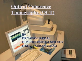
By  DR TAPAN JAKKAL  DEPT OF OPHTHALMOLOGY G.M.C.H AURANGABAD Optical Coherence Tomography (OCT)
Optical coherence tomography ,[object Object],[object Object],[object Object],[object Object],[object Object]
INTRODUCTION ,[object Object],[object Object]
Optical coherence tomography- The process is similar to that of ultrasonography, except that light is used instead of sound waves.   Analog to ultrasound
OCT (TD – principle) single reflection site
Construction of tomographic image Transverse Scanning Backscatter Intensity Axial Scanning (Depth)
[object Object],[object Object],[object Object]
Difficulties and limitations ,[object Object]
OCT Image of Normal Fovea  The OCT image above can be compared to what we know about retinal anatomy from conventional microscopic sections.  The vitreous is the black space on the top of the image.  We can identify the fovea by the normal depression.  The nerve fiber layer (NFL) and the retinal pigment epithelium (RPE) are easily identifiable.  These layers are more highly reflective than the other layers of the retina.  This higher reflectivity is represented by the "hotter" colors (red, yellow, orange, white) in the false color representation of the OCT .  The middle layers of the retina, between the NFL and RPE, are much less easily identifiable in the scan. 
Qualitative and Quantitative Analysis ,[object Object],[object Object],[object Object]
Regions ,[object Object],[object Object],[object Object],[object Object],[object Object]
Profiles ,[object Object],[object Object],[object Object],[object Object],[object Object]
The pre-retinal profile ,[object Object]
Anomalous structures that have been observed in the pre-retinal profile include the following:  1. pre-retinal membrane 2. epi-retinal membrane 3. vitreo-retinal strands 4. vitreo-retinal traction 5. pre-retinal neovascular membrane 6. pre-papillary neovascular membrane A pre-retinal membrane with traction on the fovea is pictured below.
The over-all retinal profile The normal over-all retinal profile has a slightly concave curvature that you would expect from observing the surface of a globe.  Abnormal profiles would include exaggerated concavity and convexity.  Retinal folds would also result in an abnormal over-all profile.
The following OCT image demonstrates an abnormal convexity in the over-all retinal profile.  In this case, a pigment epithelial detachment is causing the convexity.
The image below demonstrates an abnormal concavity to the over-all retinal profile.  Aside from the retinal detachment, notice the underlying concave curvature of the retina, suggesting the long eye of a significant myope.
The foveal profile The normal foveal profile is a slight depression in the surface of the retina, as pictured below.
Deformations that have been observed in the foveal profile include the following:   1. macular pucker 2. macular pseudo-hole 3. macular lamellar hole 4. macular cyst 5. macular hole, stage 1 (no depression, cyst present) 6. macular hole, stage 2 (partial rupture of retina, increased thickness) 7. macular hole, stage 3 (hole extends to RPE, increased thickness, some fluid) 8. macular hole, stage 4 (complete hole, edema at margins, complete PVD)  
 
macular cyst


The macular profile                                                                                                                                         The macular profile can, and often does,  include the fovea as it's center.  Therefore, a common OCT scan length of 6 mm would include 3 mm of the macula on each side of the fovea. 
Deformations that have been observed in the macular profile include the following:   1. serous retinal detachment (RD) 2. serous retinal pigment epithelial detachment (PED) 3. hemorrhagic pigment epithelial detachment   A serous PED is pictured below.  We know that it is a PED because the fluid (black space around the arrow) is pushing up underneath the retinal pigment epithelium, identified by the relatively highly reflective (red and orange) line (arrow).  
Intra-retinal anomalies that have been identified in the macular profile include:   1. choroidal neovascular membrane 2. diffuse intra-retinal edema 3. cystoid macular edema 4. drusen 5. hard exudates 6. scar tissue 7. atrophic degeneration 8. sub-retinal fibrosis 9. RPE tear
Cystoid Macular Edema OCT is capable of detecting small, fluid-filled, cystic spaces within the macula.
Central Serous Chorioretinopathy Central serous chorioretinopathy is characterized by the presence of fluid between the RPE and neurosensory retina.
Diabetic Retinopathy Exudates appear as accumulation of dense material within the neurosensory retina.
Artifacts ,[object Object],[object Object]
The scan below has waves in the retinal contour.  These are not retinal folds, but rather movement of the eye during the scan pass .
Scanning Tips ,[object Object],[object Object],[object Object],[object Object],[object Object]
6.Use the two buttons near the joystick for freezing and saving scans.  This saves you from having to juggle the joystick and the mouse. 7.Minimize patient fatigue by keeping scan time to a minimum.  Never scan an eye for more than 10 minutes (FDA regulation). 8.Keep the cornea lubricated.  Use artificial tears and have the patient blink when you are not saving a scan pass. 9.Move the instrument on the x and y axis (using the joystick) to work around opacities.
Anterior segment optical coherence tomography (OCT) ,[object Object],[object Object]
On the leading edge of anterior segment imaging: ,[object Object],[object Object],[object Object],[object Object],[object Object],[object Object]
Image shows an anterior-chamber angle as viewed with gonioscopy and the  OCT   The latter replaces subjective evaluation with objective measurement.
A narrow angle is apparent with OCT imaging, in this case 9.5°.
With the increase in popularity of anterior chamber imaging, and anterior segment OCT proving to be the best tool for high resolution biometry,  Anterior Segment Optical Coherence Tomography  is a  must-have  for anterior segment, refractive, cornea, and glaucoma surgeons.

More Related Content

What's hot

Intra Oular Lenses
Intra Oular LensesIntra Oular Lenses
Intra Oular Lenses
slidenka
 
Optical coherence tomography in glaucoma - Dr Shylesh Dabke
Optical coherence tomography in glaucoma - Dr Shylesh DabkeOptical coherence tomography in glaucoma - Dr Shylesh Dabke
Optical coherence tomography in glaucoma - Dr Shylesh Dabke
Shylesh Dabke
 

What's hot (20)

Intra Oular Lenses
Intra Oular LensesIntra Oular Lenses
Intra Oular Lenses
 
Keratoconus
KeratoconusKeratoconus
Keratoconus
 
Slit lamp biomicroscopy
Slit lamp biomicroscopySlit lamp biomicroscopy
Slit lamp biomicroscopy
 
Slit lamp techniques.pptx
Slit lamp techniques.pptxSlit lamp techniques.pptx
Slit lamp techniques.pptx
 
Entoptic phenomena
Entoptic phenomenaEntoptic phenomena
Entoptic phenomena
 
Low vision aids
Low vision aidsLow vision aids
Low vision aids
 
AS-OCT
AS-OCTAS-OCT
AS-OCT
 
OCT
OCTOCT
OCT
 
Clinical examination of squint
Clinical examination of squintClinical examination of squint
Clinical examination of squint
 
GDx
GDxGDx
GDx
 
Optical coherence tomography in glaucoma - Dr Shylesh Dabke
Optical coherence tomography in glaucoma - Dr Shylesh DabkeOptical coherence tomography in glaucoma - Dr Shylesh Dabke
Optical coherence tomography in glaucoma - Dr Shylesh Dabke
 
Optical coherence tomography(OCT) --macula
Optical coherence tomography(OCT) --maculaOptical coherence tomography(OCT) --macula
Optical coherence tomography(OCT) --macula
 
OCT
OCTOCT
OCT
 
Corneal drawings
Corneal drawingsCorneal drawings
Corneal drawings
 
Introduction to binocular single vision (BSV)
Introduction to binocular single vision (BSV)Introduction to binocular single vision (BSV)
Introduction to binocular single vision (BSV)
 
B SCAN
B SCAN B SCAN
B SCAN
 
Optical Coherence Tomography
Optical Coherence TomographyOptical Coherence Tomography
Optical Coherence Tomography
 
HRT and GDx VCC
HRT and GDx VCCHRT and GDx VCC
HRT and GDx VCC
 
Corneal topography
Corneal topographyCorneal topography
Corneal topography
 
Gonioscopy
GonioscopyGonioscopy
Gonioscopy
 

Viewers also liked

İnterpretation of optic coherence tomography images
İnterpretation of optic coherence tomography imagesİnterpretation of optic coherence tomography images
İnterpretation of optic coherence tomography images
Sinan çalışkan
 
Optical Coherence Tomography
Optical Coherence TomographyOptical Coherence Tomography
Optical Coherence Tomography
Manoj Aryal
 
The basics of retinal oct ophso.net
The basics of retinal oct ophso.netThe basics of retinal oct ophso.net
The basics of retinal oct ophso.net
kebaplik
 

Viewers also liked (12)

Optical coherence tomography
Optical coherence tomographyOptical coherence tomography
Optical coherence tomography
 
Optical Coherence Tomography dr md toufiqur rahman cardiologist
Optical Coherence Tomography dr md toufiqur rahman cardiologistOptical Coherence Tomography dr md toufiqur rahman cardiologist
Optical Coherence Tomography dr md toufiqur rahman cardiologist
 
OCT For Everyone..
OCT For Everyone..OCT For Everyone..
OCT For Everyone..
 
OCT - few basic facts
OCT - few basic factsOCT - few basic facts
OCT - few basic facts
 
Anatomy of retina
Anatomy of retinaAnatomy of retina
Anatomy of retina
 
OCT – Introduction and Macular disorders
OCT – Introduction and Macular disordersOCT – Introduction and Macular disorders
OCT – Introduction and Macular disorders
 
Optical coherence tomography
Optical coherence tomographyOptical coherence tomography
Optical coherence tomography
 
İnterpretation of optic coherence tomography images
İnterpretation of optic coherence tomography imagesİnterpretation of optic coherence tomography images
İnterpretation of optic coherence tomography images
 
Optical Coherence Tomography
Optical Coherence TomographyOptical Coherence Tomography
Optical Coherence Tomography
 
The basics of retinal oct ophso.net
The basics of retinal oct ophso.netThe basics of retinal oct ophso.net
The basics of retinal oct ophso.net
 
Spectralis oct normal anatomy & systematic interpretation.
Spectralis oct normal anatomy & systematic interpretation.Spectralis oct normal anatomy & systematic interpretation.
Spectralis oct normal anatomy & systematic interpretation.
 
SlideShare 101
SlideShare 101SlideShare 101
SlideShare 101
 

Similar to Optical Coherence Tomography - principle and uses in ophthalmology

Optical coherence tomography
Optical coherence tomographyOptical coherence tomography
Optical coherence tomography
Samuel Ponraj
 
Focal choroidal excavation in eyes with central serous chorioretinopathy
Focal choroidal excavation in eyes with central serous chorioretinopathy Focal choroidal excavation in eyes with central serous chorioretinopathy
Focal choroidal excavation in eyes with central serous chorioretinopathy
Abdallah Ellabban
 
Management of adult cataract II.ppt
Management of adult cataract II.pptManagement of adult cataract II.ppt
Management of adult cataract II.ppt
docsuleman
 
Management of adult cataract II.ppt
Management of adult cataract II.pptManagement of adult cataract II.ppt
Management of adult cataract II.ppt
docsuleman
 

Similar to Optical Coherence Tomography - principle and uses in ophthalmology (20)

Optical coherence tomography
Optical coherence tomographyOptical coherence tomography
Optical coherence tomography
 
Optical Coherence Tomography(OCT) in posterior segment diseases
Optical Coherence Tomography(OCT) in posterior segment diseasesOptical Coherence Tomography(OCT) in posterior segment diseases
Optical Coherence Tomography(OCT) in posterior segment diseases
 
oct-ujjval solanki
oct-ujjval solankioct-ujjval solanki
oct-ujjval solanki
 
Optical Coherence Tomography (OCT)
Optical Coherence Tomography (OCT)Optical Coherence Tomography (OCT)
Optical Coherence Tomography (OCT)
 
Oct demystified
Oct demystified  Oct demystified
Oct demystified
 
OCT in Ophthalmology
OCT in OphthalmologyOCT in Ophthalmology
OCT in Ophthalmology
 
Final oct
Final octFinal oct
Final oct
 
Role of oct in ophthalmology
Role of oct in ophthalmologyRole of oct in ophthalmology
Role of oct in ophthalmology
 
Oct introduction
Oct  introductionOct  introduction
Oct introduction
 
FOCAL CHOROIDAL EXCAVATION IN EYES WITH CENTRAL SEROUS ‎CHORIORETINOPATHY ‎
FOCAL CHOROIDAL EXCAVATION IN EYES WITH CENTRAL SEROUS ‎CHORIORETINOPATHY ‎FOCAL CHOROIDAL EXCAVATION IN EYES WITH CENTRAL SEROUS ‎CHORIORETINOPATHY ‎
FOCAL CHOROIDAL EXCAVATION IN EYES WITH CENTRAL SEROUS ‎CHORIORETINOPATHY ‎
 
Focal choroidal excavation in eyes with central serous chorioretinopathy
Focal choroidal excavation in eyes with central serous chorioretinopathy Focal choroidal excavation in eyes with central serous chorioretinopathy
Focal choroidal excavation in eyes with central serous chorioretinopathy
 
Oct and visual field by dr abdul basir safi
Oct and visual field by dr abdul basir safiOct and visual field by dr abdul basir safi
Oct and visual field by dr abdul basir safi
 
3D Tomgraphic features in dome shaped macula by swept source OCT
3D Tomgraphic features in dome shaped macula by swept source OCT3D Tomgraphic features in dome shaped macula by swept source OCT
3D Tomgraphic features in dome shaped macula by swept source OCT
 
Inferior posterior staphyloma: choroidal maps and macular complications
Inferior posterior staphyloma: choroidal maps and  macular complications Inferior posterior staphyloma: choroidal maps and  macular complications
Inferior posterior staphyloma: choroidal maps and macular complications
 
Optical/ocular coherence tomography OCT All in one Presentation
Optical/ocular coherence tomography OCT All in one PresentationOptical/ocular coherence tomography OCT All in one Presentation
Optical/ocular coherence tomography OCT All in one Presentation
 
Focal choroidal excavation in eyes with central serous chorioretinopathy
Focal choroidal excavation in eyes with central serous chorioretinopathyFocal choroidal excavation in eyes with central serous chorioretinopathy
Focal choroidal excavation in eyes with central serous chorioretinopathy
 
Dome shaped macula
Dome shaped macula Dome shaped macula
Dome shaped macula
 
Macular function tests
Macular function testsMacular function tests
Macular function tests
 
Management of adult cataract II.ppt
Management of adult cataract II.pptManagement of adult cataract II.ppt
Management of adult cataract II.ppt
 
Management of adult cataract II.ppt
Management of adult cataract II.pptManagement of adult cataract II.ppt
Management of adult cataract II.ppt
 

Recently uploaded

Call Girls Bhubaneswar Just Call 9907093804 Top Class Call Girl Service Avail...
Call Girls Bhubaneswar Just Call 9907093804 Top Class Call Girl Service Avail...Call Girls Bhubaneswar Just Call 9907093804 Top Class Call Girl Service Avail...
Call Girls Bhubaneswar Just Call 9907093804 Top Class Call Girl Service Avail...
Dipal Arora
 
College Call Girls in Haridwar 9667172968 Short 4000 Night 10000 Best call gi...
College Call Girls in Haridwar 9667172968 Short 4000 Night 10000 Best call gi...College Call Girls in Haridwar 9667172968 Short 4000 Night 10000 Best call gi...
College Call Girls in Haridwar 9667172968 Short 4000 Night 10000 Best call gi...
perfect solution
 
Call Girls in Gagan Vihar (delhi) call me [🔝 9953056974 🔝] escort service 24X7
Call Girls in Gagan Vihar (delhi) call me [🔝  9953056974 🔝] escort service 24X7Call Girls in Gagan Vihar (delhi) call me [🔝  9953056974 🔝] escort service 24X7
Call Girls in Gagan Vihar (delhi) call me [🔝 9953056974 🔝] escort service 24X7
9953056974 Low Rate Call Girls In Saket, Delhi NCR
 

Recently uploaded (20)

Night 7k to 12k Navi Mumbai Call Girl Photo 👉 BOOK NOW 9833363713 👈 ♀️ night ...
Night 7k to 12k Navi Mumbai Call Girl Photo 👉 BOOK NOW 9833363713 👈 ♀️ night ...Night 7k to 12k Navi Mumbai Call Girl Photo 👉 BOOK NOW 9833363713 👈 ♀️ night ...
Night 7k to 12k Navi Mumbai Call Girl Photo 👉 BOOK NOW 9833363713 👈 ♀️ night ...
 
Best Rate (Hyderabad) Call Girls Jahanuma ⟟ 8250192130 ⟟ High Class Call Girl...
Best Rate (Hyderabad) Call Girls Jahanuma ⟟ 8250192130 ⟟ High Class Call Girl...Best Rate (Hyderabad) Call Girls Jahanuma ⟟ 8250192130 ⟟ High Class Call Girl...
Best Rate (Hyderabad) Call Girls Jahanuma ⟟ 8250192130 ⟟ High Class Call Girl...
 
Best Rate (Patna ) Call Girls Patna ⟟ 8617370543 ⟟ High Class Call Girl In 5 ...
Best Rate (Patna ) Call Girls Patna ⟟ 8617370543 ⟟ High Class Call Girl In 5 ...Best Rate (Patna ) Call Girls Patna ⟟ 8617370543 ⟟ High Class Call Girl In 5 ...
Best Rate (Patna ) Call Girls Patna ⟟ 8617370543 ⟟ High Class Call Girl In 5 ...
 
Call Girls in Delhi Triveni Complex Escort Service(🔝))/WhatsApp 97111⇛47426
Call Girls in Delhi Triveni Complex Escort Service(🔝))/WhatsApp 97111⇛47426Call Girls in Delhi Triveni Complex Escort Service(🔝))/WhatsApp 97111⇛47426
Call Girls in Delhi Triveni Complex Escort Service(🔝))/WhatsApp 97111⇛47426
 
Call Girls Coimbatore Just Call 9907093804 Top Class Call Girl Service Available
Call Girls Coimbatore Just Call 9907093804 Top Class Call Girl Service AvailableCall Girls Coimbatore Just Call 9907093804 Top Class Call Girl Service Available
Call Girls Coimbatore Just Call 9907093804 Top Class Call Girl Service Available
 
VIP Hyderabad Call Girls Bahadurpally 7877925207 ₹5000 To 25K With AC Room 💚😋
VIP Hyderabad Call Girls Bahadurpally 7877925207 ₹5000 To 25K With AC Room 💚😋VIP Hyderabad Call Girls Bahadurpally 7877925207 ₹5000 To 25K With AC Room 💚😋
VIP Hyderabad Call Girls Bahadurpally 7877925207 ₹5000 To 25K With AC Room 💚😋
 
Call Girls Bhubaneswar Just Call 9907093804 Top Class Call Girl Service Avail...
Call Girls Bhubaneswar Just Call 9907093804 Top Class Call Girl Service Avail...Call Girls Bhubaneswar Just Call 9907093804 Top Class Call Girl Service Avail...
Call Girls Bhubaneswar Just Call 9907093804 Top Class Call Girl Service Avail...
 
Top Rated Bangalore Call Girls Richmond Circle ⟟ 9332606886 ⟟ Call Me For Ge...
Top Rated Bangalore Call Girls Richmond Circle ⟟  9332606886 ⟟ Call Me For Ge...Top Rated Bangalore Call Girls Richmond Circle ⟟  9332606886 ⟟ Call Me For Ge...
Top Rated Bangalore Call Girls Richmond Circle ⟟ 9332606886 ⟟ Call Me For Ge...
 
Call Girls Dehradun Just Call 9907093804 Top Class Call Girl Service Available
Call Girls Dehradun Just Call 9907093804 Top Class Call Girl Service AvailableCall Girls Dehradun Just Call 9907093804 Top Class Call Girl Service Available
Call Girls Dehradun Just Call 9907093804 Top Class Call Girl Service Available
 
Russian Call Girls Service Jaipur {8445551418} ❤️PALLAVI VIP Jaipur Call Gir...
Russian Call Girls Service  Jaipur {8445551418} ❤️PALLAVI VIP Jaipur Call Gir...Russian Call Girls Service  Jaipur {8445551418} ❤️PALLAVI VIP Jaipur Call Gir...
Russian Call Girls Service Jaipur {8445551418} ❤️PALLAVI VIP Jaipur Call Gir...
 
Premium Call Girls In Jaipur {8445551418} ❤️VVIP SEEMA Call Girl in Jaipur Ra...
Premium Call Girls In Jaipur {8445551418} ❤️VVIP SEEMA Call Girl in Jaipur Ra...Premium Call Girls In Jaipur {8445551418} ❤️VVIP SEEMA Call Girl in Jaipur Ra...
Premium Call Girls In Jaipur {8445551418} ❤️VVIP SEEMA Call Girl in Jaipur Ra...
 
Call Girls Cuttack Just Call 9907093804 Top Class Call Girl Service Available
Call Girls Cuttack Just Call 9907093804 Top Class Call Girl Service AvailableCall Girls Cuttack Just Call 9907093804 Top Class Call Girl Service Available
Call Girls Cuttack Just Call 9907093804 Top Class Call Girl Service Available
 
Call Girls Ludhiana Just Call 9907093804 Top Class Call Girl Service Available
Call Girls Ludhiana Just Call 9907093804 Top Class Call Girl Service AvailableCall Girls Ludhiana Just Call 9907093804 Top Class Call Girl Service Available
Call Girls Ludhiana Just Call 9907093804 Top Class Call Girl Service Available
 
College Call Girls in Haridwar 9667172968 Short 4000 Night 10000 Best call gi...
College Call Girls in Haridwar 9667172968 Short 4000 Night 10000 Best call gi...College Call Girls in Haridwar 9667172968 Short 4000 Night 10000 Best call gi...
College Call Girls in Haridwar 9667172968 Short 4000 Night 10000 Best call gi...
 
Call Girls Gwalior Just Call 8617370543 Top Class Call Girl Service Available
Call Girls Gwalior Just Call 8617370543 Top Class Call Girl Service AvailableCall Girls Gwalior Just Call 8617370543 Top Class Call Girl Service Available
Call Girls Gwalior Just Call 8617370543 Top Class Call Girl Service Available
 
Pondicherry Call Girls Book Now 9630942363 Top Class Pondicherry Escort Servi...
Pondicherry Call Girls Book Now 9630942363 Top Class Pondicherry Escort Servi...Pondicherry Call Girls Book Now 9630942363 Top Class Pondicherry Escort Servi...
Pondicherry Call Girls Book Now 9630942363 Top Class Pondicherry Escort Servi...
 
Call Girls Bareilly Just Call 8250077686 Top Class Call Girl Service Available
Call Girls Bareilly Just Call 8250077686 Top Class Call Girl Service AvailableCall Girls Bareilly Just Call 8250077686 Top Class Call Girl Service Available
Call Girls Bareilly Just Call 8250077686 Top Class Call Girl Service Available
 
Call Girls in Gagan Vihar (delhi) call me [🔝 9953056974 🔝] escort service 24X7
Call Girls in Gagan Vihar (delhi) call me [🔝  9953056974 🔝] escort service 24X7Call Girls in Gagan Vihar (delhi) call me [🔝  9953056974 🔝] escort service 24X7
Call Girls in Gagan Vihar (delhi) call me [🔝 9953056974 🔝] escort service 24X7
 
All Time Service Available Call Girls Marine Drive 📳 9820252231 For 18+ VIP C...
All Time Service Available Call Girls Marine Drive 📳 9820252231 For 18+ VIP C...All Time Service Available Call Girls Marine Drive 📳 9820252231 For 18+ VIP C...
All Time Service Available Call Girls Marine Drive 📳 9820252231 For 18+ VIP C...
 
Top Rated Bangalore Call Girls Ramamurthy Nagar ⟟ 9332606886 ⟟ Call Me For G...
Top Rated Bangalore Call Girls Ramamurthy Nagar ⟟  9332606886 ⟟ Call Me For G...Top Rated Bangalore Call Girls Ramamurthy Nagar ⟟  9332606886 ⟟ Call Me For G...
Top Rated Bangalore Call Girls Ramamurthy Nagar ⟟ 9332606886 ⟟ Call Me For G...
 

Optical Coherence Tomography - principle and uses in ophthalmology

  • 1. By DR TAPAN JAKKAL DEPT OF OPHTHALMOLOGY G.M.C.H AURANGABAD Optical Coherence Tomography (OCT)
  • 2.
  • 3.
  • 4. Optical coherence tomography- The process is similar to that of ultrasonography, except that light is used instead of sound waves.   Analog to ultrasound
  • 5. OCT (TD – principle) single reflection site
  • 6. Construction of tomographic image Transverse Scanning Backscatter Intensity Axial Scanning (Depth)
  • 7.
  • 8.
  • 9. OCT Image of Normal Fovea The OCT image above can be compared to what we know about retinal anatomy from conventional microscopic sections.  The vitreous is the black space on the top of the image.  We can identify the fovea by the normal depression.  The nerve fiber layer (NFL) and the retinal pigment epithelium (RPE) are easily identifiable.  These layers are more highly reflective than the other layers of the retina.  This higher reflectivity is represented by the "hotter" colors (red, yellow, orange, white) in the false color representation of the OCT .  The middle layers of the retina, between the NFL and RPE, are much less easily identifiable in the scan. 
  • 10.
  • 11.
  • 12.
  • 13.
  • 14. Anomalous structures that have been observed in the pre-retinal profile include the following:  1. pre-retinal membrane 2. epi-retinal membrane 3. vitreo-retinal strands 4. vitreo-retinal traction 5. pre-retinal neovascular membrane 6. pre-papillary neovascular membrane A pre-retinal membrane with traction on the fovea is pictured below.
  • 15. The over-all retinal profile The normal over-all retinal profile has a slightly concave curvature that you would expect from observing the surface of a globe.  Abnormal profiles would include exaggerated concavity and convexity.  Retinal folds would also result in an abnormal over-all profile.
  • 16. The following OCT image demonstrates an abnormal convexity in the over-all retinal profile.  In this case, a pigment epithelial detachment is causing the convexity.
  • 17. The image below demonstrates an abnormal concavity to the over-all retinal profile.  Aside from the retinal detachment, notice the underlying concave curvature of the retina, suggesting the long eye of a significant myope.
  • 18. The foveal profile The normal foveal profile is a slight depression in the surface of the retina, as pictured below.
  • 19. Deformations that have been observed in the foveal profile include the following:   1. macular pucker 2. macular pseudo-hole 3. macular lamellar hole 4. macular cyst 5. macular hole, stage 1 (no depression, cyst present) 6. macular hole, stage 2 (partial rupture of retina, increased thickness) 7. macular hole, stage 3 (hole extends to RPE, increased thickness, some fluid) 8. macular hole, stage 4 (complete hole, edema at margins, complete PVD)  
  • 20.  
  • 22.  
  • 23.  
  • 24.  
  • 25. The macular profile                                                                                                                                         The macular profile can, and often does,  include the fovea as it's center.  Therefore, a common OCT scan length of 6 mm would include 3 mm of the macula on each side of the fovea. 
  • 26. Deformations that have been observed in the macular profile include the following:   1. serous retinal detachment (RD) 2. serous retinal pigment epithelial detachment (PED) 3. hemorrhagic pigment epithelial detachment   A serous PED is pictured below.  We know that it is a PED because the fluid (black space around the arrow) is pushing up underneath the retinal pigment epithelium, identified by the relatively highly reflective (red and orange) line (arrow).  
  • 27. Intra-retinal anomalies that have been identified in the macular profile include:   1. choroidal neovascular membrane 2. diffuse intra-retinal edema 3. cystoid macular edema 4. drusen 5. hard exudates 6. scar tissue 7. atrophic degeneration 8. sub-retinal fibrosis 9. RPE tear
  • 28. Cystoid Macular Edema OCT is capable of detecting small, fluid-filled, cystic spaces within the macula.
  • 29. Central Serous Chorioretinopathy Central serous chorioretinopathy is characterized by the presence of fluid between the RPE and neurosensory retina.
  • 30. Diabetic Retinopathy Exudates appear as accumulation of dense material within the neurosensory retina.
  • 31.
  • 32. The scan below has waves in the retinal contour.  These are not retinal folds, but rather movement of the eye during the scan pass .
  • 33.
  • 34. 6.Use the two buttons near the joystick for freezing and saving scans.  This saves you from having to juggle the joystick and the mouse. 7.Minimize patient fatigue by keeping scan time to a minimum.  Never scan an eye for more than 10 minutes (FDA regulation). 8.Keep the cornea lubricated.  Use artificial tears and have the patient blink when you are not saving a scan pass. 9.Move the instrument on the x and y axis (using the joystick) to work around opacities.
  • 35.
  • 36.
  • 37. Image shows an anterior-chamber angle as viewed with gonioscopy and the OCT The latter replaces subjective evaluation with objective measurement.
  • 38. A narrow angle is apparent with OCT imaging, in this case 9.5°.
  • 39. With the increase in popularity of anterior chamber imaging, and anterior segment OCT proving to be the best tool for high resolution biometry, Anterior Segment Optical Coherence Tomography is a must-have for anterior segment, refractive, cornea, and glaucoma surgeons.