Mbj female genitalia pictures
•Als PPT, PDF herunterladen•
0 gefällt mir•7,018 views

Imágenes sobre tipos de mutilación genital femenina

Empfohlen
Weitere ähnliche Inhalte
Kürzlich hochgeladen
Kürzlich hochgeladen (20)
Empfohlen
Empfohlen (20)
Mbj female genitalia pictures
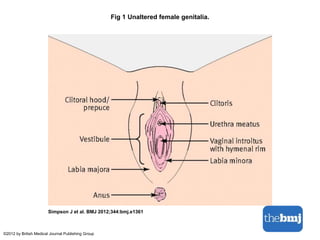
- 1. Fig 1 Unaltered female genitalia. Simpson J et al. BMJ 2012;344:bmj.e1361 ©2012 by British Medical Journal Publishing Group
- 2. Fig 2 Tissue removed in type 1 female genital mutilation (top) and type 2 female genital mutilation (bottom). Simpson J et al. BMJ 2012;344:bmj.e1361 ©2012 by British Medical Journal Publishing Group
- 3. Fig 4 Clinical appearance of type 2 female genital mutilation. Simpson J et al. BMJ 2012;344:bmj.e1361 ©2012 by British Medical Journal Publishing Group
- 4. Fig 3 Appearance of type 2 female genital mutilation (top) and type 3 female genital mutilation (bottom). Simpson J et al. BMJ 2012;344:bmj.e1361 ©2012 by British Medical Journal Publishing Group
Hinweis der Redaktion
- Fig 1 Unaltered female genitalia
- Fig 2 Tissue removed in type 1 female genital mutilation (top) and type 2 female genital mutilation (bottom)
- Fig 4 Clinical appearance of type 2 female genital mutilation. The large arrow indicates scar tissue superior to the vagina and the smaller arrow indicates the vestibule with no narrowing of the vaginal orifice
- Fig 3 Appearance of type 2 female genital mutilation (top) and type 3 female genital mutilation (bottom)