This document discusses the anatomy of the muscles of the pelvis and hip. It describes the origin, insertion, action, and innervation of key hip muscles including the gluteals, hamstrings, hip adductors, and hip flexors. It also covers topics like plexus formation, reversal of muscle function, and the roles of certain muscles in stabilizing the pelvis. The document provides a comprehensive overview of the major muscles involved in movement and control of the hip joint.
34. Nervous System
Highly
complex
Controls, stimulates, and coordinates all
other body systems
Can be divided:
– Central Nervous System (CNS)
– Peripheral Nervous System (PNS)
– Autonomic Nervous System (ANS)
36. Peripheral Nervous System
(PNS)
Composed
of all the nervous tissue outside
the vertebral canal
Begins at the anterior horn of the spinal
cord
37. Branches of the Spinal Nerves
Posterior
primary rami
innervate the deep
muscles of the back and
the skin covering these
muscles
Spinal nerve continues as
the anterior primary
ramus
38. Branches of the Spinal Nerves
(cont’d)
Anterior
primary
rami innervate all
muscles and skin not
innervated by the
posterior primary rami
39. Plexus Formation
Anterior
branches of
spinal nerves in
thoracic region give
rise to intercostal
nerves (not a plexus)
Spinal nerves in other
regions combine to
form complex
networks called
plexuses
42. Iliopsoas
Actually two muscles
– Separate origin
– Common insertion
O: Iliacus - iliac fossa
Psoas major - anterior
and lateral surfaces
T12-L5
I: Lesser trochanter
A:
Hip flexion
N:
Iliacus - Femoral Nerve
Psoas Major - L2-3
43. Rectus Femoris
Part of the quadriceps
Two-joint muscle crosses hip
and knee anteriorly
O: Anterior inferior iliac spine
I: Tibial tuberosity
A: Hip flexion, Knee extension
N: Femoral Nerve, L2-L4
44. Sartorius
Longest muscle in the body
Straplike muscle
“Tailor’s Muscle”
O: Anterior superior iliac spine
I: Proximal medial aspect of tibia
A: Hip flexion, abduction, lateral
rotation
N: Femoral Nerve, L2-L3
45. Pectineus
Located
– Medial to the iliopsoas
– Lateral to the adductor
longus
O:
I:
A:
N:
Superior ramus of pubis
Pectineal line of femur
Hip flexion and adduction
Femoral Nerve, L2-L4
46. Adductor Longus (A)
Most
superficial
– Tendon palpable in anterior-
medial groin
O:
I:
A:
N:
Pubis
Middle 1/3 linea
aspera
Hip adduction
Obturator Nerve,
L3-L4
47. Adductor Brevis (B)
Shorter
Deep to adductor longus
Superficial to adductor magnus
O: Pubis
I: Pectineal line and proximal
linea aspera
A: Hip adduction
N: Obturator Nerve, L3-4
48.
49.
50. Adductor Magnus (C)
Largest, most massive, and
deepest
Opening or hiatus in distal
attachment between linea
aspera and adductor tubercle
– Femoral artery and vein pass
through opening
– After femoral artery and vein
pass through, their names
change to popliteal artery and
vein
51. Adductor Magnus (C)
(cont’d)
O: Ischium and pubis
I: Entire linea aspera and
adductor tubercle
A: Hip adduction, extension
N: Obturator and sciatic nerve
(L3-L4)
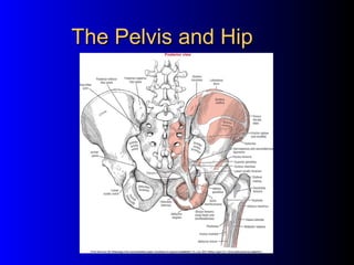
54. Gluteus Maximus
Large,
one-joint, quadrilateral-shaped,
thick, superficial muscle
Some fibers attach to iliotibial band
O:Posterior sacrum and ilium
I:
Posterior femur distal to
greater trochanter
A:
Hip extension, hyperextension,
lateral rotation
N:
Inferior gluteal nerve, L5, S1, S2
55.
56. Deep Rotator
Muscles
Number = 6, deep, mostly
posterior muscles spanning
the hip joint in a horizontal
direction
Laterally rotate the hip
O:
Posterior sacrum,
ischium, pubis
I:
Greater trochanter
area
A:
Hip lateral rotation
57. Deep Rotator Muscles
Muscle
Proximal Attachment
Obturator externus Rami of pubis and ischium
Obturator internus Rami of pubis and ischium
Quadratus femoris Ischial tuberosity
Piriformis
Sacrum
Gemellus superior Ischium
Gemellus inferior
Ischial tuberosity
Distal Attachment
Trochanteric fossa
Greater trochanter
Innervation
Obturator nerve
Nerve to obturator
internus
Intertrochanteric crest
Nerve to quadratus
femoris
Greater trochanter
S1, S2 segments
Greater trochanter
Nerve to obturator
internus
Greater trochanter
Nerve to quadratus
femoris
58. Deep Rotator Muscles (cont’d)
“Piece Goods Often Go On Quilts”
Anatomical order superior to inferior
– Piriformis
– Gemellus superior
– Obturator internus
– Gemellus inferior
– Obturator externus
– Quadratus femoris
59. Hamstrings
Posterior
thigh muscles
Two joint muscles except
short head of biceps femoris
Ischial tuberosity - common
origin
A.
B.
C.
Semitendinosus
Biceps femoris
Semimembranosus
63. Biceps Femoris (B)
O:
I:
A:
N:
Long head: Ischial tuberosity
Short head: lateral lip linea aspera
Fibular head
Long head: Extend hip, flex knee
Short head: Flex knee
Long head: Sciatic nerve, S1-S3
Short head: Common peroneal
nerve, L5-S2
66. Gluteus Minimus
Deep
and inferior to the gluteus
medius
O:
I:
Lateral ilium
Anterior surface of the
greater trochanter
A:
Hip abduction, internal
rotation
N:
Superior gluteal nerve
L4, L5, S1
67. Reversal of Muscle Function
When
you stand on one leg:
– Distal segment (femur) becomes more stable than the
proximal segment (os coxae)
– Origin moves toward insertion
Without
contracting - pelvis would drop on opposite
side
Weakness “Trendelenberg gait”
– If weak right hip abductors, stand on right, left side drops
68. Reversal of Muscle
Function (cont’d)
A. In reversal of muscle
function, the right hip
abductors contract to
keep the pelvis
steady when the left
leg is lifted.
B. When right hip
abductors are weak,
the left side of the
pelvis drops.
69. Tensor Fascia Latae
Short muscle
Long tendinous attachment
O: Anterior superior iliac spine
I: Lateral condyle of tibia
A: Combined hip flexion and
abduction
N: Superior gluteal nerve
L4-L5