
Empfohlen
Weitere ähnliche Inhalte
Was ist angesagt?
Was ist angesagt? (20)
Andere mochten auch
Andere mochten auch (19)
Ähnlich wie Orbital lymphangioma
Ähnlich wie Orbital lymphangioma (20)
Mehr von Ritesh Mahajan
Mehr von Ritesh Mahajan (20)
Kürzlich hochgeladen
Kürzlich hochgeladen (20)
Orbital lymphangioma
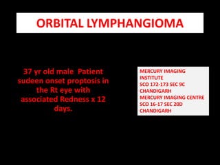
- 1. ORBITAL LYMPHANGIOMA 37 yr old male Patient sudeen onset proptosis in the Rt eye with associated Redness x 12 days. MERCURY IMAGING INSTITUTE SCO 172-173 SEC 9C CHANDIGARH MERCURY IMAGING CENTRE SCO 16-17 SEC 20D CHANDIGARH
- 2. PRESENT CASE Large macrolobulatedintraconal / extraconal mass In the RtRetroorbitalregion with following features : Fluid signal levels with acute / subacute stage haemorrhage Minimal post contrast enhancement Sudden onset proptosis .
- 3. Some facts ................................ Lymphangioma is a benign vascular tumour which is probably congenital, slowly growing and may not become clinically apparent for months and for years.Thetumour may affect the conjunctiva, the lids and the orbit. Associated similar extraorbital lesions include facial and palatal cystic lesions. Intracranial vascular anomalies have been reported to be associated with the lesion.
- 4. Intraconal / extra conal mass with macrolobulation is appreciated in the Rt orbit. Multiple fluid signal levels are appreciated . Supernatant and subnatant signal corroborates with acute / subacute stage haemorrhage.
- 5. POST CONTRAST ENHANCEMENT IS MINIMAL – SUPPORTS LYMPHANGIOMA AS POSSBILITY
- 6. FLUID LEVELS IN MULTIPLE PLANES Differentiate haemorrhagic cysts from the lymphangitic cysts . ( Haemorrhagic cysts have variable signal as per stage) ( Lymphatic cysts have hypointense signal on T1 and hyperintense signal on T2 )
- 7. GRADIENT SEQUENCE DONE TO ASSESS BLOOM IN THE FLUID SIGNAL LEVELS - Rules out chronic haemorrhage
- 8. LOBULATED CONTOUR WITH MASS EFFECT ON THE ADJACENT MUSCLE COMPARTMENTS .
- 9. PYRAMIDAL SHAPE OF THE LESION WITH BASE TOWRADS THE GLOBE
- 10. Lesson learnt........................ MR angiogram of the brain should be combined with Routine imaging of the orbital lesion to assess the connection of the orbital lesion with intracranial arterial / venous channels. Another terminology used for such kind of lesions is combined venous lymphatic -vascular malformations. Types of lymphangioma – Orbital , Superficial , Combined, Superficial lymphangioma – Preseptal compartment. Orbital lymphangioma – Post septal compartment. ( Both intraconal / extra conal components ). Combined lymphangioma – Both preseptal and post septal compartments.