
Empfohlen
Empfohlen
Weitere ähnliche Inhalte
Was ist angesagt?
Was ist angesagt? (20)
Ähnlich wie Perianal Fistula MRI Classification
Ähnlich wie Perianal Fistula MRI Classification (20)
Mehr von Ritesh Mahajan
Mehr von Ritesh Mahajan (20)
Kürzlich hochgeladen
Kürzlich hochgeladen (20)
Perianal Fistula MRI Classification
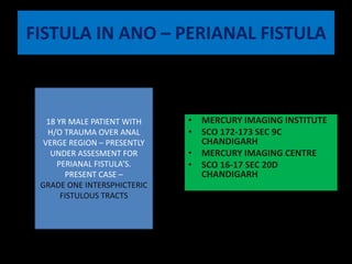
- 1. FISTULA IN ANO – PERIANAL FISTULA 18 YR MALE PATIENT WITH H/O TRAUMA OVER ANAL VERGE REGION – PRESENTLY UNDER ASSESMENT FOR PERIANAL FISTULA’S. PRESENT CASE – GRADE ONE INTERSPHICTERIC FISTULOUS TRACTS MERCURY IMAGING INSTITUTE SCO 172-173 SEC 9C CHANDIGARH MERCURY IMAGING CENTRE SCO 16-17 SEC 20D CHANDIGARH
- 2. Frederick Salmon STARTED 'The Infirmary for the Relief of the Poor afflicted with Fistula and other Diseases of the Rectum'. PERFORMED 3500 OPERATIONS WITH NOT EVEN SINGLE FATALITY. BENEVOLENT DISPENSARY TOST MARK’S HOSPITAL LONDON
- 3. ANATOMY LEVATOR SLING ISCHIO RECTAL FOSSA ISCHIOANAL FOSSA
- 4. IMPORTANT POINTS EXTERNAL ANAL SPHINCTER INTERNAL ANAL SPINCHTER Voluntary control Similar in signal to levator sling 15% of the anal tone. Straited muscle – continous with the puborectalis / levator muscle Involuntary Control Smooth Muscle Responsible For 85 % Of The Anal Tone. Continous With Circular Smooth Muscle Of The Rectum. Similar In Signal To Anal Musculature Relatively Hyperintense On Fat Sat Sequences.
- 5. IMPORTANT POINTS ANAL CLOCK TRANSVERSE ANAL LINE Define the cutaneous opening (Predicts the internal opening) LITHOTOMY POSITION DEFINE THE INTERNAL OPENING 12”o clock – Anterior perineum 3“O CLOCK - Left Lateral aspect 6 ‘ o clock - Natal cleft 9 ‘ o clock – Rt side of the perineum
- 6. CUTANEOUS OPENING Rt side – Posterior to the anal canal Lt side – Posterior to the anal canal
- 7. TRACT Rt side – linear longitudinal tract with no secondary ramifications –present in the intersphincteric plane Left side – linear longitudinal with no seconday ramifications- present in the intersphincteric plane
- 8. INTERNAL OPENING 6”0clock – Rt side of the anal canal 3’0clock – left lateral aspect.
- 9. 3D
- 10. BRIEF ABOUT PERIANAL FISTULA’S ETIOLOGY Crptoglandular hypothesis Crohn disease Tuberculosis Trauma during childbirth, Pelvic infection Pelvic malignancy Radiation therapy PREOPERATIVE EVALUATION OF PERIANAL FISTULAS To define relation of the fistulous tract with sphincter complex ( intersphicteric , trans-sphincteric, suprasphicteric ). Define Secondary tracts/ Ramifications. Define horseshoe tracts.
- 11. St James's University Hospital ClassificationMR Imaging Grading of Perianal Fistulas Grade 1: Simple Linear Intersphincteric Fistula.— Grade 2: Intersphincteric Fistula with Abscess or Secondary Track.— No secondary tract / ramifications Secondary tract / ramifications present but pathology is confined by the external sphincteric plane
- 12. St James's University Hospital ClassificationMR Imaging Grading of Perianal Fistulas Grade 3: Trans-sphincteric Fistula. Grade 4: Trans-sphincteric Fistula with Abscess or Secondary Track within the IschiorectalFossa. Trans-spincteric fistula with absecss / secondary infection in the ischiorectalfossa Transsphincteric extension with tract extending through the ischirectal / ischio anal region.
- 13. St James's University Hospital ClassificationMR Imaging Grading of Perianal Fistulas Grade 5: Supralevator and Translevator Disease. Suprasphincteric ---- high extension through intersphincteric plane Extrasphincteric : Outside the sphincteric mechanism Translevator / supralevator.
- 14. CONTRST MR – EXTRA EDGE DELINEATES THE TRACTS , INFECTIVE FOCI IN A BETTER WAY.