61. www.freelivedoctor.com Often small platelet aggregates or thrombi and/or thromboemboli Frequent Usually severe Sudden death Widely variable, may be absent, partial/complete, or lysed Variable Variable Subendocardial myocardial infarction Occlusive Frequent Variable Transmural myocardial infarction Nonocclusive, often with thromboemboli Frequent Variable Unstable angina No No >75% Stable angina Plaque-Associated Thrombus Plaque Disruption Stenoses Syndrome Coronary Artery Pathology in Ischemic Heart Disease
62.
63.
64. MYOCARDIAL RESPONSE www.freelivedoctor.com >1 hr Microvascular injury 20–40 min Irreversible cell injury 40 min to 10% of normal 10 min to 50% of normal ATP reduced <2 min Loss of contractility Seconds Onset of ATP depletion Time Feature
86. AORTIC STENOSIS 2X gradient pressure LVH, ischemia Cardiac decompensation, angina, CHF 50% die in 5 years if angina present 50% die in 2 years if CHF present www.freelivedoctor.com
109. Arrhythmogenic Right Ventricular Cardiomyopathy (Arrhythmogenic Right Ventricular Dysplasia) This is an uncommon dilated cardiomyopathy predominantly RIGHT ventricle. So is NAXOS syndrome. www.freelivedoctor.com
110.
111.
112.
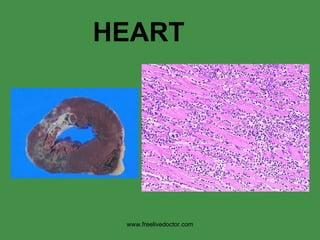
113. LYMPHOCYTIC INFILTRATES are the USUAL pattern of ALL myocarditis, but eosinophils, giant cells, and even trypanosomes can be seen occasionally www.freelivedoctor.com
This is a section from “Shotgun Histology”, in other words, the terms on the left describe the entire myocardium. Atrial natriuretic peptide (ANP), atrial natriuretic factor (ANF), atrial natriuretic hormone (ANH), or atriopeptin, is a powerful vasodilator, and a protein (polypeptide) hormone secreted by heart muscle cells. It is involved in the homeostatic control of body water, sodium, potassium and fat (adipose tissue). It is released by muscle cells in the upper chambers (atria) of the heart (atrial myocytes), in response to high blood pressure. ANP acts to reduce the water, sodium and adipose loads on the circulatory system, thereby reducing blood pressure.
The specialized myocytes of the heart’s conduction system, running sub-endocardially, have this unique appearance.
Whichever artery winds up supplying the posterior interventricular septum is said to be “DOMINANT”
The myocardial perfusion is a good test of coronary artery and myocardial function.
These features are seen so commonly in autopsies of elderly people no matter what they died from. Also keep in mind that most people who do not die ACUTELY, die in cardiac failure.
One very key philosophical question is whether atherosclerosis is part of aging or not. We can leave that for the philosophers.
The pigment which accumulates with age is called lipofucsin, and caused the heart to appear “browner” than normal. This is called “brown” atrophy of the heart.
This is the same analogy as the “straw” we talked about in the last chapter on blood vessels. You can classify cardiac diseases functionally into these 5 “pump” categories, like we had only 2 categories with the blood vessels described as straws or conduits.
Very FEW hearts of elderly people at autopsy weigh the normal 250-300 gm. Atherosclerotic or CHF hearts weigh twice as much, hypertensive hearts weight three times as much, and cardiomyopathic hearts often weigh more.
A good general diagram.
Note that not only is the FIBER thick, but so are the nuclei. Note squaring off of the nuclei, so called “BOXCAR” effect.
Can you understand why all of these findings can be related to LEFT sided heart failure? Ans: YES, primarily PULMONARY.
Can you understand why all of these findings can be related to RIGHT sided heart failure? Ans: YES, primarily STSTEMIC.
Does this look like it covers all bases? Ans: YES
Do the NAMES of these congenital heart conditions adequately describe the pathology? Ans: YES Why have I highlighted the “D”s and the “T”s? Ans: D = L shunt, T= R L shunt (cyanosis, or “blue” babies).
LEFT to RIGHT SHUNTS, NON-cyanotic
All the R L congenital shunts are CYANOTIC, and have T’s in their names.
CLASSICAL “TETROLOGY” of FALLOT: 1) VSD, large 2) OBSTRUCTION to RV flow 3) Aorta OVERRIDES the VSD 4) RVH
CHRONIC plaque PLUS acute thrombosis = Acute coronary syndromes.
Why does the necrosis spread from the endocardium to the pericardium (i.e., epicardium)?
A chamber wall is only as thick as it has to be, i.e., more pressure more thickness
Answer: owing to left atrial enlargement
Answer: lenticulostriate in basal ganglia most susceptible to hypertensive CVA
As the alveoli EXPAND in COPD, the arterioles NARROW!
A reasonably logical way of looking at COR PULMONALE, or RIGHT HEART FAILURE
Why do BOTH stenosis and regurgitation cause hypertrophy of the chamber proximal to the valve?
A GREAT classical sydenham chorea (St. Vitus “Dance”) can be seen at www.youtube.com/watch?v=RnxqqW_nH0k
![THE HEART ,[object Object],[object Object],[object Object],[object Object],[object Object],[object Object],[object Object],[object Object],[object Object],[object Object],[object Object],[object Object],www.freelivedoctor.com](data:image/gif;base64,R0lGODlhAQABAIAAAAAAAP///yH5BAEAAAAALAAAAAABAAEAAAIBRAA7)