SlideShare a Scribd company logo
1 of 11
Download to read offline
Section I – Musculoskeletal


                                                                                Figure 1B


                    Figure 1A


                                                                                Figure 1C


6.   You are shown an AP radiograph (Figure 1A), T1- (Figure 1B) and T2- (Figure 1C) weighted MR images of
     the left knee in a 14-year-old male with knee pain. Which one of the following is the MOST likely diagnosis?
     A. Osteosarcoma
     B.   Ewing’s sarcoma
     C. Giant cell tumor
     D. Chondroblastoma
     E.   Aneurysmal bone cyst

                                 Diagnostic In-Training Exam 2002                                               1
Section I – Musculoskeletal
Question 6
Findings: AP radiograph of the knee shows a lytic lesion located entirely within the epiphysis of a skeletally imma-
ture individual. Coronal T1 and T2 weighted images of the knee show a small, well-defined lesion within the
epiphysis with a low signal intensity margin on both sequences. The T1 weighted images show central decreased
signal and decreased signal in the surrounding marrow fat. T2 weighted images show mildly heterogeneous increased
T2 signal with increased T2 signal in the marrow fat. No aggressive features such as a soft tissue mass or aggressive
periostitis are seen.

Rationales:
A) Incorrect. Osteosarcoma is a common malignant tumor in this age group, and the most common location is
   at the knee. Osteosarcomas of all types are usually metaphyseal. 75% of osteosarcomas are of the conventional
   or classic type. Although most conventional osteosarcomas are lytic, they are typically more destructive in
   appearance and are usually associated with aggressive periostitis rather than the benign appearance shown here.
   Cross-sectional imaging shows best any associated soft tissue mass. MRI is useful in showing any skip lesions,
   seen in approximately 10% of cases. CT scan of the chest is part of the usual staging process, with pulmonary
   metastases seen in approximately 15% at presentation.
B) Incorrect. Ewing’s sarcoma is an aggressive malignancy of bone commonly seen in young children. It is one of
   the round blue cell tumors (like lymphoma and rhabdomyosarcoma) and is primarily a disease of the marrow.
   Ewing’s sarcoma is most commonly located in the diaphysis of a tubular bone. Radiographs show the permeative,
   moth-eaten appearance of the tumor and the malignant “star-burst” periostitis of Ewing’s. MRI best demon-
   strates the associated soft tissue mass and the extent of the tumor within the marrow. These tumors often are
   diagnosed late due to the constitutional symptoms (fever, malaise) which may be seen in the early phases of
   disease, mimicking infection.
C) Incorrect. Giant cell tumor is a tumor of bone which occurs in the epiphysis of long bones in skeletally mature
   individuals. It is most common at the knee. Giant cell tumor extends to an articular surface, and is classically
   completely lytic without mineralization within it or sclerosis around it. Although giant cell tumors may metasta-
   size to the lung, malignant giant cell tumor is extremely rare. These tumors are treated with curettage and
   exothermic methylmethacrylate. Giant cell tumors are rarely multicentric; when this occurs, they are often
   associated with Paget’s disease.
D) Correct. Chondroblastoma is a rare benign chondrogenic primary bone tumor seen in children before
   epiphyseal closure. Most occur around the knee or in the proximal femur. Radiographically, these epiphyseal
   lesions are eccentric and lucent, with well-defined sclerotic margins. Periosteal reaction is common. On MRI,
   chondroblastoma appears as well-defined areas of low to intermediate signal intensity on T1-weighted images
   and have intermediate or high signal intensity on T2-weighed images. A low-signal intensity rim, corresponding
   to a sclerotic margin, can also be seen.
E) Incorrect. Aneurysmal bone cyst (ABC) is a tumor-like lesion of bone consisting of eccentrically located in the
   metaphysis of a long bone. Pathologically the lesion has large blood filled spaces with a thin epithelial lining.
   ABC may be primary or secondary. blood filled spaces with loculation, and marked expansion of the bone.
   When secondary, they are most commonly seen in association with benign lesions such as chondroblastoma.
   MRI and CT will show the fluid-fluid levels with hemorrhage which are typical of these lesions. Soft tissue
   mass in association with ABC is extremely uncommon, and they rarely show any aggressive features

Citations:
Unni KK Dahlin’s Bone Tumors: General Aspects and Data on 11, 087 Cases (ed 5). Philadelphia PA, Lippincott- Ravin, 1996.
Turcotte RE et al. Chondroblastoma. Human Pathology 24:944-949. 1993.


2                                          American College of Radiology
Section I – Musculoskeletal


                                                 Figure 2A


                                                 Figure 2B

7.   This 22-year-old male presents with knee locking and pain after an injury while playing basketball. You are
     shown sagittal proton density (Figure 2A) and T2- (Figure 2B) weighted MR images of the knee. Which one
     of the following is the MOST likely diagnosis?
     A. Posterior cruciate ligament tear
     B.   Meniscal tear
     C. Bone bruise
     D. Patellar tendonopathy
     E.   Osteochondritis dissecans

                                 Diagnostic In-Training Exam 2002                                                  3
Section I – Musculoskeletal
Question # 7
Findings: Proton density and T2 weighted sagittal images of the knee at the level of the intercondylar notch show a
small (5mm) focus of decreased signal on both sequences just inferior and parallel to the posterior cruciate ligament.
This is the so-called “double PCL” sign, indicating a free mensical fragment. The PCL itself is normal in appearance,
as is the visualized bone marrow.

Rationales:
A) Incorrect. Tears of the posterior cruciate ligament are unusual. The PCL is an extremely strong structure, with
   twice the tensile strength of the anterior cruciate ligament. Injuries of the PCL represent only about 5-20% of
   knee injuries. The most common mechanism of injury of the PCL is a dashboard injury during a motor vehicle
   accident or contact sports such as football. Isolated injuries of the PCL are extremely rare, and there are usually
   associated injuries of the ACL, collateral ligaments or the mensici. Unlike the ACL, which consists of posterolat-
   eral and anteromedial bands, the PCL is a solid and highly organized band of collagen fibrils. On MRI, the PCL
   is solidly black on all sequences. Any increased signal within the PCL is abnormal and indicative of a partial tear.
   Rupture, when it occurs, is most common in its midportion (76%)
B) Correct. Bucket handle tears usually involve the medial meniscus in young athletes and present with locking of
   the knee. A bucket handle tear is a vertical tear with longitudinal extension from the posterior to the anterior
   horn. The inner fragment is unstable. Sagittal images show an abnormality in size of the anterior or posterior
   horns. An image through the intercondylar notch shows the displaced inner fragment of the tear paralleling the
   posterior cruciate ligament ().(the double cruciate sign). Coronal images reveal the peripheral the displaced
   fragment beneath the posterior cruciate ligament.
C) Incorrect. Bone contusions are common in knee injuries. Osteochondral impaction with “kissing” contusions
   involving the distal medial femoral condyle and lateral proximal tibial plateau are indicative of the forced valgus
   stress in external rotation which is typically the mechanism of injury in ACL tears. Bone contusions are felt to
   represent microfractures of bone trabeculae. T1 weighted MRI shows ill-defined areas of decreased signal,
   usually with no discrete fracture line. T2 weighted images, especially those with fat suppression, show diffuse
   increased T2 signal indicating edema.
D) Incorrect. Patellar tendinitis, or “”jumper’s knee”, is a chronic condition associated with repetitive trauma
   resulting in microtears in the substance of the tendon. MRI shows thickening of the patellar tendon with
   increased T2 signal. It is most commonly seen in athletes participating in sports such as volleyball where jump-
   ing and abrupt quadriceps contraction are frequent. The proximal portion of the tendon is most often involved,
   and patients have pain and tenderness at the patellar insertion of the tendon. The normal appearance of the
   highly ordered patellar tendon on MRI is decreased signal intensity on all sequences. The patellar tendon seen
   in this examination is entirely normal, with no evidence for either chronic or acute injury.
E) Incorrect. Osteochondritis dissecans (OCD) describes idiopathic osteonecrosis involving subchondral bone in
   characteristic locations, including the lateral and medial femoral condyles at the knee. The underlying process
   is avascular necrosis. The focus of osteonecrosis is typically small, but may become displaced in the joint leading
   to crepitus and pain. MRI is a sensitive examination for evaluation OCD. Early in the process, marrow edema
   may be the prominent finding. Later on, MRI can predict instability of the in situ fragment, and is well suited
   to locating loose fragments which may be radiographically difficult to see. The typical location at the knee is
   within the lateral aspect of the medial femoral condyle, but other locations, including the patella, may be seen.
   The disease commonly presents in childhood or adolescence and is more common in boys. The age and MR
   appearance in the test case are inconsistent with the diagnosis of OCD.

Citations:
Berquist TH (ed). MRI of the Musculoskeletal System (3rd edition). Philadelphis, Lippincott-Raven, 1996.
Thornton DD and Rubin DA. Magnetic Resonance Imaging of the Knee Menisci. Seminars in Roentgenology, 35:217-320, 2000.
Vande Berg BC et al. Lesions of Menisci of the Knee: Value of MRI for Recognition of Unstable Lesions. AJR 176: 771-776, 2001.


4                                           American College of Radiology
Section I – Musculoskeletal


                                                  Figure 3

8.   You are shown a lateral radiograph (Figure 3) of the left ankle of an 85-year-old woman with heel pain.
     Which one of the following is the MOST likely diagnosis?
     A. Paget’s disease
     B.   Reiter’s syndrome
     C. Insufficiency fracture
     D. Osteomyelitis
     E.   Pathologic fracture


                                  Diagnostic In-Training Exam 2002                                             5
Section I – Musculoskeletal
Question # 8
Findings: Lateral radiograph of the ankle demonstrates markedly decreased bone density. The cortices are distinct.
There is a curvilinear band of sclerosis in the posterior calcaneus.

Rationales:
A) Incorrect. Paget’s disease is an idiopathic disease of bone consisting of a process of osteolysis with marked repara-
   tive changes. It is seen in elderly individuals and is more common in the northern United States and Great
   Britain. There are three distinct radiographic phases of the disease: lytic, mixed and sclerotic. In the lytic phase,
   the most common appearance is of a long, lytic lesion with a leading edge within a long bone, the so-called
   “blade of grass” appearance. The mixed phase shows lytic and sclerotic changes and the sclerotic phase shows the
   classic trabecular and cortical thickening. Common locations include the skull, pelvis and spine. Complications
   of Paget’s disease include deformity, pseudofractures, congestive heart failure due to hypervascularity and rarely,
   sarcomatous degeneration. Paget’s disease may involve the calcaneus and the age of this patient is appropriate,
   but the appearance of the test case is not consistent with Paget’s disease.
B) Incorrect. Reiter’s syndrome is characterized by a classic triad of arthritis, urethritis, and conjunctivitis. There
   is a male predominance. Peak age at diagnosis is 15 to 35 years. Common sites of involvement are the small
   articulations of the foot, the calcaneus and the ankle, knee and sacroiliac joints. Calcaneal inflammation occurs
   along the plantar aspects and causes ill-defined erosions, often at the attachment of the plantar fascia. Hyperosto-
   sis and spur formation are later findings. The sex and age of the test patient and the location of the abnormality
   in the test images are not consistent with the diagnosis of Reiter’s syndrome.
C) Correct. Stress fractures result from repetitive muscular activity rather than direct trauma. Two basic types of
   stress fractures have been described: fatigue and insufficiency. Fatigue fractures occur in normal bone subjected
   to excessive stress. Insufficiency fractures result from normal or physiologic stress placed on bone that has less
   than normal elastic resistance, such as that seen in osteomalacia or osteoporosis. Senile osteoporosis is the loss
   of bone related to aging. The age of onset is the sixth or seventh decade for men and after menopause in women.
   Women are much more commonly affected until the ninth decade when the gender ratio equalizes. Trabecular
   bone is lost at twice the rate of cortical bone. Changes are generalized in the skeleton. Bone density may be
   measured by dual or single photon absorptiometry, quantitative computed tomography or by measuring the
   cortices of the second and third metacarpal cortices radiographically. Radiographs show markedly diminished
   bone density and also demonstrate the complications of osteoporosis, namely, insufficiency fractures. These are
   commonly located in the spine, ribs, hips and pelvis. Weightbearing bones such as the posterior calcaneus are
   also commonly affected, with the test case showing the classic features of an insufficiency fracture of the calca-
   neus.
D) Incorrect. Osteomyelitis may occur via hematogenous spread of a bacterial infection, spread from an adjacent
   source of infection, secondary to direct implantation or due to surgical manipulation or procedures. In adults
   and infants, the epiphysis of long bones may be involved due to vascular communication across the physis. In
   childhood, the physis acts as an effective barrier and most hematogenously spread infections occur in the meta-
   physis. Radiographs are insensitive in the early phases of infection. MRI and bone scan are both significantly
   more sensitive, and MRI also has increased specificity. Very early on, only soft tissue swelling and perhaps
   periostitis can be seen. As the disease progresses, radiographic changes including cortical destruction and erosions
   become more prominent. Advanced changes, including the formation of sequestra, involucra and sinus tracts
   may be seen. Soft tissue and intraosseous abcesses are best demonstrated on MRI. Chronic osteomyelitis may
   lead to squamous metaplasia, and even squamous cell carcinomas. The calcaneus could be involved with osteo-
   myelitis, either from direct penetration or extension from a heel ulcer. The test case, though, shows no periostitis
   or destructive changes to suggest osteomyelitis.
    Incorrect. Metastatic disease is the most common tumor of bone in patients over the age of 40. About 80% of
    metastatic disease to bone is from breast, prostate, kidney or lung primaries. Hematogenously spread, most
    metastatic lesions involve the marrow-rich axial and proximal appendicular skeleton. Metastases distal to the
    elbows and knees are very uncommon, and most are from breast or lung carcinoma. Rarely metastases to the


6                                        American College of Radiology
Section I – Musculoskeletal
    phalanges are seen with bronchogenic carcinoma. Pathologic fractures are common in the case of lytic metastases
    in weightbearing bones such as the proximal femur. A metastasis to the calcaneus would be extremely rare, and
    there is no underlying lesion on the radiograph to suggest that this is a pathologic fracture.

Citations:
Berquist TH. Radiology of the Foot and Ankle (ed 2). Philadelphia, PA. Lippincott Williams and Wilkins, 2000.


                                       Diagnostic In-Training Exam 2002                                          7
Section I – Musculoskeletal


                                                                              Figure 4B


                   Figure 4A


                                                                              Figure 4C

9.   You are shown an AP radiograph (Figure 4A), T1-(Figure 4B) and T2-(Figure 4C) weighted images of the
     knee of a 35-year-old woman with pain and a chronic knee effusion. There is no history of prior trauma.
     No other joints are involved. Which one of the following is the MOST likely diagnosis?
     A. Osteoarthritis
     B.   Gout
     C. Synovial chondromatosis
     D. Rheumatoid arthritis
     E.   Pigmented villonodular synovitis


8                                     American College of Radiology
Section I – Musculoskeletal
Question # 9
Findings: All of the presented images show large, well marginated erosions in the distal femur and proximal tibia.
There is a prominent synovial proliferation with decreased signal on both T1 and T2 weighting extending through-
out the spaces around the knee, including into the popliteus tendon sheath. T2 weighed image shows a moderately
large joint effusion.

Rationales:
A) Incorrect. Osteoarthritis is common in older patients, with the knee very commonly affected. Osteoarthritis has
   a mixed radiographic appearance, with hypertrophic changes including osteophyte formation and subchondral
   sclerosis, as well as subchondral cysts and even some erosive changes. The primary process in the development
   of osteoarthritis is cartilage degeneration, rather than a process involving the synovium as in the test case.
   Large joint effusions may be present, but they do not typically have any associated hemorrhage or hemosiderin
   deposition. Osteoarthritis is commonly a polyarticular disease. When a single joint is involved, it is usually due
   to antecedent trauma with damage to the joint surface and articular cartilage. This patient has not history of
   trauma, involvement of only a single joint and a relatively young age, making osteoarthritis unlikely.
B) Incorrect. The arthritis seen with gout is induced by intra-articular deposition of sodium monourate crystals.
   Aggregates of these crystals may form in the soft tissues adjacent to joints (tophi). Gout is twenty times more
   common in men than women, with age of onset usually after 40 years. Gout may be primary (related to enzy-
   matic deficiency) or secondary. Radiographic findings include well defined, periarticular erosions with sclerotic
   margins and overhanging edges. Chondrocalcinosis is a common feature, especially at the radiocarpal joint and
   the knee. The first metatarsophalangeal joint is most often affected (podagra). The radiographic findings are
   diagnostic, but uncommon in this era of early treatment of hyperuricemia.
C) Incorrect. Synovial chondromatosis is a metaplastic process of the articular cartilage resulting in nodules of
   cartilage which detach within the joint, forming loose bodies. They may or may not calcify. If calcified, the
   radiographic appearance is diagnostic showing multiple small, clacified loose bodies about a joint. The most
   commonly affected joints are the shoulder, elbow, hip and ankle. MRI shows the increased T2 and decreased
   T1 signal within the bodies that is indicative of their cartilaginous origin. MRI also shows best the associated
   large joint effusions. CT will demonstrate the small erosions on the joint surfaces which are commonly present.
   Synovial chondromatosis is usually monoarticular and is much more common in males. There is no association
   with hemorrhage or hemosiderin deposition such as is seen in the test case.
D) Incorrect. Rheumatoid arthritis frequently involves the knee. Soft tissue swelling is a common feature, and large
   popliteal cysts are a common complication. Uniform joint space loss is the most frequent finding, but marginal
   erosions may be seen peripherally, most prominent at the tibial aspect of the joint. Large erosions, such as seen
   in this case, are uncommon. Fibrinous loose bodies may form in the joint, so called “rice bodies” and large joint
   effusions and irregular synovium are commonly seen when these patients are examined with MRI. The signal
   characteristics of the synovium in rheumatoid arthritis—high T2 and low T1 signal intensity—are different
   from those seen in the test case.
E) Correct. Pigmented villonodular synovitis is a benign proliferative lesion of synovium that typically arises
   around the knee, and less frequently around other large joints. Pathologically, the lesion is characterized by
   the presence of hemosiderin-laden tissues. Plain radiographic findings include large well-defined periarticular
   erosions. MRI findings include: an intraarticular mass with a predominantly low signal intensity on T1-weighted
   and T2-weighted images, well-defined bone erosions on both sides of the joint, and a joint effusion. The
   deposition of hemosiderin produces marked signal loss on gradient echo images.

Citations:
Llauger J et al. Pigmented Villonodular Synovitis and Giant Cell Tumors of the Tendon Sheath: Radiologic and Pathologic Features.
AJR 172:1087-1091, 1999.
Lin J, Jacobson JA, Jamadar DA, Ellis JH. Pigmented Villonodular Synovitis and Related Lesions: the Spectrum of Imaging Findings.
AJR 172:191-198, 1999.


                                       Diagnostic In-Training Exam 2002                                                             9
Section I – Musculoskeletal


                                                                                Figure 5B


                   Figure 5A


                                                                                Figure 5C

10.   You are shown lateral views of the cervical spine in neutral (Figure 5A), flexion (Figure 5B) and extension
      (Figure 5C) in a 45-year-old woman with neck pain and headache. Which one of the following is the MOST
      likely diagnosis?
      A. Jacoud’s arthropathy
      B.   Rheumatoid arthritis
      C. Sarcoidosis
      D. Osteoarthritis
      E.   Ochronosis

10                                     American College of Radiology
Section I – Musculoskeletal
Question # 10
Findings: Lateral views of the cervical spine in neutral, flexion and extension show widening of the atlanto-axial
interval in neutral and flexion with reduction in extension, consistent with atlanto-axial subluxation.

Rationales:
A) Incorrect. Jacoud’s arthropathy is an infrequent sequela of rheumatic fever. Acute rheumatic fever is often associ-
   ated with migratory myalgias and arthralgias, but Jacoud’s arthropathy is the more permanent deformity result-
   ing from multiple episodes of recurrent rheumatic fever with joint involvement. The hands are invariably
   involved. Radiographic findings include easily reducible subluxations and dislocations, as well as ulnar deviation
   and swan’s neck and boutonniere deformities. Patients may be misdiagnosed with rheumatoid arthritis, but
   erosions are rarely present and the joint spaces are maintained. Jacoud’s arthropathy radiographically resembles
   systemic lupus erythematosus. Both are characterized by deformity without destruction. Changes in the feet are
   less commonly present, but similar to those in the hand. Jacoud’s arthropathy is not associated with atlanto-axial
   subluxation.
B) Correct. The most common disease associated with atlanto-axial subluxation is rheumatoid arthritis. Rheuma-
   toid arthritis is a destructive arthritis with extensive erosive changes due to inflammatory changes in the
   synovium. All synovial joints can be affected, but the disease most commonly involves the small joint of the
   hands and feet. The cervical spine is also almost invariably affected. Atlanto-axial subluxation is common. The
   atlanto-axial interval is a true synovial joint, with synovium located at anterior tubercle of C1 and the anterior
   portion of the odontoid, as well as lining the transverse ligament. The transverse ligament normally acts as a
   check to posterior translation of the odontoid. With erosion or even destruction of the transverse ligament, as
   well as erosion of the tip of the odontoid, the odontoid is free to move posteriorly which results in the radio-
   graphically observable movement and in possible impingement on the cervical spinal cord. The normal atlanto-
   axial interval should be no great than 2.2 mm in adults with no change between neutral, flexion and extension.
C) Incorrect. Sarcoidosis is a disease of unknown etiology characterized by the formation of non-caseating granulo-
   mas which may involve many organ systems simultaneously. The lung is the most common site of involvement,
   but there are skeletal manifestations in up to 15% of patients. The skeletal involvement is usually peripheral,
   with the hands, wrists and feet being the most common sites. Radiographically, there are large, well-circum-
   scribed lesions commonly involving the phalanges which have been described as “lacelike”. The distribution
   is usually bilateral but asymmetric. The spine is almost never involved and there is no association with atlanto-
   axial subluxation.
D) Incorrect. Osteoarthritis (OA) is common in elderly patients, and the spine is a common site of involvement.
   The facet joints are the target site for OA in the cervical spine, resulting in the radiographic appearance of
   spurring and sclerosis. The uncovertebral joints are also commonly involved. The combined hypertrophic
   changes of the facets and the uncovertebral joints may result in encroachment on the neural foramina leading
   to radiculopathy. The degeneration of the facet joints may result in low grade subluxations in the mid and lower
   cervical spine, but the atlanto-axial joint is not generally involved.
E) Incorrect. Ochronosis is a rare metabolic disease resulting from a deficiency of homogentisic acid oxidase,
   which leads to an accumulation of homogentisic acid in cartilage and soft tissue. This process degrades cartilage,
   leading to joint degeneration. Gross examination shows brown pigmentation of tissues. Radiographically,
   chondrocalcinosis is common as is calcification of the intervertebral discs. Ligamentous calcification may also
   be seen. The spine findings begin in the lumbar spine, ascending to the mid and upper thoracic spine. The
   cervical spine is not generally involved. Atlanto-axial subluxation is not a feature of ochronosis.

Citations:
Neva MH, Kaarela K, Kauppi M. Prevalence of radiological changes in the cervical spine - a cross sectional study after 20 years from
presentation of rheumatoid arthritis. Journal of Rheumatology 27(1):90-3, 2000.


1
                                        Diagnostic In-Training Exam 2002                                                          11

More Related Content

What's hot

Presentation1.pptx, radiological imaging of metabolic bone diseases.
Presentation1.pptx, radiological imaging of metabolic bone diseases.Presentation1.pptx, radiological imaging of metabolic bone diseases.
Presentation1.pptx, radiological imaging of metabolic bone diseases.Abdellah Nazeer
 
Imaging in arthritis
Imaging in arthritisImaging in arthritis
Imaging in arthritisNavni Garg
 
Skeletal disorders of metabolic origin
Skeletal disorders of metabolic originSkeletal disorders of metabolic origin
Skeletal disorders of metabolic originNavni Garg
 
Presentation1.pptx. imaging of the cartilage.
Presentation1.pptx. imaging of the cartilage.Presentation1.pptx. imaging of the cartilage.
Presentation1.pptx. imaging of the cartilage.Abdellah Nazeer
 
Pagets disease of bone
Pagets disease of bonePagets disease of bone
Pagets disease of bonePankaj Kaira
 
Presentation2.pptx bone tumour
Presentation2.pptx bone tumourPresentation2.pptx bone tumour
Presentation2.pptx bone tumourAbdellah Nazeer
 
Systemic approach to bone tumor radiology
Systemic approach to bone tumor radiologySystemic approach to bone tumor radiology
Systemic approach to bone tumor radiologyRamin Sadeghi
 
Aggressive & malignant bone tumours an overview
Aggressive & malignant bone tumours  an overviewAggressive & malignant bone tumours  an overview
Aggressive & malignant bone tumours an overviewvaruntandra
 
Bone tumors in the unusual locations
Bone tumors in the unusual locationsBone tumors in the unusual locations
Bone tumors in the unusual locationsRamin Sadeghi
 
Principles of management of malignant bone tumours
Principles of management of malignant bone tumoursPrinciples of management of malignant bone tumours
Principles of management of malignant bone tumoursSoliudeen Arojuraye
 
Dr.salah.radiology.bone and joints disease
Dr.salah.radiology.bone and joints diseaseDr.salah.radiology.bone and joints disease
Dr.salah.radiology.bone and joints diseasestudent
 
Hereditary multiple exostoses
Hereditary multiple exostosesHereditary multiple exostoses
Hereditary multiple exostoseszanamarques
 
Imaging findings of metabolic bone diseases
Imaging findings of metabolic bone diseases Imaging findings of metabolic bone diseases
Imaging findings of metabolic bone diseases Pankaj Kaira
 
Classification of bone tumors
Classification of bone tumorsClassification of bone tumors
Classification of bone tumorsDpt Memon
 

What's hot (20)

Presentation1.pptx, radiological imaging of metabolic bone diseases.
Presentation1.pptx, radiological imaging of metabolic bone diseases.Presentation1.pptx, radiological imaging of metabolic bone diseases.
Presentation1.pptx, radiological imaging of metabolic bone diseases.
 
Imaging in arthritis
Imaging in arthritisImaging in arthritis
Imaging in arthritis
 
Skeletal disorders of metabolic origin
Skeletal disorders of metabolic originSkeletal disorders of metabolic origin
Skeletal disorders of metabolic origin
 
Presentation1.pptx. imaging of the cartilage.
Presentation1.pptx. imaging of the cartilage.Presentation1.pptx. imaging of the cartilage.
Presentation1.pptx. imaging of the cartilage.
 
Pagets disease of bone
Pagets disease of bonePagets disease of bone
Pagets disease of bone
 
Presentation2.pptx bone tumour
Presentation2.pptx bone tumourPresentation2.pptx bone tumour
Presentation2.pptx bone tumour
 
Systemic approach to bone tumor radiology
Systemic approach to bone tumor radiologySystemic approach to bone tumor radiology
Systemic approach to bone tumor radiology
 
Bone Tumors Benign Ppt
Bone Tumors Benign PptBone Tumors Benign Ppt
Bone Tumors Benign Ppt
 
Aggressive & malignant bone tumours an overview
Aggressive & malignant bone tumours  an overviewAggressive & malignant bone tumours  an overview
Aggressive & malignant bone tumours an overview
 
Bone tumors in the unusual locations
Bone tumors in the unusual locationsBone tumors in the unusual locations
Bone tumors in the unusual locations
 
Principles of management of malignant bone tumours
Principles of management of malignant bone tumoursPrinciples of management of malignant bone tumours
Principles of management of malignant bone tumours
 
Radiology 5th year, 1st lecture (Dr. Salah Mohammad Fatih)
Radiology 5th year, 1st lecture (Dr. Salah Mohammad Fatih)Radiology 5th year, 1st lecture (Dr. Salah Mohammad Fatih)
Radiology 5th year, 1st lecture (Dr. Salah Mohammad Fatih)
 
Radiology 5th year, 3rd lecture (Dr. Salah Mohammad Fatih)
Radiology 5th year, 3rd lecture (Dr. Salah Mohammad Fatih)Radiology 5th year, 3rd lecture (Dr. Salah Mohammad Fatih)
Radiology 5th year, 3rd lecture (Dr. Salah Mohammad Fatih)
 
Dr.salah.radiology.bone and joints disease
Dr.salah.radiology.bone and joints diseaseDr.salah.radiology.bone and joints disease
Dr.salah.radiology.bone and joints disease
 
Hereditary multiple exostoses
Hereditary multiple exostosesHereditary multiple exostoses
Hereditary multiple exostoses
 
Bone tumors part 2
Bone tumors part 2Bone tumors part 2
Bone tumors part 2
 
Imaging in scurvy
Imaging in scurvyImaging in scurvy
Imaging in scurvy
 
Imaging findings of metabolic bone diseases
Imaging findings of metabolic bone diseases Imaging findings of metabolic bone diseases
Imaging findings of metabolic bone diseases
 
Classification of bone tumors
Classification of bone tumorsClassification of bone tumors
Classification of bone tumors
 
Introduction to skeletal imaging
Introduction to skeletal imagingIntroduction to skeletal imaging
Introduction to skeletal imaging
 

Viewers also liked

23205022
2320502223205022
23205022radgirl
 
23205065
2320506523205065
23205065radgirl
 
23204973
2320497323204973
23204973radgirl
 
23205001
2320500123205001
23205001radgirl
 
23204931
2320493123204931
23204931radgirl
 
23204922
2320492223204922
23204922radgirl
 
23204955
2320495523204955
23204955radgirl
 
Chapter 5 Slide Show 1
Chapter 5 Slide Show 1Chapter 5 Slide Show 1
Chapter 5 Slide Show 1marshalls1
 
Project Access Presentation final
Project Access Presentation finalProject Access Presentation final
Project Access Presentation finalandy biggin
 
Steve Morey Selected Work
Steve Morey Selected WorkSteve Morey Selected Work
Steve Morey Selected Workscmorey
 
Achieving Mastery Through Gamification
Achieving Mastery Through GamificationAchieving Mastery Through Gamification
Achieving Mastery Through GamificationPete Baikins
 
Chris Garrett Non Profit Presentation
Chris Garrett Non Profit PresentationChris Garrett Non Profit Presentation
Chris Garrett Non Profit PresentationGraham Richards
 
Application problems - Answers
Application problems - AnswersApplication problems - Answers
Application problems - AnswersCarlos Vázquez
 

Viewers also liked (20)

23205022
2320502223205022
23205022
 
23205065
2320506523205065
23205065
 
23204973
2320497323204973
23204973
 
23205001
2320500123205001
23205001
 
IncidentalomaTalk
IncidentalomaTalkIncidentalomaTalk
IncidentalomaTalk
 
Paisajes
PaisajesPaisajes
Paisajes
 
23204931
2320493123204931
23204931
 
A Present
A PresentA Present
A Present
 
Sistek Chandler Cue10
Sistek Chandler Cue10Sistek Chandler Cue10
Sistek Chandler Cue10
 
23204922
2320492223204922
23204922
 
23204955
2320495523204955
23204955
 
Chapter 5 Slide Show 1
Chapter 5 Slide Show 1Chapter 5 Slide Show 1
Chapter 5 Slide Show 1
 
Project Access Presentation final
Project Access Presentation finalProject Access Presentation final
Project Access Presentation final
 
Steve Morey Selected Work
Steve Morey Selected WorkSteve Morey Selected Work
Steve Morey Selected Work
 
E learn2016r ver_mike
E learn2016r ver_mikeE learn2016r ver_mike
E learn2016r ver_mike
 
Achieving Mastery Through Gamification
Achieving Mastery Through GamificationAchieving Mastery Through Gamification
Achieving Mastery Through Gamification
 
1.2 security 1
1.2 security 11.2 security 1
1.2 security 1
 
Micro Applicazioni Mobile
Micro Applicazioni MobileMicro Applicazioni Mobile
Micro Applicazioni Mobile
 
Chris Garrett Non Profit Presentation
Chris Garrett Non Profit PresentationChris Garrett Non Profit Presentation
Chris Garrett Non Profit Presentation
 
Application problems - Answers
Application problems - AnswersApplication problems - Answers
Application problems - Answers
 

Similar to 23204928

Muculoskeletal Pediatic Imaging..pptx
Muculoskeletal Pediatic Imaging..pptxMuculoskeletal Pediatic Imaging..pptx
Muculoskeletal Pediatic Imaging..pptxAbdellah Nazeer
 
Presentation1, radiological imaging of anterior knee pain.
Presentation1, radiological imaging of anterior knee pain.Presentation1, radiological imaging of anterior knee pain.
Presentation1, radiological imaging of anterior knee pain.Abdellah Nazeer
 
Presentation1.pptx, radiological imaging of spinal cord tumour.
Presentation1.pptx, radiological imaging of spinal cord tumour.Presentation1.pptx, radiological imaging of spinal cord tumour.
Presentation1.pptx, radiological imaging of spinal cord tumour.Abdellah Nazeer
 
joints Radiological evaluation pdf.pdf
joints Radiological evaluation pdf.pdfjoints Radiological evaluation pdf.pdf
joints Radiological evaluation pdf.pdfIhtPostPlanAll
 
23205008
2320500823205008
23205008radgirl
 
Simple and aneurysmal Bone cyst - Definition, Classfication, Investigations, ...
Simple and aneurysmal Bone cyst - Definition, Classfication, Investigations, ...Simple and aneurysmal Bone cyst - Definition, Classfication, Investigations, ...
Simple and aneurysmal Bone cyst - Definition, Classfication, Investigations, ...orthoslides
 
Transient Osteoporosis of Hip
Transient Osteoporosis of HipTransient Osteoporosis of Hip
Transient Osteoporosis of Hipvinod naneria
 
23204967
2320496723204967
23204967radgirl
 
Subchondral Events and Treatment
Subchondral Events and TreatmentSubchondral Events and Treatment
Subchondral Events and TreatmentVladimir Bobic
 
23205052
2320505223205052
23205052radgirl
 
bone tumors-1.pptx
bone tumors-1.pptxbone tumors-1.pptx
bone tumors-1.pptxkattabharath
 
Presentation1, artifacts and pitfalls of the wrist and elbow joints.
Presentation1, artifacts and pitfalls of the wrist and elbow joints.Presentation1, artifacts and pitfalls of the wrist and elbow joints.
Presentation1, artifacts and pitfalls of the wrist and elbow joints.Abdellah Nazeer
 
Bobic Subchondral Events - ICRS Wroclaw 091020
Bobic   Subchondral Events - ICRS Wroclaw 091020Bobic   Subchondral Events - ICRS Wroclaw 091020
Bobic Subchondral Events - ICRS Wroclaw 091020Vladimir Bobic
 
Orbital tumours
Orbital tumoursOrbital tumours
Orbital tumoursairwave12
 
Enchondroma with Secondary Aneurysmal Bone Cyst
Enchondroma with Secondary Aneurysmal Bone CystEnchondroma with Secondary Aneurysmal Bone Cyst
Enchondroma with Secondary Aneurysmal Bone CystI E
 
radiology.Spine 1.(dr.ameer)
radiology.Spine 1.(dr.ameer)radiology.Spine 1.(dr.ameer)
radiology.Spine 1.(dr.ameer)student
 
Foundations of Diagnostic Imaging for Physical Therapist
Foundations of Diagnostic Imaging for Physical TherapistFoundations of Diagnostic Imaging for Physical Therapist
Foundations of Diagnostic Imaging for Physical TherapistDana Tew
 

Similar to 23204928 (20)

Muculoskeletal Pediatic Imaging..pptx
Muculoskeletal Pediatic Imaging..pptxMuculoskeletal Pediatic Imaging..pptx
Muculoskeletal Pediatic Imaging..pptx
 
Presentation1, radiological imaging of anterior knee pain.
Presentation1, radiological imaging of anterior knee pain.Presentation1, radiological imaging of anterior knee pain.
Presentation1, radiological imaging of anterior knee pain.
 
Presentation1.pptx, radiological imaging of spinal cord tumour.
Presentation1.pptx, radiological imaging of spinal cord tumour.Presentation1.pptx, radiological imaging of spinal cord tumour.
Presentation1.pptx, radiological imaging of spinal cord tumour.
 
joints Radiological evaluation pdf.pdf
joints Radiological evaluation pdf.pdfjoints Radiological evaluation pdf.pdf
joints Radiological evaluation pdf.pdf
 
MRI in Tibial Fractures
MRI in Tibial FracturesMRI in Tibial Fractures
MRI in Tibial Fractures
 
23205008
2320500823205008
23205008
 
Simple and aneurysmal Bone cyst - Definition, Classfication, Investigations, ...
Simple and aneurysmal Bone cyst - Definition, Classfication, Investigations, ...Simple and aneurysmal Bone cyst - Definition, Classfication, Investigations, ...
Simple and aneurysmal Bone cyst - Definition, Classfication, Investigations, ...
 
Transient Osteoporosis of Hip
Transient Osteoporosis of HipTransient Osteoporosis of Hip
Transient Osteoporosis of Hip
 
23204967
2320496723204967
23204967
 
Subchondral Events and Treatment
Subchondral Events and TreatmentSubchondral Events and Treatment
Subchondral Events and Treatment
 
23205052
2320505223205052
23205052
 
Benign bone tumors
Benign bone tumorsBenign bone tumors
Benign bone tumors
 
International Journal of Orthopedics: Research & Therapy
International Journal of Orthopedics: Research & TherapyInternational Journal of Orthopedics: Research & Therapy
International Journal of Orthopedics: Research & Therapy
 
bone tumors-1.pptx
bone tumors-1.pptxbone tumors-1.pptx
bone tumors-1.pptx
 
Presentation1, artifacts and pitfalls of the wrist and elbow joints.
Presentation1, artifacts and pitfalls of the wrist and elbow joints.Presentation1, artifacts and pitfalls of the wrist and elbow joints.
Presentation1, artifacts and pitfalls of the wrist and elbow joints.
 
Bobic Subchondral Events - ICRS Wroclaw 091020
Bobic   Subchondral Events - ICRS Wroclaw 091020Bobic   Subchondral Events - ICRS Wroclaw 091020
Bobic Subchondral Events - ICRS Wroclaw 091020
 
Orbital tumours
Orbital tumoursOrbital tumours
Orbital tumours
 
Enchondroma with Secondary Aneurysmal Bone Cyst
Enchondroma with Secondary Aneurysmal Bone CystEnchondroma with Secondary Aneurysmal Bone Cyst
Enchondroma with Secondary Aneurysmal Bone Cyst
 
radiology.Spine 1.(dr.ameer)
radiology.Spine 1.(dr.ameer)radiology.Spine 1.(dr.ameer)
radiology.Spine 1.(dr.ameer)
 
Foundations of Diagnostic Imaging for Physical Therapist
Foundations of Diagnostic Imaging for Physical TherapistFoundations of Diagnostic Imaging for Physical Therapist
Foundations of Diagnostic Imaging for Physical Therapist
 

More from radgirl

23205062
2320506223205062
23205062radgirl
 
23205059
2320505923205059
23205059radgirl
 
23205056
2320505623205056
23205056radgirl
 
23205042
2320504223205042
23205042radgirl
 
23205045
2320504523205045
23205045radgirl
 
23205048
2320504823205048
23205048radgirl
 
23205039
2320503923205039
23205039radgirl
 
23205036
2320503623205036
23205036radgirl
 
23205032
2320503223205032
23205032radgirl
 
23205029
2320502923205029
23205029radgirl
 
23205025
2320502523205025
23205025radgirl
 
23205019
2320501923205019
23205019radgirl
 
23205016
2320501623205016
23205016radgirl
 
23205011
2320501123205011
23205011radgirl
 
23204998
2320499823204998
23204998radgirl
 
23205004
2320500423205004
23205004radgirl
 
23204995
2320499523204995
23204995radgirl
 
23204989
2320498923204989
23204989radgirl
 
23204992
2320499223204992
23204992radgirl
 
23204986
2320498623204986
23204986radgirl
 

More from radgirl (20)

23205062
2320506223205062
23205062
 
23205059
2320505923205059
23205059
 
23205056
2320505623205056
23205056
 
23205042
2320504223205042
23205042
 
23205045
2320504523205045
23205045
 
23205048
2320504823205048
23205048
 
23205039
2320503923205039
23205039
 
23205036
2320503623205036
23205036
 
23205032
2320503223205032
23205032
 
23205029
2320502923205029
23205029
 
23205025
2320502523205025
23205025
 
23205019
2320501923205019
23205019
 
23205016
2320501623205016
23205016
 
23205011
2320501123205011
23205011
 
23204998
2320499823204998
23204998
 
23205004
2320500423205004
23205004
 
23204995
2320499523204995
23204995
 
23204989
2320498923204989
23204989
 
23204992
2320499223204992
23204992
 
23204986
2320498623204986
23204986
 

Recently uploaded

See the 2,456 pharmacies on the National E-Pharmacy Platform
See the 2,456 pharmacies on the National E-Pharmacy PlatformSee the 2,456 pharmacies on the National E-Pharmacy Platform
See the 2,456 pharmacies on the National E-Pharmacy PlatformKweku Zurek
 
Call Girls Hebbal Just Call 7001305949 Top Class Call Girl Service Available
Call Girls Hebbal Just Call 7001305949 Top Class Call Girl Service AvailableCall Girls Hebbal Just Call 7001305949 Top Class Call Girl Service Available
Call Girls Hebbal Just Call 7001305949 Top Class Call Girl Service Availablenarwatsonia7
 
Call Girls Kanakapura Road Just Call 7001305949 Top Class Call Girl Service A...
Call Girls Kanakapura Road Just Call 7001305949 Top Class Call Girl Service A...Call Girls Kanakapura Road Just Call 7001305949 Top Class Call Girl Service A...
Call Girls Kanakapura Road Just Call 7001305949 Top Class Call Girl Service A...narwatsonia7
 
Low Rate Call Girls Pune Esha 9907093804 Short 1500 Night 6000 Best call girl...
Low Rate Call Girls Pune Esha 9907093804 Short 1500 Night 6000 Best call girl...Low Rate Call Girls Pune Esha 9907093804 Short 1500 Night 6000 Best call girl...
Low Rate Call Girls Pune Esha 9907093804 Short 1500 Night 6000 Best call girl...Miss joya
 
Book Call Girls in Yelahanka - For 7001305949 Cheap & Best with original Photos
Book Call Girls in Yelahanka - For 7001305949 Cheap & Best with original PhotosBook Call Girls in Yelahanka - For 7001305949 Cheap & Best with original Photos
Book Call Girls in Yelahanka - For 7001305949 Cheap & Best with original Photosnarwatsonia7
 
High Profile Call Girls Jaipur Vani 8445551418 Independent Escort Service Jaipur
High Profile Call Girls Jaipur Vani 8445551418 Independent Escort Service JaipurHigh Profile Call Girls Jaipur Vani 8445551418 Independent Escort Service Jaipur
High Profile Call Girls Jaipur Vani 8445551418 Independent Escort Service Jaipurparulsinha
 
Call Girls Service Noida Maya 9711199012 Independent Escort Service Noida
Call Girls Service Noida Maya 9711199012 Independent Escort Service NoidaCall Girls Service Noida Maya 9711199012 Independent Escort Service Noida
Call Girls Service Noida Maya 9711199012 Independent Escort Service NoidaPooja Gupta
 
Call Girls Electronic City Just Call 7001305949 Top Class Call Girl Service A...
Call Girls Electronic City Just Call 7001305949 Top Class Call Girl Service A...Call Girls Electronic City Just Call 7001305949 Top Class Call Girl Service A...
Call Girls Electronic City Just Call 7001305949 Top Class Call Girl Service A...narwatsonia7
 
Call Girls Jayanagar Just Call 7001305949 Top Class Call Girl Service Available
Call Girls Jayanagar Just Call 7001305949 Top Class Call Girl Service AvailableCall Girls Jayanagar Just Call 7001305949 Top Class Call Girl Service Available
Call Girls Jayanagar Just Call 7001305949 Top Class Call Girl Service Availablenarwatsonia7
 
College Call Girls Vyasarpadi Whatsapp 7001305949 Independent Escort Service
College Call Girls Vyasarpadi Whatsapp 7001305949 Independent Escort ServiceCollege Call Girls Vyasarpadi Whatsapp 7001305949 Independent Escort Service
College Call Girls Vyasarpadi Whatsapp 7001305949 Independent Escort ServiceNehru place Escorts
 
Call Girl Bangalore Nandini 7001305949 Independent Escort Service Bangalore
Call Girl Bangalore Nandini 7001305949 Independent Escort Service BangaloreCall Girl Bangalore Nandini 7001305949 Independent Escort Service Bangalore
Call Girl Bangalore Nandini 7001305949 Independent Escort Service Bangalorenarwatsonia7
 
Call Girls Whitefield Just Call 7001305949 Top Class Call Girl Service Available
Call Girls Whitefield Just Call 7001305949 Top Class Call Girl Service AvailableCall Girls Whitefield Just Call 7001305949 Top Class Call Girl Service Available
Call Girls Whitefield Just Call 7001305949 Top Class Call Girl Service Availablenarwatsonia7
 
Russian Call Girls in Pune Riya 9907093804 Short 1500 Night 6000 Best call gi...
Russian Call Girls in Pune Riya 9907093804 Short 1500 Night 6000 Best call gi...Russian Call Girls in Pune Riya 9907093804 Short 1500 Night 6000 Best call gi...
Russian Call Girls in Pune Riya 9907093804 Short 1500 Night 6000 Best call gi...Miss joya
 
College Call Girls Pune Mira 9907093804 Short 1500 Night 6000 Best call girls...
College Call Girls Pune Mira 9907093804 Short 1500 Night 6000 Best call girls...College Call Girls Pune Mira 9907093804 Short 1500 Night 6000 Best call girls...
College Call Girls Pune Mira 9907093804 Short 1500 Night 6000 Best call girls...Miss joya
 
Russian Call Girl Brookfield - 7001305949 Escorts Service 50% Off with Cash O...
Russian Call Girl Brookfield - 7001305949 Escorts Service 50% Off with Cash O...Russian Call Girl Brookfield - 7001305949 Escorts Service 50% Off with Cash O...
Russian Call Girl Brookfield - 7001305949 Escorts Service 50% Off with Cash O...narwatsonia7
 
Call Girls Frazer Town Just Call 7001305949 Top Class Call Girl Service Avail...
Call Girls Frazer Town Just Call 7001305949 Top Class Call Girl Service Avail...Call Girls Frazer Town Just Call 7001305949 Top Class Call Girl Service Avail...
Call Girls Frazer Town Just Call 7001305949 Top Class Call Girl Service Avail...narwatsonia7
 
Call Girls Hosur Just Call 7001305949 Top Class Call Girl Service Available
Call Girls Hosur Just Call 7001305949 Top Class Call Girl Service AvailableCall Girls Hosur Just Call 7001305949 Top Class Call Girl Service Available
Call Girls Hosur Just Call 7001305949 Top Class Call Girl Service Availablenarwatsonia7
 
Call Girl Service Bidadi - For 7001305949 Cheap & Best with original Photos
Call Girl Service Bidadi - For 7001305949 Cheap & Best with original PhotosCall Girl Service Bidadi - For 7001305949 Cheap & Best with original Photos
Call Girl Service Bidadi - For 7001305949 Cheap & Best with original Photosnarwatsonia7
 
Kolkata Call Girls Services 9907093804 @24x7 High Class Babes Here Call Now
Kolkata Call Girls Services 9907093804 @24x7 High Class Babes Here Call NowKolkata Call Girls Services 9907093804 @24x7 High Class Babes Here Call Now
Kolkata Call Girls Services 9907093804 @24x7 High Class Babes Here Call NowNehru place Escorts
 
Call Girls ITPL Just Call 7001305949 Top Class Call Girl Service Available
Call Girls ITPL Just Call 7001305949 Top Class Call Girl Service AvailableCall Girls ITPL Just Call 7001305949 Top Class Call Girl Service Available
Call Girls ITPL Just Call 7001305949 Top Class Call Girl Service Availablenarwatsonia7
 

Recently uploaded (20)

See the 2,456 pharmacies on the National E-Pharmacy Platform
See the 2,456 pharmacies on the National E-Pharmacy PlatformSee the 2,456 pharmacies on the National E-Pharmacy Platform
See the 2,456 pharmacies on the National E-Pharmacy Platform
 
Call Girls Hebbal Just Call 7001305949 Top Class Call Girl Service Available
Call Girls Hebbal Just Call 7001305949 Top Class Call Girl Service AvailableCall Girls Hebbal Just Call 7001305949 Top Class Call Girl Service Available
Call Girls Hebbal Just Call 7001305949 Top Class Call Girl Service Available
 
Call Girls Kanakapura Road Just Call 7001305949 Top Class Call Girl Service A...
Call Girls Kanakapura Road Just Call 7001305949 Top Class Call Girl Service A...Call Girls Kanakapura Road Just Call 7001305949 Top Class Call Girl Service A...
Call Girls Kanakapura Road Just Call 7001305949 Top Class Call Girl Service A...
 
Low Rate Call Girls Pune Esha 9907093804 Short 1500 Night 6000 Best call girl...
Low Rate Call Girls Pune Esha 9907093804 Short 1500 Night 6000 Best call girl...Low Rate Call Girls Pune Esha 9907093804 Short 1500 Night 6000 Best call girl...
Low Rate Call Girls Pune Esha 9907093804 Short 1500 Night 6000 Best call girl...
 
Book Call Girls in Yelahanka - For 7001305949 Cheap & Best with original Photos
Book Call Girls in Yelahanka - For 7001305949 Cheap & Best with original PhotosBook Call Girls in Yelahanka - For 7001305949 Cheap & Best with original Photos
Book Call Girls in Yelahanka - For 7001305949 Cheap & Best with original Photos
 
High Profile Call Girls Jaipur Vani 8445551418 Independent Escort Service Jaipur
High Profile Call Girls Jaipur Vani 8445551418 Independent Escort Service JaipurHigh Profile Call Girls Jaipur Vani 8445551418 Independent Escort Service Jaipur
High Profile Call Girls Jaipur Vani 8445551418 Independent Escort Service Jaipur
 
Call Girls Service Noida Maya 9711199012 Independent Escort Service Noida
Call Girls Service Noida Maya 9711199012 Independent Escort Service NoidaCall Girls Service Noida Maya 9711199012 Independent Escort Service Noida
Call Girls Service Noida Maya 9711199012 Independent Escort Service Noida
 
Call Girls Electronic City Just Call 7001305949 Top Class Call Girl Service A...
Call Girls Electronic City Just Call 7001305949 Top Class Call Girl Service A...Call Girls Electronic City Just Call 7001305949 Top Class Call Girl Service A...
Call Girls Electronic City Just Call 7001305949 Top Class Call Girl Service A...
 
Call Girls Jayanagar Just Call 7001305949 Top Class Call Girl Service Available
Call Girls Jayanagar Just Call 7001305949 Top Class Call Girl Service AvailableCall Girls Jayanagar Just Call 7001305949 Top Class Call Girl Service Available
Call Girls Jayanagar Just Call 7001305949 Top Class Call Girl Service Available
 
College Call Girls Vyasarpadi Whatsapp 7001305949 Independent Escort Service
College Call Girls Vyasarpadi Whatsapp 7001305949 Independent Escort ServiceCollege Call Girls Vyasarpadi Whatsapp 7001305949 Independent Escort Service
College Call Girls Vyasarpadi Whatsapp 7001305949 Independent Escort Service
 
Call Girl Bangalore Nandini 7001305949 Independent Escort Service Bangalore
Call Girl Bangalore Nandini 7001305949 Independent Escort Service BangaloreCall Girl Bangalore Nandini 7001305949 Independent Escort Service Bangalore
Call Girl Bangalore Nandini 7001305949 Independent Escort Service Bangalore
 
Call Girls Whitefield Just Call 7001305949 Top Class Call Girl Service Available
Call Girls Whitefield Just Call 7001305949 Top Class Call Girl Service AvailableCall Girls Whitefield Just Call 7001305949 Top Class Call Girl Service Available
Call Girls Whitefield Just Call 7001305949 Top Class Call Girl Service Available
 
Russian Call Girls in Pune Riya 9907093804 Short 1500 Night 6000 Best call gi...
Russian Call Girls in Pune Riya 9907093804 Short 1500 Night 6000 Best call gi...Russian Call Girls in Pune Riya 9907093804 Short 1500 Night 6000 Best call gi...
Russian Call Girls in Pune Riya 9907093804 Short 1500 Night 6000 Best call gi...
 
College Call Girls Pune Mira 9907093804 Short 1500 Night 6000 Best call girls...
College Call Girls Pune Mira 9907093804 Short 1500 Night 6000 Best call girls...College Call Girls Pune Mira 9907093804 Short 1500 Night 6000 Best call girls...
College Call Girls Pune Mira 9907093804 Short 1500 Night 6000 Best call girls...
 
Russian Call Girl Brookfield - 7001305949 Escorts Service 50% Off with Cash O...
Russian Call Girl Brookfield - 7001305949 Escorts Service 50% Off with Cash O...Russian Call Girl Brookfield - 7001305949 Escorts Service 50% Off with Cash O...
Russian Call Girl Brookfield - 7001305949 Escorts Service 50% Off with Cash O...
 
Call Girls Frazer Town Just Call 7001305949 Top Class Call Girl Service Avail...
Call Girls Frazer Town Just Call 7001305949 Top Class Call Girl Service Avail...Call Girls Frazer Town Just Call 7001305949 Top Class Call Girl Service Avail...
Call Girls Frazer Town Just Call 7001305949 Top Class Call Girl Service Avail...
 
Call Girls Hosur Just Call 7001305949 Top Class Call Girl Service Available
Call Girls Hosur Just Call 7001305949 Top Class Call Girl Service AvailableCall Girls Hosur Just Call 7001305949 Top Class Call Girl Service Available
Call Girls Hosur Just Call 7001305949 Top Class Call Girl Service Available
 
Call Girl Service Bidadi - For 7001305949 Cheap & Best with original Photos
Call Girl Service Bidadi - For 7001305949 Cheap & Best with original PhotosCall Girl Service Bidadi - For 7001305949 Cheap & Best with original Photos
Call Girl Service Bidadi - For 7001305949 Cheap & Best with original Photos
 
Kolkata Call Girls Services 9907093804 @24x7 High Class Babes Here Call Now
Kolkata Call Girls Services 9907093804 @24x7 High Class Babes Here Call NowKolkata Call Girls Services 9907093804 @24x7 High Class Babes Here Call Now
Kolkata Call Girls Services 9907093804 @24x7 High Class Babes Here Call Now
 
Call Girls ITPL Just Call 7001305949 Top Class Call Girl Service Available
Call Girls ITPL Just Call 7001305949 Top Class Call Girl Service AvailableCall Girls ITPL Just Call 7001305949 Top Class Call Girl Service Available
Call Girls ITPL Just Call 7001305949 Top Class Call Girl Service Available
 

23204928

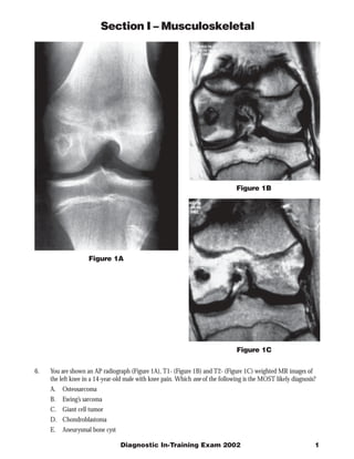
  • 1. Section I – Musculoskeletal Figure 1B Figure 1A Figure 1C 6. You are shown an AP radiograph (Figure 1A), T1- (Figure 1B) and T2- (Figure 1C) weighted MR images of the left knee in a 14-year-old male with knee pain. Which one of the following is the MOST likely diagnosis? A. Osteosarcoma B. Ewing’s sarcoma C. Giant cell tumor D. Chondroblastoma E. Aneurysmal bone cyst Diagnostic In-Training Exam 2002 1
  • 2. Section I – Musculoskeletal Question 6 Findings: AP radiograph of the knee shows a lytic lesion located entirely within the epiphysis of a skeletally imma- ture individual. Coronal T1 and T2 weighted images of the knee show a small, well-defined lesion within the epiphysis with a low signal intensity margin on both sequences. The T1 weighted images show central decreased signal and decreased signal in the surrounding marrow fat. T2 weighted images show mildly heterogeneous increased T2 signal with increased T2 signal in the marrow fat. No aggressive features such as a soft tissue mass or aggressive periostitis are seen. Rationales: A) Incorrect. Osteosarcoma is a common malignant tumor in this age group, and the most common location is at the knee. Osteosarcomas of all types are usually metaphyseal. 75% of osteosarcomas are of the conventional or classic type. Although most conventional osteosarcomas are lytic, they are typically more destructive in appearance and are usually associated with aggressive periostitis rather than the benign appearance shown here. Cross-sectional imaging shows best any associated soft tissue mass. MRI is useful in showing any skip lesions, seen in approximately 10% of cases. CT scan of the chest is part of the usual staging process, with pulmonary metastases seen in approximately 15% at presentation. B) Incorrect. Ewing’s sarcoma is an aggressive malignancy of bone commonly seen in young children. It is one of the round blue cell tumors (like lymphoma and rhabdomyosarcoma) and is primarily a disease of the marrow. Ewing’s sarcoma is most commonly located in the diaphysis of a tubular bone. Radiographs show the permeative, moth-eaten appearance of the tumor and the malignant “star-burst” periostitis of Ewing’s. MRI best demon- strates the associated soft tissue mass and the extent of the tumor within the marrow. These tumors often are diagnosed late due to the constitutional symptoms (fever, malaise) which may be seen in the early phases of disease, mimicking infection. C) Incorrect. Giant cell tumor is a tumor of bone which occurs in the epiphysis of long bones in skeletally mature individuals. It is most common at the knee. Giant cell tumor extends to an articular surface, and is classically completely lytic without mineralization within it or sclerosis around it. Although giant cell tumors may metasta- size to the lung, malignant giant cell tumor is extremely rare. These tumors are treated with curettage and exothermic methylmethacrylate. Giant cell tumors are rarely multicentric; when this occurs, they are often associated with Paget’s disease. D) Correct. Chondroblastoma is a rare benign chondrogenic primary bone tumor seen in children before epiphyseal closure. Most occur around the knee or in the proximal femur. Radiographically, these epiphyseal lesions are eccentric and lucent, with well-defined sclerotic margins. Periosteal reaction is common. On MRI, chondroblastoma appears as well-defined areas of low to intermediate signal intensity on T1-weighted images and have intermediate or high signal intensity on T2-weighed images. A low-signal intensity rim, corresponding to a sclerotic margin, can also be seen. E) Incorrect. Aneurysmal bone cyst (ABC) is a tumor-like lesion of bone consisting of eccentrically located in the metaphysis of a long bone. Pathologically the lesion has large blood filled spaces with a thin epithelial lining. ABC may be primary or secondary. blood filled spaces with loculation, and marked expansion of the bone. When secondary, they are most commonly seen in association with benign lesions such as chondroblastoma. MRI and CT will show the fluid-fluid levels with hemorrhage which are typical of these lesions. Soft tissue mass in association with ABC is extremely uncommon, and they rarely show any aggressive features Citations: Unni KK Dahlin’s Bone Tumors: General Aspects and Data on 11, 087 Cases (ed 5). Philadelphia PA, Lippincott- Ravin, 1996. Turcotte RE et al. Chondroblastoma. Human Pathology 24:944-949. 1993. 2 American College of Radiology
  • 3. Section I – Musculoskeletal Figure 2A Figure 2B 7. This 22-year-old male presents with knee locking and pain after an injury while playing basketball. You are shown sagittal proton density (Figure 2A) and T2- (Figure 2B) weighted MR images of the knee. Which one of the following is the MOST likely diagnosis? A. Posterior cruciate ligament tear B. Meniscal tear C. Bone bruise D. Patellar tendonopathy E. Osteochondritis dissecans Diagnostic In-Training Exam 2002 3
  • 4. Section I – Musculoskeletal Question # 7 Findings: Proton density and T2 weighted sagittal images of the knee at the level of the intercondylar notch show a small (5mm) focus of decreased signal on both sequences just inferior and parallel to the posterior cruciate ligament. This is the so-called “double PCL” sign, indicating a free mensical fragment. The PCL itself is normal in appearance, as is the visualized bone marrow. Rationales: A) Incorrect. Tears of the posterior cruciate ligament are unusual. The PCL is an extremely strong structure, with twice the tensile strength of the anterior cruciate ligament. Injuries of the PCL represent only about 5-20% of knee injuries. The most common mechanism of injury of the PCL is a dashboard injury during a motor vehicle accident or contact sports such as football. Isolated injuries of the PCL are extremely rare, and there are usually associated injuries of the ACL, collateral ligaments or the mensici. Unlike the ACL, which consists of posterolat- eral and anteromedial bands, the PCL is a solid and highly organized band of collagen fibrils. On MRI, the PCL is solidly black on all sequences. Any increased signal within the PCL is abnormal and indicative of a partial tear. Rupture, when it occurs, is most common in its midportion (76%) B) Correct. Bucket handle tears usually involve the medial meniscus in young athletes and present with locking of the knee. A bucket handle tear is a vertical tear with longitudinal extension from the posterior to the anterior horn. The inner fragment is unstable. Sagittal images show an abnormality in size of the anterior or posterior horns. An image through the intercondylar notch shows the displaced inner fragment of the tear paralleling the posterior cruciate ligament ().(the double cruciate sign). Coronal images reveal the peripheral the displaced fragment beneath the posterior cruciate ligament. C) Incorrect. Bone contusions are common in knee injuries. Osteochondral impaction with “kissing” contusions involving the distal medial femoral condyle and lateral proximal tibial plateau are indicative of the forced valgus stress in external rotation which is typically the mechanism of injury in ACL tears. Bone contusions are felt to represent microfractures of bone trabeculae. T1 weighted MRI shows ill-defined areas of decreased signal, usually with no discrete fracture line. T2 weighted images, especially those with fat suppression, show diffuse increased T2 signal indicating edema. D) Incorrect. Patellar tendinitis, or “”jumper’s knee”, is a chronic condition associated with repetitive trauma resulting in microtears in the substance of the tendon. MRI shows thickening of the patellar tendon with increased T2 signal. It is most commonly seen in athletes participating in sports such as volleyball where jump- ing and abrupt quadriceps contraction are frequent. The proximal portion of the tendon is most often involved, and patients have pain and tenderness at the patellar insertion of the tendon. The normal appearance of the highly ordered patellar tendon on MRI is decreased signal intensity on all sequences. The patellar tendon seen in this examination is entirely normal, with no evidence for either chronic or acute injury. E) Incorrect. Osteochondritis dissecans (OCD) describes idiopathic osteonecrosis involving subchondral bone in characteristic locations, including the lateral and medial femoral condyles at the knee. The underlying process is avascular necrosis. The focus of osteonecrosis is typically small, but may become displaced in the joint leading to crepitus and pain. MRI is a sensitive examination for evaluation OCD. Early in the process, marrow edema may be the prominent finding. Later on, MRI can predict instability of the in situ fragment, and is well suited to locating loose fragments which may be radiographically difficult to see. The typical location at the knee is within the lateral aspect of the medial femoral condyle, but other locations, including the patella, may be seen. The disease commonly presents in childhood or adolescence and is more common in boys. The age and MR appearance in the test case are inconsistent with the diagnosis of OCD. Citations: Berquist TH (ed). MRI of the Musculoskeletal System (3rd edition). Philadelphis, Lippincott-Raven, 1996. Thornton DD and Rubin DA. Magnetic Resonance Imaging of the Knee Menisci. Seminars in Roentgenology, 35:217-320, 2000. Vande Berg BC et al. Lesions of Menisci of the Knee: Value of MRI for Recognition of Unstable Lesions. AJR 176: 771-776, 2001. 4 American College of Radiology
  • 5. Section I – Musculoskeletal Figure 3 8. You are shown a lateral radiograph (Figure 3) of the left ankle of an 85-year-old woman with heel pain. Which one of the following is the MOST likely diagnosis? A. Paget’s disease B. Reiter’s syndrome C. Insufficiency fracture D. Osteomyelitis E. Pathologic fracture Diagnostic In-Training Exam 2002 5
  • 6. Section I – Musculoskeletal Question # 8 Findings: Lateral radiograph of the ankle demonstrates markedly decreased bone density. The cortices are distinct. There is a curvilinear band of sclerosis in the posterior calcaneus. Rationales: A) Incorrect. Paget’s disease is an idiopathic disease of bone consisting of a process of osteolysis with marked repara- tive changes. It is seen in elderly individuals and is more common in the northern United States and Great Britain. There are three distinct radiographic phases of the disease: lytic, mixed and sclerotic. In the lytic phase, the most common appearance is of a long, lytic lesion with a leading edge within a long bone, the so-called “blade of grass” appearance. The mixed phase shows lytic and sclerotic changes and the sclerotic phase shows the classic trabecular and cortical thickening. Common locations include the skull, pelvis and spine. Complications of Paget’s disease include deformity, pseudofractures, congestive heart failure due to hypervascularity and rarely, sarcomatous degeneration. Paget’s disease may involve the calcaneus and the age of this patient is appropriate, but the appearance of the test case is not consistent with Paget’s disease. B) Incorrect. Reiter’s syndrome is characterized by a classic triad of arthritis, urethritis, and conjunctivitis. There is a male predominance. Peak age at diagnosis is 15 to 35 years. Common sites of involvement are the small articulations of the foot, the calcaneus and the ankle, knee and sacroiliac joints. Calcaneal inflammation occurs along the plantar aspects and causes ill-defined erosions, often at the attachment of the plantar fascia. Hyperosto- sis and spur formation are later findings. The sex and age of the test patient and the location of the abnormality in the test images are not consistent with the diagnosis of Reiter’s syndrome. C) Correct. Stress fractures result from repetitive muscular activity rather than direct trauma. Two basic types of stress fractures have been described: fatigue and insufficiency. Fatigue fractures occur in normal bone subjected to excessive stress. Insufficiency fractures result from normal or physiologic stress placed on bone that has less than normal elastic resistance, such as that seen in osteomalacia or osteoporosis. Senile osteoporosis is the loss of bone related to aging. The age of onset is the sixth or seventh decade for men and after menopause in women. Women are much more commonly affected until the ninth decade when the gender ratio equalizes. Trabecular bone is lost at twice the rate of cortical bone. Changes are generalized in the skeleton. Bone density may be measured by dual or single photon absorptiometry, quantitative computed tomography or by measuring the cortices of the second and third metacarpal cortices radiographically. Radiographs show markedly diminished bone density and also demonstrate the complications of osteoporosis, namely, insufficiency fractures. These are commonly located in the spine, ribs, hips and pelvis. Weightbearing bones such as the posterior calcaneus are also commonly affected, with the test case showing the classic features of an insufficiency fracture of the calca- neus. D) Incorrect. Osteomyelitis may occur via hematogenous spread of a bacterial infection, spread from an adjacent source of infection, secondary to direct implantation or due to surgical manipulation or procedures. In adults and infants, the epiphysis of long bones may be involved due to vascular communication across the physis. In childhood, the physis acts as an effective barrier and most hematogenously spread infections occur in the meta- physis. Radiographs are insensitive in the early phases of infection. MRI and bone scan are both significantly more sensitive, and MRI also has increased specificity. Very early on, only soft tissue swelling and perhaps periostitis can be seen. As the disease progresses, radiographic changes including cortical destruction and erosions become more prominent. Advanced changes, including the formation of sequestra, involucra and sinus tracts may be seen. Soft tissue and intraosseous abcesses are best demonstrated on MRI. Chronic osteomyelitis may lead to squamous metaplasia, and even squamous cell carcinomas. The calcaneus could be involved with osteo- myelitis, either from direct penetration or extension from a heel ulcer. The test case, though, shows no periostitis or destructive changes to suggest osteomyelitis. Incorrect. Metastatic disease is the most common tumor of bone in patients over the age of 40. About 80% of metastatic disease to bone is from breast, prostate, kidney or lung primaries. Hematogenously spread, most metastatic lesions involve the marrow-rich axial and proximal appendicular skeleton. Metastases distal to the elbows and knees are very uncommon, and most are from breast or lung carcinoma. Rarely metastases to the 6 American College of Radiology
  • 7. Section I – Musculoskeletal phalanges are seen with bronchogenic carcinoma. Pathologic fractures are common in the case of lytic metastases in weightbearing bones such as the proximal femur. A metastasis to the calcaneus would be extremely rare, and there is no underlying lesion on the radiograph to suggest that this is a pathologic fracture. Citations: Berquist TH. Radiology of the Foot and Ankle (ed 2). Philadelphia, PA. Lippincott Williams and Wilkins, 2000. Diagnostic In-Training Exam 2002 7
  • 8. Section I – Musculoskeletal Figure 4B Figure 4A Figure 4C 9. You are shown an AP radiograph (Figure 4A), T1-(Figure 4B) and T2-(Figure 4C) weighted images of the knee of a 35-year-old woman with pain and a chronic knee effusion. There is no history of prior trauma. No other joints are involved. Which one of the following is the MOST likely diagnosis? A. Osteoarthritis B. Gout C. Synovial chondromatosis D. Rheumatoid arthritis E. Pigmented villonodular synovitis 8 American College of Radiology
  • 9. Section I – Musculoskeletal Question # 9 Findings: All of the presented images show large, well marginated erosions in the distal femur and proximal tibia. There is a prominent synovial proliferation with decreased signal on both T1 and T2 weighting extending through- out the spaces around the knee, including into the popliteus tendon sheath. T2 weighed image shows a moderately large joint effusion. Rationales: A) Incorrect. Osteoarthritis is common in older patients, with the knee very commonly affected. Osteoarthritis has a mixed radiographic appearance, with hypertrophic changes including osteophyte formation and subchondral sclerosis, as well as subchondral cysts and even some erosive changes. The primary process in the development of osteoarthritis is cartilage degeneration, rather than a process involving the synovium as in the test case. Large joint effusions may be present, but they do not typically have any associated hemorrhage or hemosiderin deposition. Osteoarthritis is commonly a polyarticular disease. When a single joint is involved, it is usually due to antecedent trauma with damage to the joint surface and articular cartilage. This patient has not history of trauma, involvement of only a single joint and a relatively young age, making osteoarthritis unlikely. B) Incorrect. The arthritis seen with gout is induced by intra-articular deposition of sodium monourate crystals. Aggregates of these crystals may form in the soft tissues adjacent to joints (tophi). Gout is twenty times more common in men than women, with age of onset usually after 40 years. Gout may be primary (related to enzy- matic deficiency) or secondary. Radiographic findings include well defined, periarticular erosions with sclerotic margins and overhanging edges. Chondrocalcinosis is a common feature, especially at the radiocarpal joint and the knee. The first metatarsophalangeal joint is most often affected (podagra). The radiographic findings are diagnostic, but uncommon in this era of early treatment of hyperuricemia. C) Incorrect. Synovial chondromatosis is a metaplastic process of the articular cartilage resulting in nodules of cartilage which detach within the joint, forming loose bodies. They may or may not calcify. If calcified, the radiographic appearance is diagnostic showing multiple small, clacified loose bodies about a joint. The most commonly affected joints are the shoulder, elbow, hip and ankle. MRI shows the increased T2 and decreased T1 signal within the bodies that is indicative of their cartilaginous origin. MRI also shows best the associated large joint effusions. CT will demonstrate the small erosions on the joint surfaces which are commonly present. Synovial chondromatosis is usually monoarticular and is much more common in males. There is no association with hemorrhage or hemosiderin deposition such as is seen in the test case. D) Incorrect. Rheumatoid arthritis frequently involves the knee. Soft tissue swelling is a common feature, and large popliteal cysts are a common complication. Uniform joint space loss is the most frequent finding, but marginal erosions may be seen peripherally, most prominent at the tibial aspect of the joint. Large erosions, such as seen in this case, are uncommon. Fibrinous loose bodies may form in the joint, so called “rice bodies” and large joint effusions and irregular synovium are commonly seen when these patients are examined with MRI. The signal characteristics of the synovium in rheumatoid arthritis—high T2 and low T1 signal intensity—are different from those seen in the test case. E) Correct. Pigmented villonodular synovitis is a benign proliferative lesion of synovium that typically arises around the knee, and less frequently around other large joints. Pathologically, the lesion is characterized by the presence of hemosiderin-laden tissues. Plain radiographic findings include large well-defined periarticular erosions. MRI findings include: an intraarticular mass with a predominantly low signal intensity on T1-weighted and T2-weighted images, well-defined bone erosions on both sides of the joint, and a joint effusion. The deposition of hemosiderin produces marked signal loss on gradient echo images. Citations: Llauger J et al. Pigmented Villonodular Synovitis and Giant Cell Tumors of the Tendon Sheath: Radiologic and Pathologic Features. AJR 172:1087-1091, 1999. Lin J, Jacobson JA, Jamadar DA, Ellis JH. Pigmented Villonodular Synovitis and Related Lesions: the Spectrum of Imaging Findings. AJR 172:191-198, 1999. Diagnostic In-Training Exam 2002 9
  • 10. Section I – Musculoskeletal Figure 5B Figure 5A Figure 5C 10. You are shown lateral views of the cervical spine in neutral (Figure 5A), flexion (Figure 5B) and extension (Figure 5C) in a 45-year-old woman with neck pain and headache. Which one of the following is the MOST likely diagnosis? A. Jacoud’s arthropathy B. Rheumatoid arthritis C. Sarcoidosis D. Osteoarthritis E. Ochronosis 10 American College of Radiology
  • 11. Section I – Musculoskeletal Question # 10 Findings: Lateral views of the cervical spine in neutral, flexion and extension show widening of the atlanto-axial interval in neutral and flexion with reduction in extension, consistent with atlanto-axial subluxation. Rationales: A) Incorrect. Jacoud’s arthropathy is an infrequent sequela of rheumatic fever. Acute rheumatic fever is often associ- ated with migratory myalgias and arthralgias, but Jacoud’s arthropathy is the more permanent deformity result- ing from multiple episodes of recurrent rheumatic fever with joint involvement. The hands are invariably involved. Radiographic findings include easily reducible subluxations and dislocations, as well as ulnar deviation and swan’s neck and boutonniere deformities. Patients may be misdiagnosed with rheumatoid arthritis, but erosions are rarely present and the joint spaces are maintained. Jacoud’s arthropathy radiographically resembles systemic lupus erythematosus. Both are characterized by deformity without destruction. Changes in the feet are less commonly present, but similar to those in the hand. Jacoud’s arthropathy is not associated with atlanto-axial subluxation. B) Correct. The most common disease associated with atlanto-axial subluxation is rheumatoid arthritis. Rheuma- toid arthritis is a destructive arthritis with extensive erosive changes due to inflammatory changes in the synovium. All synovial joints can be affected, but the disease most commonly involves the small joint of the hands and feet. The cervical spine is also almost invariably affected. Atlanto-axial subluxation is common. The atlanto-axial interval is a true synovial joint, with synovium located at anterior tubercle of C1 and the anterior portion of the odontoid, as well as lining the transverse ligament. The transverse ligament normally acts as a check to posterior translation of the odontoid. With erosion or even destruction of the transverse ligament, as well as erosion of the tip of the odontoid, the odontoid is free to move posteriorly which results in the radio- graphically observable movement and in possible impingement on the cervical spinal cord. The normal atlanto- axial interval should be no great than 2.2 mm in adults with no change between neutral, flexion and extension. C) Incorrect. Sarcoidosis is a disease of unknown etiology characterized by the formation of non-caseating granulo- mas which may involve many organ systems simultaneously. The lung is the most common site of involvement, but there are skeletal manifestations in up to 15% of patients. The skeletal involvement is usually peripheral, with the hands, wrists and feet being the most common sites. Radiographically, there are large, well-circum- scribed lesions commonly involving the phalanges which have been described as “lacelike”. The distribution is usually bilateral but asymmetric. The spine is almost never involved and there is no association with atlanto- axial subluxation. D) Incorrect. Osteoarthritis (OA) is common in elderly patients, and the spine is a common site of involvement. The facet joints are the target site for OA in the cervical spine, resulting in the radiographic appearance of spurring and sclerosis. The uncovertebral joints are also commonly involved. The combined hypertrophic changes of the facets and the uncovertebral joints may result in encroachment on the neural foramina leading to radiculopathy. The degeneration of the facet joints may result in low grade subluxations in the mid and lower cervical spine, but the atlanto-axial joint is not generally involved. E) Incorrect. Ochronosis is a rare metabolic disease resulting from a deficiency of homogentisic acid oxidase, which leads to an accumulation of homogentisic acid in cartilage and soft tissue. This process degrades cartilage, leading to joint degeneration. Gross examination shows brown pigmentation of tissues. Radiographically, chondrocalcinosis is common as is calcification of the intervertebral discs. Ligamentous calcification may also be seen. The spine findings begin in the lumbar spine, ascending to the mid and upper thoracic spine. The cervical spine is not generally involved. Atlanto-axial subluxation is not a feature of ochronosis. Citations: Neva MH, Kaarela K, Kauppi M. Prevalence of radiological changes in the cervical spine - a cross sectional study after 20 years from presentation of rheumatoid arthritis. Journal of Rheumatology 27(1):90-3, 2000. 1 Diagnostic In-Training Exam 2002 11