3. A mesenchymal
malignancy that
differentiates to
produce osteoid is an
osteosarcoma no
matter how much
osteoid is produced Osteoid
Deposition
4. Malignant tumor of
mesenchymal origin
having Spindle shaped
cells that produce osteoid
Second most common
primary malignancy of
bone
15% of all biopsied
primary bone tumors
5. In 1805, the French surgeon Alexis Boyer first
used the term "osteosarcoma’’
In 1847, the Baron Guillaume Dupuytren described
gross pathologic appearance of osteosarcoma
Jaffe and Lichtenstein established histologic
criteria to diagnose common bone tumors
6. In the 1970s, Dr Norman Jaffe used variety of
effective chemotherapeutic agents
Dr William F. Enneking introduced his surgical
staging system for musculoskeletal sarcomas.
7. In the United States, the incidence of
osteosarcoma is 400 cases per year (4.8 per
million population <20 y).
higher in blacks than in whites
higher in males than in females
The overall 5-year survival rate is 63%
8.
9. Sites:
Metaphysis: 90%
Diaphysis: 8-10%
- can occur in any bone
Long Bones: 70%-80%
Distal Femur (40%)
Proximal Tibia (20%)
Proximal Humerus
(15%)
Axial Skeleton
Pelvis
10. The exact cause of osteosarcoma is unknown
Risk factors –
Rapid bone growth
Environmental
Genetic
Pre-existing benign /malignant lesions
11. Rapid bone growth
- increased incidence during the
adolescent growth spurt
- location in the metaphyseal area adjacent to the
growth plate (physis) of long bones
Genetic
Li-Fraumeni syndrome (p53 mutation)
Rothmund-Thomson syndrome
Mutation of the RB gene (retinoblastoma)
12. Environmental
Radiation – localised radiation >2000 rads
latent period -4 years to 40 years
alpha rays>beta rays
Chemicals – methylcholanthrene
acetylaminofluorene
beryllium compounds
Virus - RNA :mouse sarcoma virus
DNA :polyoma ,SV 40 virus
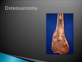
14. Gross appearance
Sclerosing or osteolytic
Typical features
metaphyseal location
variable consistency and
colour
subperiosteal spread
periosteum,epiphysis,
cartilage act as barrier
15. Gross appearance
Greyish white colour
Consistency varies
from
Soft and gritty to hard
Erodes inner cortex
Invades subperiosteal
space
Penetrate periosteum
and invades soft tissue
16. Microscopic features
Extensive irregular
sheets of new osteoid
Peripheral hypercellular
stroma with anaplastic
cells forming osteoid
Central cells entraped
by osseous tissue
become small and
rounded
17. Gross appearance
Very vascular tumour
Consistency soft and
friable
Necrotic and
hemorrhagic cavities
interspersed with
fibrous tissue
Pathological fracture
common
18. Microscopic features
Blood containing
spaces without
endothelial lining
Sparse osseous tissue
Spindle cells,
anaplastic osteoblast,
giant cells are seen
27. Very rare
High grade
confined to the cortex
Sites: Diaphysis of
femur or tibia
28. Synchronous Osteosarcoma:
Lesions that affect multiple bones
discovered within 6 months of each other
Metachronous Osteosarcoma:
Lesions involving multiple bones
discovered more than 6 months apart
29. Malignant transformation of benign condition
Paget disease
fibrous dysplasia
bone infarct
Arising in dedifferentiated chondrosarcoma
30. A mesenchymal malignancy that differentiates to
produce osteoid bone
Second most common primary malignant tumor of
bone
Site - metaphysis of long bone
Age – adolescent (15 – 25 years)
31. Etiology not known
Associated risk factors –
rapid bone growth in young
pre-existing bone lesion in old age
Most common - Conventional Osteosarcoma